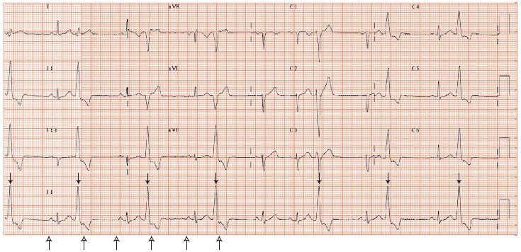
Fig. 47.2 Left bundle branch block (LBBB) pattern ventricular premature contractions (VPCs). Rhythm strip (bottom) shows some beats clearly sinus (P waves, illustrated by open arrows from below, followed by narrow complex QRS), others (large bold arrow above) occur early, starting before the previous sinus beats have finished, have no preceding P wave, are broad, and have inverted T waves. These are VPCs. They have a fixed relationship to the preceding sinus beat, i.e. are triggered by that beat. The VPCs have a LBBB pattern, i.e. they arise from the right ventricle, high up as the VPC axis is directed towards lead III – i.e. from the RV outflow tract, usually idiopathic, but if there is a family history of sudden death/cardiomyopathy, or history of syncope, consider arrhythmogenic right ventricular dysplasia, a rare serious condition. The P waves march through at a regular rate (small arrows below), appearing on the downslope of the VPC T wave as a small indentation. Ventricular premature contractions are followed by a full compensatory pause. The underlying ECG is normal.

Fig. 47.3 Twenty-four hour Holter monitor. Top heart rate. The patient falls asleep when the heart rate falls, between 10 and 11 PM, and wakes around 7 AM. During the day the heart rate is much higher than at night. The bottom trace shows ventricular premature contraction (VPC) frequency (right-hand scale); VPC rate is much higher at night, when the heart rate is low, than during the day. This is the common pattern, and reflects VPC dependency on long QT intervals.

Ventricular ectopic beats (also termed ventricular premature contractions [VPCs], ventricular premature beats [VPBs], ventricular extrasystoles [VEs]) are common and produce symptoms from:
Stay updated, free articles. Join our Telegram channel

Full access? Get Clinical Tree


