Fig. 56.2 (a) Mobitz type I (otherwise known as Wenckebach) second degree heart block. If one looks at the rhythm strip, one can see gradual prolongation of the PR interval over three cardiac cycles, then a nonconducted beat (i.e. one not transmitted to the ventricle). The sequence then repeats itself, though with two beats occurring before the P wave fails to get to the ventricle. (b) Type II Mobitz heart block. Every second P wave is transmitted to the ventricle (see arrows on the rhythm strip showing the P waves). The PR interval in the transmitted beats is normal. The underling ECG is fairly unremarkable aside from mild T wave inversion in the inferior leads – this raises the possibility that the heart block is the result of right coronary artery territory ischaemia.
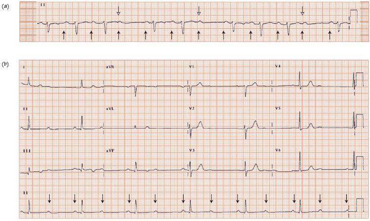
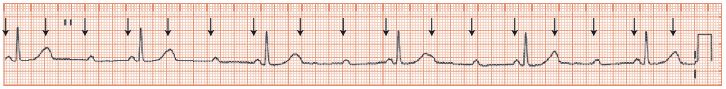
Fig. 56.3 Mobitz type II heart block showing 3 to 1 (3 : 1) block. The arrows on the rhythm strip show where the P waves are; the P wave rate is fairly unremarkable, but only every third P wave is transmitted to the ventricle. It is relatively easy to miss the ‘third’ P wave, hidden in the T wave. As the severity of the heart block is greater here than in the ECG in Fig. 56.2b, there is greater concern that this patient will go on to develop third degree heart block, despite the narrow QRS complex on the transmitted beats.
Stay updated, free articles. Join our Telegram channel

Full access? Get Clinical Tree


