Fig. 12.1
Graphical representation of auscultation findings in a patient with the Austin Flint murmur
Test Results
Color M-mode and 2-D echo (Fig. 12.2a–d).
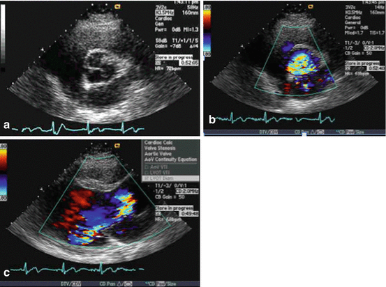
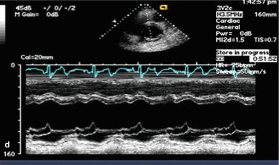
Fig. 12.2
(a–d) Echocardiograms. (a) An abnormal valve with deformed left coronary leaflet. (b) Severe aortic regurgitation on color Doppler. (c) The AR jet courses along the anterior mitral leaflet. (d) Fluttering and early closure of the mitral valve is shown on M-mode
Backflow evident in thoracic aorta, demonstrating severe regurgitation.
Anterior mitral valve leaflet (AMVL) flutters due to turbulent flow impinging on mitral valve in proto-diastole, mid-diastole and during atrial contraction [1].
Clinical Basics
Normal Anatomy
Normal anatomy of the aortic valve can be reviewed in chapters on aortic stenosis and aortic regurgitation.
Definitions
Though the Austin Flint murmur presents like a mitral murmur, it does not indicate the presence of any mitral valve lesions. Rather, AF accompanies severe aortic lesions coupled with considerable AR.
Etiology
The etiology behind AF is not completely understood. However, it is agreed that AF is NOT due to functional MS.
Some commonly accepted mechanisms for AF include:
Functional mitral stenosis during ventricular filling. This results from the regurgitant AR jet impinging on the AMVL and restricting atrial-ventricular flow [1].
The aortic valve stream contacts the mitral valve stream, with the AMVL vibrating between them. This causes turbulence in the apical area [1].
Turbulence caused by the apical irradiation of the regurgitant aortic jet could cause the murmur [1].
The regurgitant jet’s impact on the left ventricular imaging studies suggests a combined etiology including turbulent flow, mitral valve fluttering, and septal bulging [1].
The direction of AR flow seems unrelated to the presence of AF.
While AF is correlated with increased peak mitral inflow velocity there is no temporal relationship between AF and mitral flow (phonocardiography).
Presentation
Prevalence
One study found that out of 51 patients with AR and a normal MV, 30 exhibited findings suggestive of the AF murmur.< div class='tao-gold-member'>Only gold members can continue reading. Log In or Register to continue
Stay updated, free articles. Join our Telegram channel

Full access? Get Clinical Tree


