Fig. 11.1
Typical auscultatory findings associated with mitral valve stenosis include loud S1 and decrescendo-type diastolic rumble. Note that MS is best heard with the patient in the left lateral decubitus position (LLDP) after exercise. The LLDP bring the apex of the heart closer to the chest wall and exercising increases cardiac output and thus the volume of blood being forcibly pumped through the stenotic mitral valve
Loud S1 at the left sternal border.
Opening snap following S2 at the left lower sternal border and apex.
Decrescendo-type diastolic rumble, best heard when the patient expires.
Auscultation.
Test Results
Echocardiography (Fig. 11.2a, b) shows mild right ventricular enlargement, no aortic stenosis. There is moderate MR with a right ventricular systolic pressure of 34 mmHg. The mitral valve gradient was 4 mmHg, and the mitral valve area was 1.5 cm2.
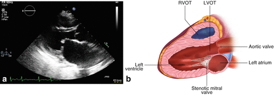
Fig. 11.2
(a) Echocardiogram (parasternal long axis view) showing the mitral valve with hallmarks of mitral stenosis including thickening of the valve and tethering of the anterior mitral valve leaflet. (b) Artist rendering of a parasternal long axis view of the myocardium on echocardiogram. Note the thickened calcified valves characteristic of mitral valve stenosis. Symptoms typically occur once the MV area measures between 1.2 and 1.6 cm2
The Doppler echocardiography demonstrates an increased left atrioventricular pressure gradient (Fig. 11.3).
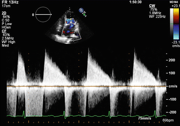
Fig. 11.3
The thickened valves MS causes seen on 2D echocardiography lead to persistently increased left atrioventricular pressure which is seen in the continuous wave Doppler profile showing high diastolic velocity with a reduced rate of decay (i.e., the velocity remains high throughout diastole). The left atrial pressure, for which the A2-opening snap interval is a surrogate, is an important prognostic indicator of the severity of the disease
Clinical Basics
Normal Anatomy Valve
Bileaflet valve with the aortic leaflet in fibrous continuity with the noncoronary cusp of the aortic valve.
Supported by two papillary muscle groups: anterolateral and posteromedial.
There is considerable variation in the morphology of the papillary muscles.
Definition
Occurs from left ventricular inflow obstruction due to thickening and immobilization of the mitral valve leaflets.
Results in increased left atrial pressure, pulmonary HTN, right ventricle enlargement.
Left ventricle is unaffected in isolated MS.
Etiology (Fig. 11.4a, b)
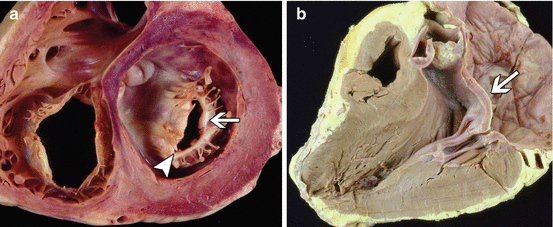
Fig. 11.4
(a) The heart of a 44 year old woman with rheumatic mitral stenosis. Fibrous thickening of the mitral leaflets and fusion of the commissures create a fish mouth appearance (arrowhead). (b) A three chamber view of a 55 year old woman’s heart with rheumatic mitral stenosis. The mitral valve leaflets and chordae show significant thickening. The arrow depicts the bowing of the anterior leaflet (With permission of Dr. William D. Edwards, Department of Pathology, Mayo Clinic, Rochester, MN.)
The predominant cause of MS is rheumatic fever.
Rare complication of.
Malignant carcinoid disease, SLE, RA, Fabry disease, and Whipple disease.
25 % of all patients with rheumatic heart disease have isolated MS.
40 % have combined MS and MR.
Multivalve involvement occurs in 38 % of MS patients.
Aortic valve affected in about 35 %.
Tricuspid valve in about 6 %.
Pulmonic valve is rarely affected.
Rheumatic fever results in characteristic changes of the mitral valve.
Thickening at the leaflet edges.
Fusion of the commissures.
Chordal shortening and fusion.
“Fishmouth” appearance.
Congenital MS is uncommon and typically is diagnosed in infancy or early childhood.
Estimated risk of endocarditis in patients with MS is 0.17/1000 patient-years.
Signs and Symptoms
Common symptoms of mitral stenosis include:
Exertional dyspnea.
Fatigue.
Orthopnea.
Paroxysmal nocturnal dyspnea.
Pulmonary edema or right-sided heart failure.
Complications occurring with increased age include:
Atrial fibrillation (increasing probability).
Heavy calcification, fibrosis of valve leaflets.
Subvalvular fusion.
Prevalence
United States and Western Europe have low incidence due to low prevalence of rheumatic fever.
Lack of primary and secondary prevention methods in developing countries elevates statistics of MS development and its severity.
2/3 of all patients with rheumatic MS are female.
Key Auscultation Findings (Fig. 11.5)
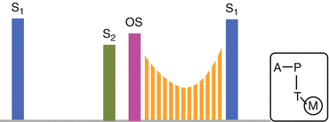
Fig. 11.5
Graphic depiction of the auscultation features of mitral stenosis. Notice the decrescendo-crescendo diastolic murmur with a preceding opening snap (Source: Based on a figure in http://morningreporttgh.blogspot.com/2010/03/mitral-stenosis.html)
Low pitched decrescendo-crescendo diastolic murmur.
Heard best at the apex with patient in left lateral decubitus position.
Loud, accentuated S1.
Opening snap heard after S2.
Short A2-OS interval indicates worse prognosis.
Loudness of murmur does not correlate with severity of MS.
Length of murmur correlates with severity of MS.
Diastolic rumbling murmur at apex due to low pitch.
Best heard with patient in left lateral decubitus position after limited exercise.
Listen with the bell at apex.
May radiate to axilla or LLSB.
Low frequency caused by large volume of blood going through MV under low levels of pressure, leading to turbulence after OS.
Will feature a presystolic crescendo to the loud M1, usually at a higher frequency than the diastolic rumble.
Mild exercise will increase cardiac output, and may help to enhance detection of the murmur.
Increased volume flow through a stenosed valve will cause more turbulence.
There is no murmur in the systolic phase.
Loud S1 from stenotic leaflet closing.
Loud S1 is not specific for MS; may be palpated.
Calcification or thickening reduces the intensity of S1.
Prolonged Q-S1 is indicative and represents increased LA pressure.
S2/P2 loud with pulmonary HTN.
Heard in both mitral and aortic areas.
Heard best at cardiac base.
Evolves to single S2 in cases of pulmonary HTN.
Opening snap following S2.
OS high-pitched and literally snapping; do not confuse with P2.
Between LLSB and apex.
Most audible in expiration, at or just medial to apex, with diaphragm of stethoscope.
Due to sudden deceleration/tensing of opening mitral leaflets; mobility necessary.
OS will disappear when stenosis immobilizes leaflets.
S2-OS will shorten as the severity of MS progresses.< div class='tao-gold-member'>Only gold members can continue reading. Log In or Register to continue

Stay updated, free articles. Join our Telegram channel

Full access? Get Clinical Tree


