Fig. 5.1
Representation of the patient’s auscultation exam. A systolic crescendo-decrescendo murmur grade 3 and diastolic decrescendo murmur grade 1 is present. S1 normal, however the aortic component of the S2 is diminished, with an intensity beneath that of P2
Text Results
Echo: Aortic Valve Area (AVA) 0.8 cm2, AV gradient 2.84 m/s (Fig. 5.2).
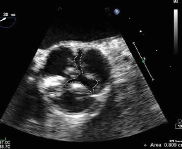
Fig. 5.2
Echocardiogram showing a parasternal short axis view of the aortic valve with an aortic valve area by planimetry of 0.8 cm2
ECG: LV hypertrophy, left atrial hypertrophy, T wave inversion and ST segment depression (in leads I, AVL, V5, V6) (Fig. 5.3).
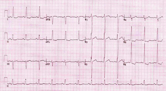
Fig. 5.3
ECG shows LV hypertrophy, ST depression
Clinical Basics
Normal Anatomy
The normal aortic valve has three leaflets, the right, left and noncoronary leaflet. The right coronary artery arises from the right cusp, and the left coronary artery arises from the left cusp.
The normal aortic valve has an area of 3–4 cm2.
Definition
Aortic stenosis is a pathologic narrowing of the aortic valve.
Causes
Causes of AS include congenital aortic stenosis, bicuspid calcific aortic stenosis, tricuspid aortic valve sclerosis and calcific stenosis.
Prevalence
The prevalence of AS increases with age:
0.02 % at age 18–44 years.
0.1 % at age 45–54 years.
0.2 % at age 55–64 years.
1.3 % at age 65–74 years.
2.8 % at age 75 years and older.
Pathophysiology
Stenosis of valve usually occurs gradually over time due to calcification.
The narrowing of the valve causes an increased pressure gradient across the valve leading to high velocity, turbulent ejection of blood.
The increased pressure gradient leads to decreased ejection fraction which subsequently causes hypertrophic changes of the heart, specifically, left ventricular hypertrophy which maintain ejection fraction.
The increased muscle mass of the heart puts it at an increased risk for left ventricular failure, reduced coronary blood flow per gram of muscle, and limited coronary vasodilator reserve.
Symptoms
Hallmark symptoms of aortic stenosis include:
Angina.
Syncope.
Heart failure.
Key Auscultation Features
Crescendo-decrescendo systolic ejection murmur with ejection click that is heard best at the base of the heart and may radiate to the carotids. The underlying physiologic cause is turbulent flow through a narrowed aortic valve.
Murmur intensity increases following a post-extrasystolic pause (post-extrasystolicaccentuation) and during the release phase of the Valsalva maneuver.
Loss of splitting of S2 (loss of A2) due to inflexible aortic leaflets closing inaudibly.
Presence of S4 due to vigorous atrial contraction in the face of a partially closed mitral valve during presystole.
Auscultation examples of aortic stenosis.
Click here to listen to an example of moderate aortic stenosis and see an image of the phonocardiogram (Video 5.1). The peak to peak gradient was 60 mmHg.
Click here to listen to an example of severe aortic stenosis as described by Dr. W. Proctor Harvey (Video 5.2).
Differential Diagnosis of Key Auscultation Features
A systolic murmur with ejection may arise from:
pulmonary valve stenosis.
atrial septal defect.
mitral regurgitation.
tricuspid regurgitation.
high cardiac output state.
hypertrophic obstructive cardiomyopathy.
High-pitched decrescendo diastolic murmur due to regurgitation through a compromised aortic valve.
Differential diagnosis for a diastolic murmur.
aortic regurgitation.
pulmonary regurgitation.
mitral valve stenosis.
tricuspid valve stenosis.
atrial septal defect.
Clinical Clues to the Detection of the Lesion
Other clinical findings to aid in the diagnosis of aortic stenosis are below:
Carotid pulsus parvus et tardus on palpation caused by reduced stroke volume and a prolonged ejection phase.
Echocardiography.
Reduced aortic valve area (AVA) indicative of a calcified aortic valve unable to open fully.
Increased velocity through the aortic valve due to elevated blood flow through a narrowed aortic valve.
Increased systolic pressure gradient between the left ventricle and the aorta (AV gradient) caused by left ventricular outflow obstruction in the face of a narrowed aortic valve.
ECG.
Left ventricular hypertrophy caused by the prolonged elevation of left ventricular contractile force required to eject blood through a stenotic aortic valve.
T wave inversion and ST segment depression (leads I, AVL, V5, V6) indicative of myocardial ischemia due to left ventricular dysfunction and decreased myocardial O2 supply.
Left atrial hypertrophy secondary to the elevated contractile force necessary to eject blood into an over-filled and hypertrophic left ventricle.
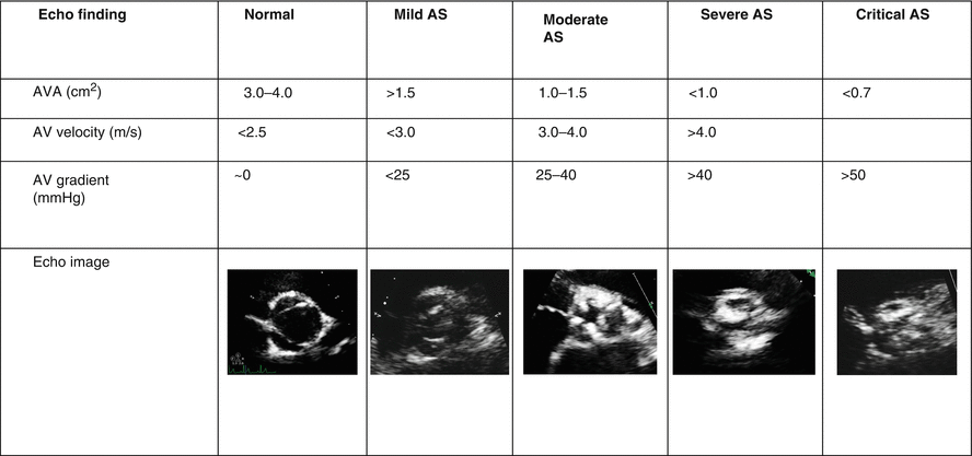
Classification of the Severity of Aortic Stenosis
Diagnostic Implications of the Auscultation Features
Auscultation findings aid in the diagnosis of the severity of AS.
Increased intensity of murmur indicative of a higher pressure gradient.< div class='tao-gold-member'>Only gold members can continue reading. Log In or Register to continue

Stay updated, free articles. Join our Telegram channel

Full access? Get Clinical Tree


