Fig. 1.1
Operative field for left scalene lymph node biopsy. The main anatomic landmarks are drawn: (a) suprasternal notch; (b) left clavicle; (c) left sternocleidomastoid muscle with protruding left external jugular vein; interrupted line shows the site of the supraclavicular incision
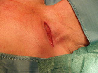
Fig. 1.2
Left supraclavicular incision
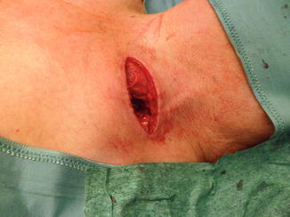
Fig. 1.3
External margin of the clavicular insertion of the sternocleidomastoid muscle
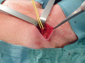
Fig. 1.4
Medial retraction of the clavicular insertion of the sternocleidomastoid muscle and identification of the omohyoid muscle
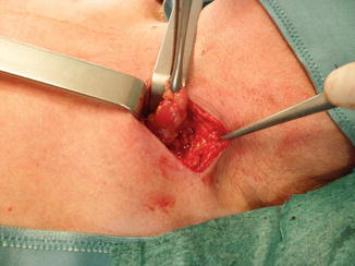
Fig. 1.5
Excision of the scalene fat pad
1.5.3 Mediastinoscopic Technique
Mediastinoscopic biopsy of the scalene lymph nodes is performed under general anaesthesia after mediastinoscopy has proved nodal disease. Its objective is to determine if there is extrathoracic nodal spread. The patient is in the supine decubitus with hyperextended neck and is prepared and draped as for mediastinoscopy. During mediastinoscopy, biopsies of mediastinal lymph nodes are sent for frozen section, and if pathological study reveals nodal disease, either N2 or N3, then the scalene lymph nodes are explored and biopsied. From the same collar incision, finger dissection creates a passage behind the sterno-clavicular insertions of the sternocleidomastoid muscle (Fig. 1.6). The mediastinoscope is then passed behind the muscle insertions. At this point, the tip of the mediastinoscope is directed towards the scalene fat pad (Fig. 1.7). Biopsies of nodes are performed with the same biopsy forceps used for mediastinoscopy. Biopsies are sent for pathological study and for culture if an infectious disease is suspected.
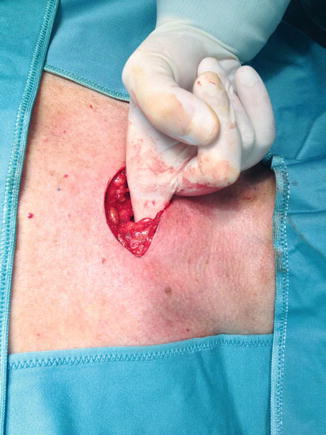
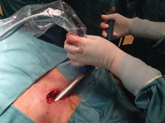

Fig. 1.6
Finger dissection creates a passage behind the sterno-clavicular insertions of the sternocleidomastoid muscle on the left side for mediastinoscopic biopsy of scalene lymph nodes

Fig. 1.7
The mediastinoscope is passed behind the sternocleidomastoid muscle to reach the scalene fat pad
1.6 Results
In thoracic oncology, the most recent publication on the topic is the one by Lee and Ginsberg [4]. In a series of 81 patients with lung cancer who had undergone staging mediastinoscopy and were found to have N2 or intrathoracic N3 disease, scalene lymph node biopsy on the side of the tumour was performed from the same collar incision using the mediastinoscope. Six (15 %) of 39 patients with N2 disease and 13 (68 %) of 19 patients with intrathoracic N3 disease at mediastinoscopy had positive scalene lymph nodes. From a total of 58 patients with positive mediastinoscopy, 19 (32 %) had nodal disease in the scalene fat pad. All of them had centrally located non-squamous cell carcinomas [4]. These results should be taken into consideration when including patients with mediastinal nodal disease in clinical trials. If scalene lymph node biopsy is not performed, unnoticed N3 disease contaminates the series and influences in a negative way the results of the trial.
Regarding gynaecological malignancies, a relatively recent publication described the results of left scalene lymph node biopsy in a series of 49 patients: 33 with cervix carcinoma and 16 with corpus uteri carcinoma. A total of 9 (18 %) were positive: 7 (21 %) in the cervix carcinoma group and 2 (12 %) in the corpus uteri carcinoma group. When the results were analysed according to the status of the periaortic lymph nodes, the positive results were found mainly among patients with grossly positive and pathologically confirmed periaortic lymph nodes: 7 (44 %) out of 16 in the group of cervix carcinoma and 2 (33 %) out of 6 in the group of corpus carcinoma [7]. Other series of patients with carcinoma of the cervix who underwent scalene lymph node biopsy have shown positivity rates of between 8 and 30 %, with the consistent finding that most of the positive cases have involved high iliac or para-aortic lymph nodes [12–17].
Stay updated, free articles. Join our Telegram channel

Full access? Get Clinical Tree


