Controversy exists concerning the relation between diagonal ear lobe crease (DELC) and coronary artery disease (CAD). We examined whether DELC is associated with CAD using coronary computed tomography (CT) angiography. We studied 430 consecutive patients without a history of coronary artery intervention who underwent CT angiography on a dual-source scanner. Presence of DELC was agreed by 2 blinded observers. Two blinded readers evaluated CT angiography images for presence of CAD and for significant CAD (≥50% stenosis). Chi-square and t tests were used to assess demographic differences between subgroups with and without DELC and the relation of DELC to 4 measurements of CAD: any CAD, significant CAD, multivessel disease (cutoff ≥2), and number of segments with plaque (cutoff ≥3). Multivariable logistic regression was performed to adjust for CAD confounders: age, gender, symptoms, and CAD risk factors. Mean age was 61 ± 13 and 61% were men. DELC was found in 71%, any CAD in 71%, and significant CAD in 17% of patients. After adjusting for confounders, DELC remained a significant predictor of all 4 measurements of CAD (odds ratio 1.8 to 3.3, p = 0.002 to 0.017). Sensitivity, specificity, and positive and negative predictive values for DELC in detecting any CAD were 78%, 43%, 77%, and 45%. Test accuracy was calculated at 67%. Area under the receiver operator characteristic curve was 61% (p = 0.001). In conclusion, in this study of patients imaged with CT angiography, finding DELC was independently and significantly associated with increased prevalence, extent, and severity of CAD.
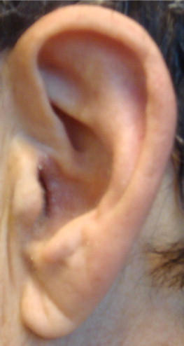
Diagonal ear lobe crease (DELC; Figure 1 ) , also known as “Frank sign,” is a wrinkle-like line extending diagonally from the tragus across the lobule to the rear edge of the auricle of the ear and is not related to sleeping position or wearing earrings. It was first associated with coronary artery disease (CAD) in an article by Frank published in 1973. Since then, the association between DELC and CAD and how they are linked have remained controversial, with some research echoing Frank’s work and others disputing it. Practically, finding of DELC is not used routinely in physical examination. The aim of our study was to assess whether presence of DELC correlates to measurements of the presence, extent, and severity of CAD as determined by coronary computed tomography (CT) angiography.

Methods
We studied 459 consecutive patients who underwent CT angiography during a 9-month period in 1 medical center. After excluding those with previous coronary artery bypass surgery (n = 23), previous coronary artery stenting (n = 2), or an uninterpretable scan (n = 4), 430 patients formed the study population in which 52% were white, 22% were Hispanic, 10% were Asian, 8% were African-American, and 8% did not respond the question about ethnicity. Indications for undergoing CT angiography were chest pain in 46%, previous equivocal results from other imaging modalities in 34%, screening before noncardiac operations in 11%, and screening because of multiple and unbalanced coronary risk factors in 9%. Written informed consent was obtained and the study was approved by our institutional review board.
Immediately before performance of CT angiography, patients were examined for DELC by 2 trained observers who determined by consensus whether DELC was present in both ear lobes. Observers were blinded to patients’ histories and to results of any previously performed CT angiography or coronary calcium scan.
CT angiography was performed on a SOMATOM Definition dual-source scanner (Siemens Medical Systems, Forchheim, Germany). The imaging protocol has been described previously in detail. Beta blockade with oral or intravenous metoprolol was used whenever the heart rate at rest was >70 beats/min. All patients received sublingual nitroglycerin 0.4 mg 3 to 5 minutes before scanning. Patients were scanned after injection of iodinated contrast 80 ml at 5 to 6 ml/s, triggered by >100 HU in the ascending aorta, in a single breath hold. Acquired CT angiography data were reconstructed in mid-diastole and end-systole or only in mid-diastole if performed prospectively using a slice thickness of 0.6 mm (0.75 mm for body mass index >35 kg/m 2 ). Reconstructed data were transferred to a Siemens workstation (Leonardo; Siemens Medical Systems) for interpretation.
CT angiography interpretation was performed by 2 expert readers who were blinded to the presence or absence of DELC using a scale of 0 to 4 for the number of diseased vessels (including left main, left anterior descending, left circumflex, and right coronary arteries) and the modified American Heart Association 15-segment coronary artery tree model. Diameter stenosis severity (scale of 0 to 6: 0%, 1% to 24%, 25% to 49%, 50% to 69%, 70% to 89%, 90% to 99% and 100%) was recorded for each coronary artery segment. Consensus interpretation was obtained to resolve discrepancies between readers.
Four different measurements of CAD presence, extent, and severity were tested as separate binary outcomes. We defined “any CAD” as the presence of any coronary artery plaque on CT angiography. For CAD extent, we evaluated the number of coronary artery vessels with plaque (cutoff ≥2) and the number of coronary artery segments with plaque (cutoff ≥3). For CAD severity, we evaluated the presence of angiographically significant CAD (cutoff ≥50% stenosis).
Continuous variables were described as mean ± SD and compared using Student’s t test. Binary variables were described as frequencies and categorical variables were described as medians, and these were compared by chi-square test. Regression analysis was performed while controlling for the following potential confounders: age (binary, cutoff >70 years old ), male gender, presence of hypertension, hyperlipidemia, diabetes mellitus, family history of premature CAD, smoking, and any symptoms of chest pain. Hypertension was defined as a previously established diagnosis, a systolic blood pressure >140 mm Hg, a diastolic blood pressure >90 mm Hg, or antihypertensive drug use. Smoking was defined as current smoker or previous heavy smoker (>20 package-years). Diabetes mellitus was defined as a previously established diagnosis, insulin or oral hypoglycemic therapy, fasting glucose level >126 mg/dl, or nonfasting glucose level >200 mg/dl. Family CAD history was defined as myocardial infarction, coronary revascularization, or sudden cardiac death in a first-degree man relative <55 years old or woman relative <65 years old. Odds ratios with 95% confidence intervals were calculated while adjusting for the listed confounders as binary variables.
For quality control, 2 observers were obliged to have consensus on the presence of DELC when present bilaterally. Study interpretation was conducted by 2 expert readers who reached consensus on severity of coronary artery stenosis. All participating investigators who gathered data for the study were blinded to patient clinical information. Artifactual studies were excluded.
Results
Demographic characteristics of the 430 participants are presented in Table 1 . Prevalence of any CAD was 71% and that of significant CAD was 17%. Prevalence of DELC was 71%. Overall interobserver agreement for presence of DELC was 97.4%. There was no difference in prevalence of any chest pain between patients with and without DELC (47% vs 46%, p = 0.844).
| Variable | Entire Cohort (n = 430) | With DELC (n = 307, 71%) | Without DELC (n = 123, 29%) | p Value ⁎ † |
|---|---|---|---|---|
| Men | 262 (61%) | 190 (62%) | 72 (59%) | 0.581 |
| Age (years) | 61 ± 13 | 64 ± 12 | 53 ± 13 | <0.001 |
| Hypertension ‡ | 245 (57%) | 188 (61%) | 57 (46%) | 0.006 |
| Hyperlipidemia ‡ | 270 (63%) | 205 (67%) | 65 (53%) | 0.007 |
| Smoker ‡ | 163 (38%) | 125 (41%) | 38 (31%) | 0.064 |
| Diabetes mellitus ‡ | 65 (15%) | 48 (16%) | 17 (14%) | 0.639 |
| Medications | ||||
| β Blockers | 132 (31%) | 98 (32%) | 34 (28%) | 0.602 |
| Statin | 177 (41%) | 132 (43%) | 45 (37%) | 0.188 |
| Angiotensin-converting enzyme inhibitor/angiotensin receptor blocker | 123 (29%) | 92 (30%) | 31 (25%) | 0.587 |
| Aspirin | 70 (16%) | 50 (16%) | 20 (16%) | 0.977 |
| Blood tests § | ||||
| Glucose (mg/dl) | 94 ± 29 | 95 ± 32 | 92 ± 18 | 0.259 |
| Total cholesterol (mg/dl) | 166 ± 40 | 167 ± 42 | 163 ± 36 | 0.415 |
| Low-density lipoprotein cholesterol (mg/dl) | 96 ± 33 | 96 ± 33 | 96 ± 31 | 0.902 |
| High-density lipoprotein cholesterol (mg/dl) | 48 ± 18 | 49 ± 18 | 47 ± 18 | 0.400 |
| Triglycerides (mg/dl) | 114 ± 83 | 117 ± 85 | 106 ± 78 | 0.228 |
| Other | ||||
| Any chest pain | 199 (46%) | 143 (47%) | 56 (46%) | 0.844 |
⁎ Between subgroups with and without diagonal ear lobe crease. Statistically significant difference (2-tailed p <0.05).
† All numbers represent percent prevalence unless otherwise specified.
‡ Risk factors were determined according to patient’s medical records and guidelines; see text.
§ Blood tests were drawn at the time of computed tomography angiography or results were present in recent (<1 month) medical records.
Patients with DELC were significantly older (64 ± 12 vs 53 ± 13 years, p <0.001) and had a significantly higher prevalence of hypertension (61% vs 46%, p = 0.006) and hyperlipidemia (67% vs 53%, p = 0.007) than patients without DELC.
All 4 measurements of CAD (any CAD, significant CAD, ≥2 diseased vessels, and ≥3 diseased segments) were significantly more prevalent in the group with DELC than in the group without DELC ( Table 2 ). Sensitivity, specificity, and positive and negative predictive values for DELC to diagnose any CAD were 78%, 43%, 77%, and 45%, with an overall accuracy of 67% and an area under the receiver operator characteristic curve of 0.61 (95% confidence interval 0.55 to 0.67, p = 0.001).
| CAD Measurement | All (n = 430) | With DELC (n = 307, 71%) | Without DELC (n = 123, 29%) | p Value ⁎ |
|---|---|---|---|---|
| Any coronary artery disease | 303 (71%) | 235 (77%) | 68 (55%) | <0.001 |
| Significant coronary artery disease | 71 (17%) | 64 (21%) | 7 (6%) | <0.001 |
| Number of diseased vessels | 1.6 | 1.7 ± 0.1 | 1.1 ± 0.1 | <0.0001 |
| ≥2 diseased vessels | 224 (52%) | 182 (59%) | 42 (34%) | <0.001 |
| Number of diseased segments | 3.4 | 3.9 ± 0.2 | 2.2 ± 0.3 | <0.0001 |
| ≥3 diseased segments | 202 (47%) | 168 (55%) | 34 (28%) | <0.001 |
Stay updated, free articles. Join our Telegram channel

Full access? Get Clinical Tree


