


Atrial flutter and atrial fibrillation are at the two extremes of a spectrum. At the flutter end of the spectrum, the causative reentering impulse cycles around a single circuit (usually within the right atrium), producing slower, regular, uniform, and sharp (“sawtooth-like”) waves (F waves). At the fibrillation end of the spectrum, the causative reentering impulse cycles around multiple circuits, producing more rapid, irregular, multiform, and rounded waves (f waves). As a result of the macro–re-entry mechanism responsible for both atrial flutter and fibrillation, P waves are replaced either by F waves, representing the continuous activation within the flutter circuit, or by f waves, representing the continuous activation within the fibrillation circuits.
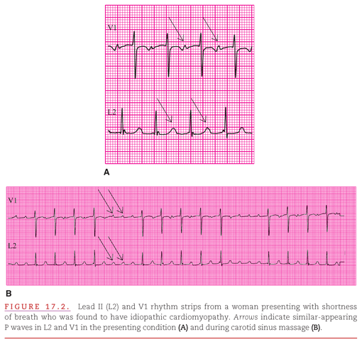
Although atrial micro–re-entrant tachycardia (paroxysmal atrial tachycardia [PAT]) occurs rarely, it is useful to present an example because of its clearly identifiable characteristics (Fig. 17.2). In Figure 17.2A, a micro–re-entrant tachycardia masquerades as a typically appearing sinus tachycardia (110 beats per minute) with first-degree atrioventricular (AV) block (0.26 second). However, in Figure 17.2B, the true atrial rate of 220 beats per minute is revealed by the vagal stimulation provided by carotid sinus massage. This atrial tachyarrhythmia could possibly be atrial flutter, but the discrete P waves rather than a sawtooth-like morphology in both leads V1 and L2 (II) makes flutter unlikely. In the patient presented in Figure 17.2, sinus rhythm emerged after electrical cardioversion, confirming the reentrant mechanism of the tachycardia. The similar P-wave morphologies observed during PAT and sinus rhythm indicate that the micro–re-entrant circuit was in or near the sinus node, high in the right atrium.
ATRIAL RATE AND REGULARITY IN ATRIAL FLUTTER/FIBRILLATION
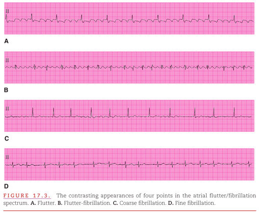
The F waves of atrial flutter typically occur at rates between 200 and 350 beats per minute (Fig. 17.3A).1,2 At an atrial rate of >350 beats per minute, either the atrial waves have some characteristics of both flutter and fibrillation at a single point in time (see Fig. 17.3B) or there are alternations between F and f waves, with the appropriate term being atrial flutter-fibrillation. Fibrillation varies from coarse to fine. In coarse fibrillation, prominent f waves are clearly visible in many leads of the electrocardiogram (ECG; see Fig. 17.3C); in fine fibrillation, either there are small f waves or no visible atrial activity (see Fig. 17.3D).
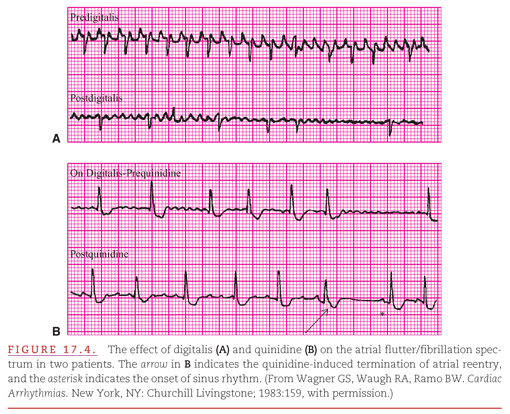
Some patients’ atrial tachyarrhythmias may spontaneously undergo a change from flutter to fibrillation, whereas others have such variation only when certain drugs are administered. Digitalis increases the atrial rate toward the fibrillation end of the spectrum by shortening the refractory periods of atrial myocardial cells within the re-entry circuit (Fig. 17.4A). Conversely, drugs such as quinidine and procainamide (Vaughn Williams class IA) and flecainide (class IC) decrease the atrial rate toward the flutter end of the spectrum by lengthening the refractory periods of the atrial myocardial cells (see Fig. 17.4B).
VENTRICULAR RATE AND REGULARITY IN ATRIAL FLUTTER/FIBRILLATION
Atrial flutter produces a ventricular rhythm that varies from precisely regular to irregularly irregular; however, fibrillation always produces an irregularly irregular ventricular rhythm. Therefore, when atrial fibrillation is accompanied by a regular ventricular rate, there is dissociation between the atrial and ventricular rhythms. A pacemaking Purkinje cell in the common bundle or bundle branches is initiating the regular ventricular rhythm.
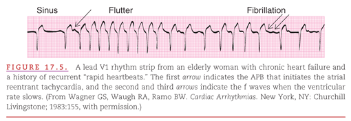
Because the atrial rate may vary dramatically within the flutter/fibrillation spectrum, the ventricular rate may also vary. At times, it may change abruptly from rapid and regular to slow and irregular, incorrectly suggesting a change in the basic underlying atrial rhythm (Fig. 17.5). After two beats of sinus rhythm, an atrial premature beat (APB) initiates a supraventricular tachyarrhythmia that is initially regular and then becomes irregular. The regular phase is most likely caused by flutter at a rate of 200 beats per minute with 1:1 AV conduction, after which the irregular phase occurs because the atrial rate accelerates into the flutter/fibrillation spectrum at 300 beats per minute, with a resultant slower, irregular ventricular rate.
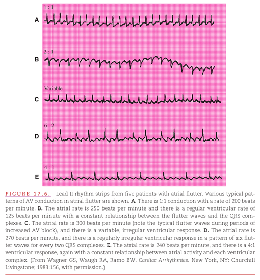
During atrial flutter, the ratio of atrial to ventricular waveforms may vary from 1:1 to 2:1 to 6:2 to 4:1 (Fig. 17.6A–E) so that the ventricles may or may not have a rapid rate. The ratio of atrial-to-ventricular waveforms depends on the capability of the slowly conducting AV node to transport the atrial impulses to the common bundle. When ratios of atrial-to-ventricular waveforms of 1:1, 2:1, or 4:1 remain constant (see Fig. 17.6A, B, and E), the ventricular rhythm is regular. When the ratio is 6:2 (because the atrial impulses are blocked at two levels within the AV node), the ventricular rhythm is regularly irregular. When the AV conduction ratio is variable (e.g., switching between 2:1 and 6:2 conduction), the ventricular rhythm is irregularly irregular.
When atrial fibrillation is present, the innumerable f waves compete for penetration of the AV node, making it most difficult for any impulse to reach the common bundle. Therefore, the ventricular rhythm is slower at the fibrillation end of the flutter/fibrillation spectrum (Fig. 17.7).3–5 Typically, a 1:1 AV relationship persists to the upper limit of the atrial rate during exercise or with other conditions that enhance sympathetic stimulation. However, when the atrial rate increases through other mechanisms (such as artificial atrial pacing or a reentrant tachyarrhythmia), the 1:1 AV relationship persists only until the atrial rate reaches 150 to 160 beats per minute. Above this atrial rate, the physiologic delay in AV nodal conduction prevents some atrial impulses from reaching the ventricles. As the atrial rate increases further, the ventricular rate decreases because of the competition within the AV node. The nonconducted atrial impulses are blocked only after they have been able to penetrate some distance into the node. This concealed conduction depolarizes a part of the AV node, making it refractory to the following atrial impulse.6 Changes in the sympathetic-to-parasympathetic balance can either facilitate (sympathetic) or further inhibit (parasympathetic) AV nodal conduction, as indicated by the directions of the arrows at the right side of the figure.

ONSET OF ATRIAL FLUTTER/FIBRILLATION
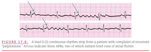
Both spontaneous and electrically induced atrial flutter/fibrillation may typically be produced when APBs occur within a narrow range of the atrial refractory period. Thus, flutter/fibrillation is typically sudden in onset, as are all reentrant arrhythmias (Fig. 17.8).
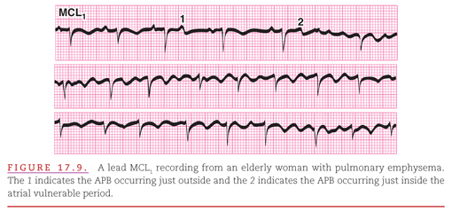
Like the ventricles, the atria have a vulnerable period: a point in the atrial cycle at which an APB is most likely to precipitate atrial flutter/fibrillation (Fig. 17.9). Killip and Gault7 have formulated the situation as follows: If the interval from the normal P wave to the premature P wave is less than half of the preceding interval between normal P waves, the premature P wave is within the atrial vulnerable period and may induce flutter/fibrillation.
TERMINATION OF ATRIAL FLUTTER/FIBRILLATION
As illustrated in Figure 17.8, atrial flutter/fibrillation may terminate spontaneously. Presumably, the reason for this is that the recycling impulse fails to encounter receptive cells and thus has “nowhere to go.” When the reentry persists and creates either acute cardiac dysfunction or a chronic clinical problem, some medical intervention may be required. There are two possible treatment strategies:
1. Enhance the AV block to slow the ventricular rate.
2. Terminate the flutter/fibrillation.
Terminating or “breaking” this atrial reentrant tachycardia may be accomplished either with drugs or through electrical stimulation. A drug can effect termination of a tachyarrhythmia either by increasing the speed of the recycling impulse so that it encounters only cells that are still refractory or by prolonging the refractory periods of the involved cells. No drugs yet exist that both speed conduction and prolong the refractory period. The primary therapeutic effect of available drugs is prolongation of refractoriness. An electrical stimulation can terminate a tachyarrhythmia by suddenly depolarizing all cardiac cells that are not already in the depolarized state (Fig. 17.10A). This eliminates the receptivity to reentry that is required to maintain the tachyarrhythmia. As illustrated in Figure 17.10B, such an electrical stimulation cannot “break” a tachyarrhythmia produced by accelerated automaticity; instead, the rapidly discharging site continues to maintain the tachyarrhythmia after a brief interruption.


When the atrial re-entry is orderly, as at the flutter end of the flutter/fibrillation spectrum, it can be suddenly terminated by intra-atrial electrical stimulation from a pacemaker. Maintenance of the orderly reentry of the impulse requires that areas of atrial myocardium have completed their refractory periods and are receptive to the advancing wave of depolarization. A properly timed stimulus, delivered via a pacing electrode, produces premature activation of receptive areas, and the advancing wavefront encounters no cells that it can depolarize.
Stay updated, free articles. Join our Telegram channel

Full access? Get Clinical Tree


