Typical Subendocardial Ischemia
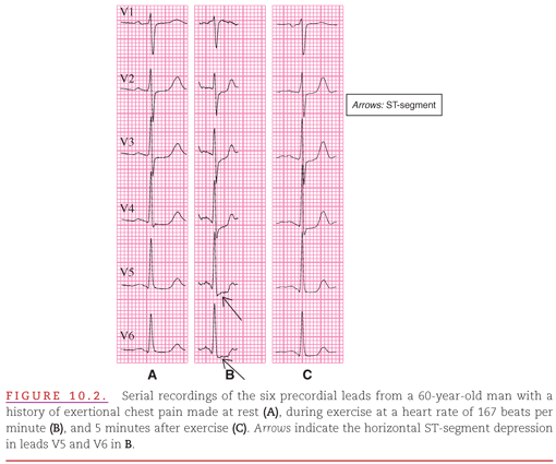
Partial obstructions within the coronary arteries do not produce insufficient blood supply and therefore cannot be detected on the resting ECG (Fig. 10.2A). However, when such a partial obstruction prevents myocardial blood flow from increasing enough to meet the increased metabolic demand during stress, the resulting ischemia (limited to the subendocardial layer of the left ventricle) is manifested by horizontal (see Fig. 10.2B) or downsloping ST-segment depression.3 The ST-segment depression typically disappears within several minutes after the demand on the myocardium is returned to baseline levels by stopping the exercise (see Fig. 10.2C) because the myocardial cells have been only reversibly ischemic.
A combination of two diagnostic criteria in at least one ECG lead is typically required for diagnosing left-ventricular subendocardial ischemia (Fig. 10.3):
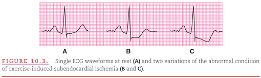
1. ST-segment depression of ≥1 mm (0.10 mV) at the J point.
2. Either a horizontal or a downward slope toward the end of the ST segment at its junction with the T wave.
The terminal part of the T wave typically remains positive (see Fig. 10.3A–C) but with progressively diminished amplitude from Figure 10.3B and C.
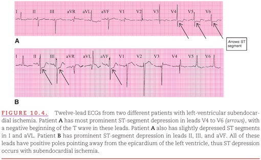
As indicated in Chapter 2, the positive poles of most of the standard limb and precordial ECG leads are directed toward the left ventricle. Subendocardial ischemia causes the ST segment to move generally away from the left ventricle (see Fig. 9.6A). The changes in the ST segment appear negative or depressed in the leftward (I, aVL, or V4 to V6) and inferiorly oriented leads (II, III, and aVF), which have their positive poles directed toward the left ventricle (Fig. 10.4A, B). As previously stated in Chapter 9, the location of the ECG leads showing ST-segment depression is not indicative of the involved region of left-ventricular subendocardial ischemia. In leads that have their positive poles directed away from the left ventricle (limb lead aVR and precordial leads V1 and V2), ST elevation is typically present (see Fig. 10.4A, B).
It may be useful to now return to the Video 9.2 (QR code below) which demonstrates how ischemic action potentials in the subendocardium create the ECG pattern of subendocardial ischemia.

The change in the ST segments that occurs with left-ventricular subendocardial ischemia (Fig. 10.5A) is similar to that described in Chapter 5 for left-ventricular strain. However, left-ventricular strain also causes the T waves to be directed away from the left ventricle and is accompanied by the QRS changes of left-ventricular hypertrophy (LVH; see Fig. 10.5B
Stay updated, free articles. Join our Telegram channel

Full access? Get Clinical Tree


