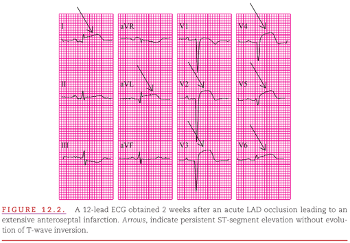
In some patients, the ST-segment elevation does not completely resolve and T-wave inversion fails to occur during the reperfusing phase of an MI (Fig. 12.2). This condition more commonly occurs with anterior infarcts than with those in the other locations in the left ventricle (LV).6 The lack of ST-segment resolution has been associated acutely with failure to reperfuse and chronically with thinning of the left-ventricular wall caused by infarct expansion.7,8 The most extreme manifestation of infarct expansion is the development of a ventricular aneurysm, but this is almost always prevented by successful reperfusion therapy.
T-Wave Migration: Toward to Away from the Infarct
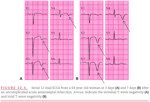
The movement of the T waves toward the involved region of the LV also resolves as the jeopardized myocardium either recovers or infarcts. Unlike the ST segment, however, T waves do not typically return only to their original positions. The T waves typically migrate past the isoelectric baseline until they are directed away from the involved region.9 They assume an appearance identical to that described in Chapter 9 as “postischemic T waves,” indicating that there is no ongoing myocardial ischemia. This evolution of the T wave is illustrated in serial ECGs at 3 and 7 days post anterior infarction (Fig. 12.3A, B). Typically, the terminal portion of the T wave is the first to become inverted, followed by the middle and initial portions.
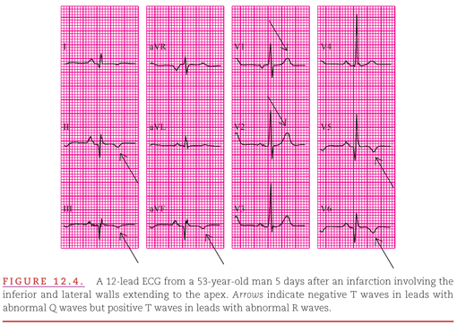
Similarly, when the lateral wall of the LV is involved, the T waves eventually become markedly positive in the leads in which the injury current was represented by ST depression. Figure 12.4 illustrates the prominent positive T waves (arrows) with abnormal R waves in leads V1 and V2 that accompany the negative T waves (arrows) with abnormal Q waves in other leads (II, III, aVF, V5, and V6) during the healing phase of an extensive inferior infarction.
Evolving QRS Complex Away from the Infarct
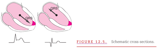
The QRS complex is the key waveform for evaluating the presence, location, and extent of a healing or chronic MI. As indicated in Chapter 11, almost immediately after a complete coronary artery occlusion, the QRS complex deviates toward the involved myocardial region, primarily because of delay in myocardial activation and secondarily because of the ischemia current of injury. Because the process of infarction begins in the most poorly perfused subendocardial layer of the myocardium, the deviation of the terminal QRS waveforms toward the ischemic region is replaced by deviation of the initial QRS waveforms away from the infarcted region (Fig. 12.5).10 Absence of any electrical activation of the infarcted myocardium has replaced the delayed activation of the severely ischemic myocardium, as illustrated in Figure 12.5.
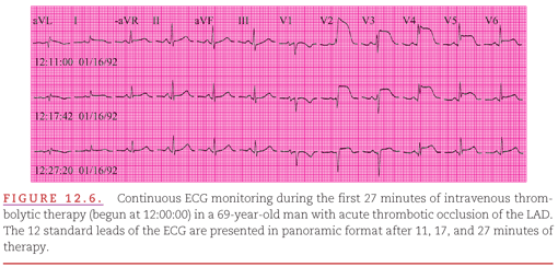
The evolving appearance of the abnormalities of the QRS complex produced by an anterior infarction during the first few minutes of intravenous thrombolytic therapy with continuous monitoring for ischemia is shown in Figure 12.6. Secondary changes in the morphology of the QRS complex during transmural myocardial ischemia have shifted the axis of the complex toward the anterior left-ventricular wall. Myocardial reperfusion is accompanied by rapid resolution of the transmural myocardial ischemia and a shift of the QRS complex away from the anterior left-ventricular wall. Although it may appear that the reperfusion itself has caused the infarction, it is much more likely that the infarction occurred before initiation of the therapy that led to reperfusion and that its detection on the ECG was obscured by the secondary changes of the QRS complex caused by the ischemic injury currents.
Transmural myocardial ischemia involving the thin right-ventricular free wall may be manifested on the ECG by deviation of the ST segment (see Chapter 11), but right-ventricular infarction is not manifested by significant alteration of the QRS complex. This is because activation of the right-ventricular free wall is insignificant in comparison with activation of the thicker interventricular septum and left-ventricular free wall. MI evolves from transmural myocardial ischemia in the distal aspects of the left-ventricular myocardium supplied by one of the three major coronary arteries (see Fig. 11.6).11
QRS Complex for Diagnosing
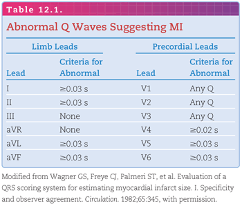
The initial portion of the axis of the QRS complex deviates most prominently away from the area of infarction and is represented on the ECG by a prolonged Q-wave duration. As presented in Figure 3.6, the initial QRS waveform may normally be negative in all leads except V1 to V3. The presence of any Q wave is considered abnormal only in these 3 of the 12 standard ECG leads. Table 12.1 indicates the upper limits of normal Q-wave duration in the various ECG leads.12 The duration of the Q wave should be the primary measurement used in the definition of abnormality, because the amplitudes of the individual QRS waveforms vary with the overall amplitude of the QRS complex. As discussed in the next section, Q-wave amplitude should be considered abnormal only in relation to the amplitude of the following R wave.
Many cardiac conditions other than MI are capable of producing abnormal initial QRS waveforms. As indicated in Chapters 5 through 7, abnormalities that commonly prolong the duration of the Q wave are:
1. ventricular hypertrophy;
2. intraventricular conduction abnormalities; and
3. ventricular preexcitation.
The term “Q wave” as used here also refers to the Q-wave equivalent of abnormal R waves in leads V1 and V2. Therefore, the following steps should be considered in the evaluation of Q waves for the presence of MI:
1. Are abnormal Q waves present in any lead?
2. Are criteria present for other cardiac conditions that can produce abnormal Q waves?
3. Does the extent of Q-wave abnormality exceed that which could have been produced by some other cardiac condition?
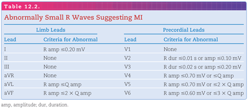
The deviation of the QRS axis away from the area of an MI may, in the absence of abnormal Q waves, be represented by diminished R waves. Table 12.2 indicates the leads in which R waves of less than a certain amplitude or duration may be indicative of MI.2
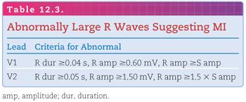
Infarction in the lateral wall of the LV is represented by a positive rather than a negative deviation of the QRS complex. This results in an increased rather than a decreased R-wave duration and amplitude in precordial leads VI and V213 (Table 12.3).
QRS Complex for Localizing
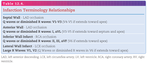
Table 12.4 indicates the relationships among the coronary arteries, left-ventricular walls, and ECG leads that provide a basis for localizing MIs.14 It may also be helpful to refer to Figure 11.6 as this learning unit is read.
Stay updated, free articles. Join our Telegram channel

Full access? Get Clinical Tree


