Prosthetic Valves


Prosthetic Valves


The era of valve surgery preceded the development of echocardiography by only a few years. It is therefore not surprising that one of the earliest applications of echocardiography was the study of prosthetic valve function. With the tremendous advances in surgical techniques over the past four decades, the role of echocardiography has evolved and broadened in this important field. Because neither the perfect valve repair nor the perfect prosthesis yet exists, ongoing assessment of valve function is a key aspect of the management of patients after valve surgery. Echocardiography, with its noninvasive ability to evaluate both anatomy and function, has become the diagnostic modality of choice for this purpose.

The echocardiographic assessment of prosthetic valves is complex. Flow dynamics are different through prosthetic valves compared with native valves. Both the size and type of the prosthesis influence the range of expected flow velocities and thus the definition of normal versus abnormal function. The echocardiographer must determine the specific type of prosthetic valve and whether the structural and functional parameters exceed the limits of normal for a given size and type. Despite these challenges, the combination of echocardiography and Doppler imaging techniques is ideally suited to assessing prosthetic valves. Whether monitoring valve function over time or detecting the specific cause of prosthesis dysfunction, echocardiographic techniques have become indispensable in this important clinical area.


Types of Prosthetic Valves

The two major categories of prosthetic valves are mechanical valves and tissue valves or bioprostheses (Table 15.1). The mechanical prosthetic valves can be further divided into caged ball and tilting disk designs. The caged ball prosthesis was the first type of artificial heart valve and the Starr-Edwards valve is by far the most common (Fig. 15.1). It consists of a circular sewing ring on which is mounted a U-shaped cage that contains a silastic ball occluder. To open, the ball moves forward into the cage, allowing blood flow around the entire circumference. To occlude, the ball is driven back into the sewing ring to prevent backflow.

Several tilting disk prostheses are currently in use (Fig. 15.2). The single disk prosthesis consists of a round sewing ring and a circular disk fixed eccentrically to the ring via a hinge. The disk moves through an arc of less than 90° (usually 55°-85°), thereby allowing antegrade flow in the open position and seating within the sewing ring to prevent backflow in the closed position. The Bjök-Shiley, Omnicarbon, and Medtronic-Hall are examples of single tilting disk prostheses. Because the hinge is eccentrically positioned within the sewing ring and the disk opens less than 90°, major and minor orifices are created and some stagnation of flow occurs behind the disk. Bileaflet tilting disk valves consist of two semicircular disks that open and close on a hinge mechanism within the sewing ring. The opening angle is generally more vertical (approximately 80°) than the single disk prosthesis and results in three distinct orifices: two larger ones on either side and a smaller central rectangular-shaped orifice. Examples of bileaflet titling disks include the St. Jude Medical and CarboMedics valves.

Unlike mechanical valves, bioprostheses are constructed from either human or animal tissue (Fig. 15.3). Among the most commonly used are the porcine bioprostheses, including the Hancock and Carpentier-Edwards valves. These are porcine aortic valves that have been preserved and fixed within a polypropylene mount attached to a Dacron sewing ring. Pericardial prostheses are also in use today. Because the tissue has been preserved, it is less pliable than native valve tissue. The leaflets themselves are supported by stents, which vary in number and design and arise vertically from the sewing ring. More recently, “stentless” bioprostheses have been developed for use in the aortic position. They consist of porcine aortic valves that include the annulus, valve, and root preserved intact. Stentless aortic valves have neither a prosthetic sewing ring nor supporting stents. Instead, the porcine leaflets are supported via a flexible cuff. They are often customized by the surgeon in the operating room at the time of implantation.


Table 15.1 Types of Prosthetic Valvesa


Mechanical


Caged ball


Starr-Edwards


Single disc


Bjök-Shiley


Medtronic-Hall


Omnicarbon


Lillehei-Kaster


Bileaflet disc


St. Jude Medical


CarboMedics


On-X


ATS Open Pivot


Tissue


Porcine


Carpentier-Edwards


Hancock II


SJM Biocor


Bovine


Hancock


Pericardial


Carpentier-Edwards


CarboMedics Mitroflow


Ionescu-Shiley


Hancock


Stentless


St. Jude Toronto


Medtronic Freestyle


Edwards Prima Plus


Homografts


aIncludes valves no longer being implanted.


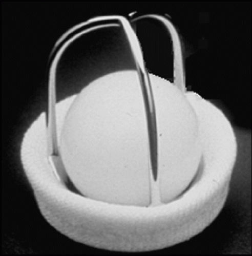
FIGURE 15.1. A Starr-Edwards prosthesis.

Homograft valves are derived from human aortic or pulmonary valve tissue that has undergone cryopreservation and may be either stented or unstented. They are most often used in the aortic position. Here, they are either implanted in the subcoronary position (called a “free hand” valve), as a miniroot procedure (implanted within the native aortic root), or as part of a full root and valve replacement procedure. Another example of their use is the Ross procedure, which involves autotransplantation of the pulmonary valve into the aortic position and placement of homograft in the pulmonary position. Homografts are also used in valved conduits but are rarely used to replace a mitral or tricuspid valve.

Valve repair, although not involving a prosthetic valve, usually requires the use of prosthetic material. Aortic valve repair has been performed successfully in a limited number of centers. It may be useful in the treatment of regurgitant bicuspid valves or in the setting of regurgitation due to aortic root pathology. Mitral valve repair is performed more widely and with more consistently successful results. It is generally undertaken in the setting of a myxomatous valve or when mitral regurgitation is due to left ventricular dilation or dysfunction. Both surgical and percutaneous approaches are available. In most cases, mitral repair involves use of a ring to reduce the effective size of the valve orifice.


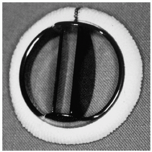
FIGURE 15.2. A St. Jude prosthetic valve.


FIGURE 15.3. A porcine bioprosthetic valve.

Most recently, percutaneous approaches to valve replacement have been developed. These generally involve the aortic valve and remain investigational but have shown substantial promise in clinical trials.


Normal Prosthetic Valve Function

The indications for echocardiography in patients with prosthetic valves are summarized in Table 15.2. Visualization of
prosthetic valves often requires a combination of transthoracic and transesophageal imaging. Although the role of threedimensional imaging continues to evolve, the improved spatial orientation provided by modern equipment provides a unique and potentially valuable perspective. Two-dimensional echocardiography is used to determine the type of valve and to evaluate its structure and function. Using this modality, the stability of the sewing ring is assessed. Rocking or independent motion of the prosthesis is often an indication of dehiscence. The presence of abnormal masses, such as thrombi or vegetations, should be determined. Shadowing from the prosthesis may obscure such pathology and multiple imaging windows may be required for a complete evaluation. Motion of the leaflets, disks, or occluder mechanism should also be assessed from the two-dimensional study. An important early step in the echocardiographic assessment of prosthetic valves is recognizing the range of normal findings. Figure 15.4 is a normally functioning porcine aortic prosthesis. Leaflet opening during systole resembles that of a normal native valve. The overall appearance is so similar, in fact, that normally functioning aortic bioprostheses are occasionally mistaken for “normal” aortic valves when historical information is not available. When examined carefully, however, the sewing ring and struts are more echogenic than normal and tend to shadow the leaflets, a clue to the presence of prosthetic material. A normal porcine mitral prosthesis, assessed using three-dimensional echocardiography, is shown in Figure 15.5. Note how this technique permits the valve to be visualized from opposite perspectives, the left atrial side and the ventricular aspect.


Table 15.2 Indications for Echocardiography in Interventions for Valvular Heart Disease and Prosthetic Valves


Class I


1.


Assessment of the timing of valvular intervention based on ventricular compensation, function, and/or severity of primary and secondary lesions


2.


Selection of alternative therapies for mitral valve disease (such as balloon valvuloplasty, operative valve repair, valve replacement)a


3.


Use of echocardiography (especially TEE) in guiding the performance of interventional techniques and surgery (e.g., balloon valvotomy and valve repair) for valvular disease


4.


Postintervention baseline studies for valve function (early) and ventricular remodeling (late)


5.


Reevaluation of patients with valve replacement with changing clinical signs and symptoms; suspected prosthetic dysfunction (stenosis, regurgitation) or thrombosisa


6.


Transthoracic and Doppler echocardiography is indicated in patients with suspected prosthetic valve thrombosis to assess hemodynamic severity


7.


Transesophageal echocardiography is indicated in patients with suspected valve thrombosis to assess valve motion and clot burden


Class IIa


8.


Routine reevaluation study after baseline studies of patients with valve replacements with mild to moderate ventricular dysfunction without changing clinical signs or symptoms


Class IIb


9.


Routine reevaluation at the time of increased failure rate of a bioprosthesis without clinical evidence of prosthetic dysfunction


Class III


10.


Routine reevaluation of patients with valve replacements without suspicion of valvular dysfunction and unchanged clinical signs and symptoms


11.


Patients whose clinical status precludes therapeutic interventions


a TEE may provide incremental value in addition to information obtained by TTE. TEE, transesophageal echocardiography; TTE, transthoracic echocardiography. Adapted from Cheitlin MD, Alpert JS, Armstrong WF, et al. ACC/AHA Guidelines for the Clinical Application of Echocardiography: a report of the American College of Cardiology/American Heart Association Task Force on Practice Guidelines (Committee on Clinical Application of Echocardiography) developed in collaboration with the American Society of Echocardiography. Circulation 1997;95:1686-1744, with permission.


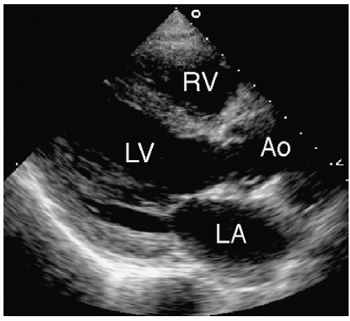
FIGURE 15.4. An echocardiogram of a normally functioning porcine bioprosthetic aortic valve.


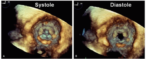
FIGURE 15.5. A three-dimensional echocardiogram of a normal porcine mitral prosthesis is shown in systole (A) and diastole (B). This view is taken from the left ventricular perspective and shows the struts pointing into the left ventricle. A view from the opposite side, the left atrium, is also possible.

Figure 15.6 shows a Starr-Edwards valve in the mitral position. The protruding, high-profile cage in the left ventricle is diagnostic. When examined in real time, the poppet can be seen moving forward and backward in the cage. These valves are highly echogenic, and small thrombi or vegetations can be easily hidden or overlooked. A normally functioning St. Jude mitral prosthesis is presented in Figures 15.7 and 15.8. In Figure 15.7, the two hemidisks open and close in synchrony, although it is often difficult to distinguish both on transthoracic imaging. Significant shadowing occurs, and the left atrium is not well seen in most cases. In Figure 15.8, three-dimensional echocardiography is used to more completely visualize the hemidisks. This approach also provides a thorough circumferential recording of the sewing ring. Figure 15.9 shows a stable aortic St. Jude valve. In this example, the disks are obscured by the walls of the aorta. A distinct shadow from the sewing ring is apparent, extending into the left atrium. Stentless aortic valves are the most recent option in prostheses and are being implanted with increasing frequency. An example of a normal Medtronic Freestyle valve is provided in Figure 15.10. Distinguishing a normally functioning stentless valve from a native aortic valve can be impossible.

Blood flow through normally functioning prosthetic valves differs from flow through native valves in several important ways. First, artificial heart valves are inherently stenotic. There is a variety of explanations for this consistent observation. The sewing ring of the valve may be too small relative to the flow. In young patients, what passes for an adequately sized valve in childhood may become functionally stenotic as the patient grows. More importantly, the effective orifice area is significantly smaller than the area of the sewing ring because the valve assembly (i.e., the occluder mechanism) occupies some of the central space. Leaflets of bioprostheses, by virtue of the preservation process, are stiffer and therefore these valves have
a higher resistance to forward flow compared with equivalently sized native valves. Thus, flow velocity through a normally functioning artificial valve is generally higher than would occur through a normal native valve. However, the range of velocities through a normally functioning bioprosthesis is considerable. Both valve size and type determine the pressure gradient that one can expect in the absence of dysfunction. For example, stented bioprosthetic valves may have slightly higher gradients than mechanical valves of similar size, which tend to have higher gradients than stentless valves. For all these reasons, the range of velocities that must be considered normal varies widely among prosthetic valves. This is illustrated in Figure 15.11. In Figure 15.11A, a newly implanted St. Jude aortic prosthesis is shown. Although functioning normally by clinical criteria, the Doppler study demonstrates a maximal velocity of 290 cm/sec and a mean gradient of 20 mmHg. Also note the distinctive “clicks” that correspond to the opening and closing of the disks. In contrast, Figure 15.11B illustrates flow through a normally functioning bioprosthetic aortic valve. In this case, no significant increase in velocity is present. Prosthetic valve clicks are not typically seen in normally functioning bioprostheses.


FIGURE 15.6. A normally functioning Starr-Edwards mitral prosthesis. A: During systole, the poppet is seated within the sewing ring (arrows). B: During diastole, the poppet moves forward into the cage (arrows), allowing blood flow around the occluder.


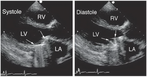
FIGURE 15.7. A normally functioning St. Jude mitral prosthesis. A: During systole, the hemidisks are shown in the closed position (arrows). B: During diastole, the two disks are recorded in the open position (arrows).

Another important difference between native and prosthetic valves is the shape and number of orifices through which forward flow occurs. As noted previously, a bileaflet tilting disk valve has three separate orifices, a rectangular-shaped central orifice surrounded by two larger semicircular orifices (Fig. 15.12). Flow velocity is highest through the central orifice, and if this flow is sampled with continuous wave Doppler imaging, an overestimation of the true gradient can occur. This is because flow through all three orifices contributes to net gradient. By only sampling the highest velocity through the central orifice and ignoring lower velocity flow through the other two, an
overestimation of true gradient occurs. Flow through a caged ball valve does not go through a well-defined orifice but rather goes around the periphery of the spherical occluder (Fig. 15.13). The variability and orientation of the flow complicate the Doppler interrogation of these valves. Flow through bioprostheses is often triangular in shape and may occur through an area that is significantly smaller than the sewing ring itself. Note in Figure 15.14 the position of the three struts and how they effectively form a triangular orifice, the area of which is considerably smaller than the surrounding sewing ring. All these factors contribute to the challenges inherent to assessing prosthetic valve function by any technique.


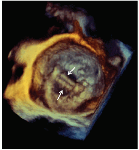
FIGURE 15.8. A three-dimensional echocardiogram of a normal St. Jude mitral prosthesis is shown from the perspective of the left atrium. In real time, the hemidisks (arrows) are seen to open and close from above.


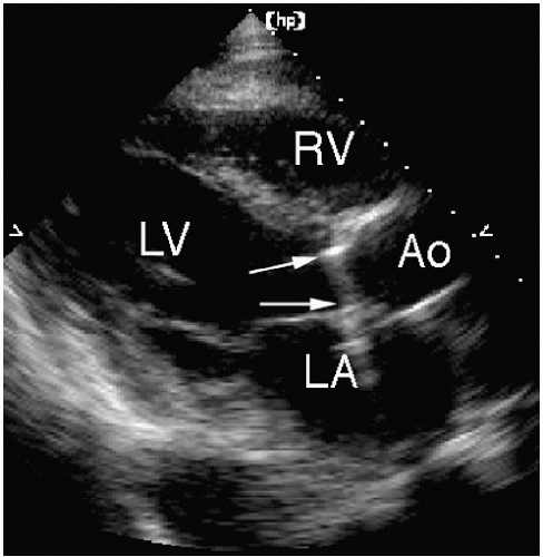
FIGURE 15.9. A normally functioning St. Jude aortic prosthesis. The sewing ring is indicated by the arrows. The walls of the aortic root often obscure the motion of the disks.

A potentially important phenomenon affecting flow through prosthetic valves involves pressure recovery. This occurs when a portion of the kinetic energy released as blood crosses the valve is recovered in the form of pressure downstream. The amount of energy that is recovered depends on how smoothly the transition of flow occurs between the valve and the downstream conduit. For this reason, pressure recovery is most clinically relevant for a St. Jude prosthesis in the aortic position, particularly in the presence of a normal sized aortic root. In this setting, the deceleration (and relaminarization) of blood downstream from the prosthesis is associated with a rise in pressure (i.e., pressure recovery). The net effect is the development of a high, but very localized, gradient through the central orifice of the prosthesis immediately distal to the disks (Fig. 15.15). Then,

as pressure recovers (or increases) downstream, the net pressure gradient diminishes. This means that Doppler imaging, by recording the maximal velocity within the vena contracta, will demonstrate a higher gradient compared to catheter-based methods, which will be lower due to pressure recovery. Although pressure recovery is one potential explanation for a discrepancy in which Doppler imaging reports a higher gradient than catheterization, it does not imply that either method is “right” or “wrong”, rather that local changes in pressure will naturally result in differences in methodology. It should be emphasized that this higher gradient value obtained by Doppler imaging is a real phenomenon, although less physiologically relevant than the net gradient between the left ventricle and the aorta. This concept of pressure recovery is further discussed in Chapter 9.


FIGURE 15.10. A normally functioning Medtronic Freestyle valve is shown in the aortic position. A: During systole, the valve is shown in the opened position. B: During diastole, the cusps are barely visible. Normally functioning stentless valves appear very similar to normal native valves.


FIGURE 15.11. Doppler evaluations of a normally functioning St. Jude bileaflet prosthesis (A) and a porcine prosthesis (B). In both cases, contour of the flow signal and maximal velocity are within the expected range. Note the opening and closing valve clicks that are associated with the mechanical but not the tissue prosthesis. AV, aortic valve.


FIGURE 15.12. A transesophageal echocardiogram from a patient with a St. Jude mitral prosthesis demonstrates the appearance of the discs during diastole (A) and systole (B). This technique is ideal to record opening and closing of the hemidisks. C: Flow through one of the larger semicircular orifices is recorded using transthoracic Doppler imaging.


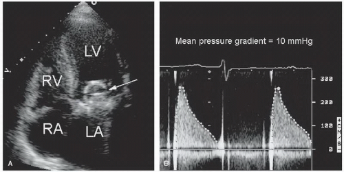
FIGURE 15.13. A: A Starr-Edwards mitral prosthesis (arrow). B: Doppler imaging demonstrates flow through the valve. The mean pressure gradient is approximately 10 mm Hg.


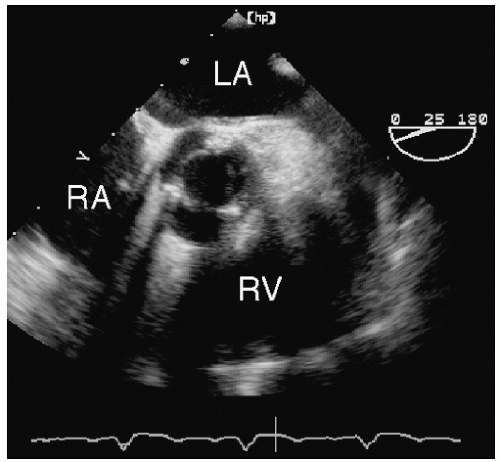
FIGURE 15.14. A short-axis view of a porcine aortic prosthesis from transesophageal echocardiography. The three struts are visualized, forming a triangular-shaped orifice.


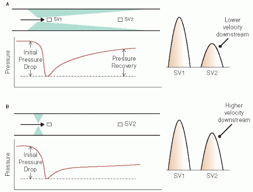
FIGURE 15.15. The concept of pressure recovery. A: In the absence of pressure recovery, different locations for sample volume (SV) measurement yield fairly similar velocities. B: Flow through a tapered stenosis results in significant pressure recovery downstream from the obstruction. In this case, sampling within the obstruction (SV1) yields a higher velocity compared with a sample site downstream (SV2) where pressure recovery has occurred. At this site, the recovery of pressure is associated with a lower velocity. See text for details.

Another unique aspect of prosthetic valve function is the presence of normal, or physiologic, regurgitation. This occurs with virtually all types of mechanical prostheses and is actually part of the design of the valve. Physiologic regurgitation can be divided into two types: closure backflow and leakage. Closure backflow occurs because of the flow reversal required to close the occluding mechanism. This results in a small amount of regurgitation that ends once the occluder mechanism is seated in the sewing ring (Fig. 15.16). Leakage backflow occurs after the prosthesis has closed and is the result of a small amount of retrograde flow between and around the occluding mechanism. It is often part of the design of the prosthesis to provide a washing mechanism and prevent thrombus formation on its upstream side. Because leakage backflow may be holosystolic (or holodiastolic, depending on valve location), it must be distinguished from pathologic regurgitation. This depends on the severity and the pattern of regurgitation. For example, leakage through a bileaflet valve often results in two symmetric narrow jets directed obliquely from the edges of the valve. This type of physiologic regurgitation is illustrated in Figure 15.17. Normal bioprosthetic valves may also exhibit mild regurgitation. For example, some pericardial valves demonstrate mild central regurgitation that resolves 4 to 6 weeks after implantation.

Despite these differences in flow characteristics, the basic Doppler principles applied to native valves are also relevant to the study of prosthetic valves. For example, Doppler imaging can be used to measure both the maximal and mean pressure gradient across prostheses (Fig. 15.18). The assumptions that are critical to the modified Bernoulli equation apply to prosthetic valves as well. Thus, the correlation between pressure gradients obtained by the Doppler technique compared with cardiac catheterization is generally very good. However, because of the existence of multiple jets through many types of prosthetic valves, more than one velocity pattern can often be recorded. As noted previously, the phenomenon of pressure recovery may also lead to overestimation of the pressure gradient. Figure 15.19 illustrates flow through different types of mitral prostheses. Note the variability in the contour and velocity among the four examples. Gradients across “normal” prosthetic valves vary across a wider range compared with native valves. For this reason, it is often helpful to obtain a baseline Doppler
imaging study in all patients at a time when the valve is known to be functioning normally, such as during the first postoperative clinic visit. This can then be used as a reference for future evaluations to help determine whether a given pressure gradient is normal or abnormal for the individual. In addition, tables have been published providing a range of normal values for different types of valves in the various positions.


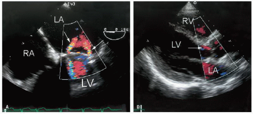
FIGURE 15.16. Physiologic regurgitation through a normally functioning St. Jude mitral prosthesis (arrows) (A) and a porcine aortic prosthesis (arrow) (B).

The continuity equation can also be used to measure the effective orifice area of prosthetic valves. The value of this measurement has the same limitations just described for pressure gradients. Finally, for prosthetic mitral and tricuspid valves, the pressure half-time technique is useful to quantify the severity of stenosis. However, pressure half-time generally overestimates the valve area in the presence of a mitral prosthesis. Again, having a baseline study and using the patient as his or her own control is essential for future management.


FIGURE 15.17. Physiologic regurgitation through a St. Jude aortic valve. The jets originate at the periphery and appear to cross just below the valve (arrow). The occurrence of this type of regurgitation is part of the design of many prosthetic valves.


Application of Echocardiography to Patients with Prosthetic Valves

In patients with prosthetic valves, the role of echocardiography begins in the operating suite at the time of surgery. A comprehensive transesophageal evaluation of the diseased valve(s), if
not performed previously, is essential for optimal intraoperative management. Thus, echocardiography is routinely used prior to valve surgery (to make decisions regarding type and size of prosthesis, feasibility of repair, etc), during surgery (to assess the success and completeness of the procedure), and following surgery (to establish a new baseline and to document a successful procedure). The specific indications for intraoperative transesophageal echocardiography are listed in Table 15.3. Its value in this setting is well documented. Clinical series indicate that intraoperative echo results change the operative plan in up to 15% of cases and identify a problem of sufficient magnitude to warrant revision in approximately 5% of patients. This is especially true in valve surgery, particularly valve repair procedures. As expected, the potential value of echocardiography is directly related to the complexity of the procedure. Valve repair, replacement of multiple valves, valve surgery involving complicated endocarditis, and valve replacement involving stentless valves or homografts are examples of technically challenging operative procedures where to value of intraoperative echocardiography is well established.


FIGURE 15.18. Doppler imaging is used to record flow through an aortic prosthesis. The peak and mean gradients are indicated. Note the presence of valve clicks at the time of opening and closing.


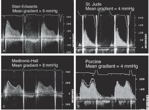
FIGURE 15.19. A-D: Doppler recording of flow through four different mitral prosthetic valves. The mean gradient across each prosthesis is indicated.


Table 15.3 Intraoperative Assessment Using Transesophageal Echocardiography


Class I


1.


Intraoperative transesophageal echocardiography is recommended for valve repair surgery. (Level of Evidence: B)


2.


Intraoperative transesophageal echocardiography is recommended for valve replacement surgery with a stentless xenograft, homograft, or autograft valve. (Level of Evidence: B)


3.


Intraoperative transesophageal echocardiography is recommended for valve surgery for infective endocarditis. (Level of Evidence: B)


Class IIa


4.


Intraoperative transesophageal echocardiography is reasonable for all patients undergoing cardiac valve surgery. (Level of Evidence: C)


From Bonow RO, Carabello BA, Chatterjee K, et al. ACC/AHA 2006 guidelines for the management of patients with valvular disease: a report of the American College of Cardiology/American Heart Association Task Force on Practice Guidelines. J Am Coll Cardiol 2006;48:14-41.


Following discharge, the role of echocardiography consists of defining baseline function and serial assessment for evidence
of dysfunction. Both American College of Cardiology/American Heart Association Management Guidelines and Appropriateness Criteria have been published to provide guidance in this area (see Table 15.4). From these documents, there is general consensus that echocardiography should be performed soon after valve surgery as part of the initial evaluation of the patient during the recovery phase. This examination should focus on an assessment of left and right ventricular function, determination of pulmonary artery pressure, and, of course, a thorough evaluation of the repaired or replaced valve. Because all prosthetic valves have some degree of obstruction, a critical part of the evaluation is to determine the pressure gradient. Careful assessment of regurgitation is also important. Mild valvular regurgitation is normally present in many prosthetic valves. On the other hand, perivalvular regurgitation is usually an abnormal finding that requires thorough assessment and follow-up. Thus, the initial postoperative echocardiogram should clearly document the presence and severity of regurgitation and differentiate normal from abnormal forms.


Table 15.4 Evidence-based Indications and Appropriateness Criteria Related to the Evaluation of Prosthetic Valves


Class I


1.


For patients with prosthetic heart valves, a history, physical examination, and appropriate tests should be performed at the first postoperative outpatient evaluation, 2-4 weeks after hospital discharge. This should include a transthoracic Doppler echocardiogram if a baseline echocardiogram was not obtained before hospital discharge. (Level of Evidence: C)


2.


For patients with prosthetic heart valves, routine follow-up visits should be conducted annually, with earlier reevaluations (with echocardiography) if there is a change in clinical status. (Level of Evidence: C)


Class IIb


3.


Patients with bioprosthetic valves may be considered for annual echocardiograms after the first 5 years in the absence of a change in clinical status. (Level of Evidence: C)


Class III


4.


Routine annual echocardiograms are not indicated in the absence of a change in clinical status in patients with mechanical heart valves or during the first 5 years after valve replacement with a bioprosthetic valve. (Level of Evidence: C)


Criteria


Appropriateness Score (1-9)


28.


Initial evaluation of prosthetic valve for establishment of baseline after placement


A (9)


30.


Reevaluation of patients with prosthetic valve with suspected dysfunction or thrombosis or a change in clinical status


A (9)


29.


Routine (yearly) evaluation of a patient with a prosthetic valve in whom there is no suspicion of valvular dysfunction and no change in clinical status


I (3)


Reprinted with permission of the ACCF from Douglas PS, Khandheria B, Stainback RF, et al. ACCF/ASE/ACEP/ASNC/SCAI/SCCT/SCMR 2007 appropriateness criteria for transthoracic and transesophageal echocardiography. J Am Coll Cardiol 2007;50(2):187-204.


Following this initial echocardiographic study, subsequent assessment must be individualized. According to the guidelines, there is agreement that echocardiography should be considered if there is a change in clinical status, evidence of infection, or reason to suspect valve dysfunction. Routine (e.g., annual) echocardiographic studies, in the absence of one of the indications listed in the guidelines, are not recommended. However, once dysfunction is documented, serial evaluation, including clinical and echocardiographic monitoring, should be undertaken. This would include, for example, patients with bioprosthetic valves who exhibit early signs of primary tissue degeneration. Finally, in children who are still growing, the possibility of developing prosthesis-patient mismatch mandates, particularly, close follow-up. This occurs because the effective orifice area of the prosthesis remains fixed while the child’s stroke volume increases with age. Monitoring for worsening hemodynamics as a result of normal growth is essential.


General Approach to Prosthetic Valves

Transthoracic two-dimensional imaging is generally adequate to distinguish among the various types of prosthetic valves. However, the high reflectance of the prosthetic material creates challenges for the echocardiographer. Because the speed of sound changes as it passes through prosthetic materials, size and appearance can be distorted. Some decrease in gain setting is generally necessary to compensate for these differences. The high reflectance also leads to shadowing behind the prostheses. Reverberations frequently appear behind the prosthetic structures, which may obscure targets of interest. To overcome these problems, multiple echocardiographic windows must be used to fully interrogate the areas around prosthetic valves. A thorough anatomic assessment is also facilitated by the use of three-dimensional techniques. For example, a properly oriented three-dimensional image will provide a complete, circumferential view of a sewing ring, so that any abnormal masses that might be present will be visualized. In other cases, transesophageal echocardiography will be necessary to provide a thorough examination. Most recently, transesophageal three-dimensional echocardiography has been applied to the assessment of prosthetic valves. Initial experience suggests that this new technique is well suited for the assessment of mitral prostheses (Fig. 15.20). By displaying en face views of the mitral apparatus from both atrial and ventricular perspectives, a very complete assessment of structure and function is feasible in most patients. Experience using transesophageal threedimensional imaging for aortic and tricuspid prostheses is limited and may present more technical challenges.

The two-dimensional echocardiographic appearance of bioprosthetic leaflets more closely approximates that of native valves. In fact, newer stentless aortic prostheses can be nearly indistinguishable from a normal native aortic valve. For stented valves, imaging is ideally performed with the ultrasound beam aligned parallel to flow to avoid the shadowing effects of the stents and sewing ring. The leaflets themselves are quite similar to native valve tissue, both in texture and excursion. Over time, bioprostheses tend to thicken and become fibrotic, leading to increased echogenicity and reduced excursion on two-dimensional imaging (Fig. 15.21). Such valves can become stenotic and/or regurgitant. This illustration demonstrates a brittle, fibrotic porcine mitral valve with partial rupture of one cusp leading to severe mitral regurgitation. In all cases, a combination of two-dimensional and Doppler imaging is required to thoroughly assess bioprosthetic valves (Fig. 15.22).


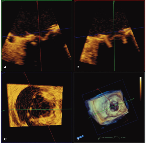
FIGURE 15.20. A porcine mitral prosthesis is evaluated with transesophageal three-dimensional echocardiography. Panels A and B are systolic frames of the prosthesis with two-dimensional echocardiography. Panel C is a short-axis view of the prosthesis. In panel D, a volume rendered three-dimensional image provides a clear circumferential view of the sewing ring.


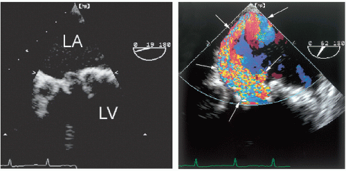
FIGURE 15.21. An example of primary tissue degeneration involving a porcine mitral valve. The leaflets are thickened and fibrotic with decreased mobility (left). Right: Color Doppler imaging demonstrates severe mitral regurgitation with an eccentric jet (arrows).


FIGURE 15.22. A: An example of a mildly thickened porcine mitral prosthesis. The structure and motion of the leaflets are often obscured by the struts. B: Doppler imaging demonstrates a mean gradient of 10 mmHg. MV, mitral valve.


FIGURE 15.23. M-mode echocardiogram of a St. Jude mitral prosthetic valve. M-mode echocardiography is ideal to record the brisk opening and closing of the disks (arrows). IVS, interventricular septum; MV, mitral valve.

For the reasons noted above, mechanical valves can be quite difficult to assess with two-dimensional echocardiography. Although gross abnormalities can be detected, more subtle changes are often missed, especially with transthoracic imaging. The primary goals of two-dimensional echocardiography in this setting are to confirm stability of the sewing ring, determine the specific type of prosthesis, confirm the opening and closing motion of the occluding mechanism, and evaluate for gross structural abnormalities such as vegetations and thrombi. Assessing the mobility of the occluding mechanism can be difficult. However, through careful interrogation, the rapid motion of the leading edge of the disk or ball generally can be recorded. In normal prostheses, the motion is brisk and consistent with each beat (Figs. 15.6, 15.7, 15.8 and 15.9). M-mode imaging can be useful in this case to more precisely define the brisk opening and closing and the degree of excursion of the occluder (Fig. 15.23). For bileaflet prostheses, it is important to search for both hemidisks, which often have slightly out of phase motion as they open and close in close proximity (Figs. 15.7 and 15.12).

As with two-dimensional imaging, the Doppler examination also faces unique challenges in the setting of a prosthetic valve. Because of the variability of flow through and around the different prostheses, color flow imaging is often helpful to define the location and direction of the various flow patterns. Some prosthetic valves have more than one orifice and, consequently, a complex flow profile. Once the desired flow patterns are localized with color flow imaging, pulsed and continuous wave Doppler imaging can be oriented to quantify flow velocity. As already noted, velocities will always tend to be higher through prosthetic valves, depending in part on the size of the specific prosthesis. Whenever velocity is higher than expected, consider the possibility of pressure recovery, as discussed previously.


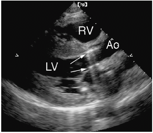
FIGURE 15.24. The presence of a St. Jude aortic prosthesis (arrows) creates a pattern of reverberations that extends into the left atrium. This creates a shadowing effect and can obscure the presence of mitral regurgitation.


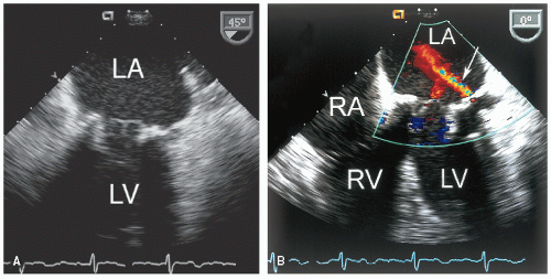
FIGURE 15.25. A: A porcine mitral prosthesis is visualized using transesophageal echocardiography. B: Color Doppler imaging demonstrates both transvalvular and perivalvular (arrow) mitral regurgitation.

Assessing valvular regurgitation is primarily limited by the shadowing effect of the prosthetic valve itself. Because the signal-to-noise ratio for Doppler imaging is lower compared with two-dimensional echocardiographic imaging, the shadowing effect is even more pronounced and the ability to record a Doppler signal “behind” a prosthetic valve is very limited. Multiple views must be used to fully interrogate the regurgitant signal. Figure 15.24 demonstrates how the shadowing effect of an aortic prosthesis obscures the left atrium from the parasternal window. It is also important to distinguish transvalvular from perivalvular regurgitation. This is best accomplished using color flow imaging to interrogate the circumference of the sewing ring on the upstream side of the valve (Fig. 15.25). With the increased sensitivity of modern equipment, a small amount of perivalvular regurgitation may be recorded in the immediate postoperative period that will often disappear or diminish over
time (Fig. 15.26). Three-dimensional transesophageal imaging will likely prove to be the most sensitive method for this determination. Figure 15.27 is an example of flow through a normally functioning mechanical mitral prosthesis recorded with real-time three-dimensional imaging. In this example, both antegrade flow and mild regurgitant flow are demonstrated. One advantage of this approach is the ability to distinguish flow through the various orifices of a mechanical prosthesis. Spectral Doppler recordings of prosthetic valve flow will also include brief, high-velocity signals referred to as “clicks.” These are intense recordings associated with both the opening and closing of the occluder mechanism. They provide useful information on timing and are particularly helpful to identify the various phases of filling and ejection. In Figure 15.28, both normal and abnormal St. Jude aortic prostheses are shown. In Figure 15.28A, note the valve clicks marking opening and closing of the normal valve. Figure 15.28B is taken from a patient with a prosthesis that is partially obstructed by a thrombus on the sewing ring. Note that the opening valve click is absent, and the closing click is very faint. The high velocity is evidence of the increased pressure gradient across the partially obstructed valve.

Only gold members can continue reading. Log In or Register to continue

Stay updated, free articles. Join our Telegram channel

Jun 22, 2016 | Posted by in CARDIOLOGY | Comments Off on Prosthetic Valves

Full access? Get Clinical Tree

Get Clinical Tree app for offline access