1. The PB occurs early in the cardiac cycle.
2. The presence of the PB prevents the occurrence of the next normal beat.
3. There is a pause following the PB until the next normal beat occurs.
A single PB is potentially the first beat of a sustained tachyarrhythmia (“tach”). It may be followed by any number of similarly appearing beats to which the terms in the following two paragraphs are applied (Table 15.1).
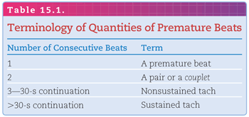
When a PB follows every normal beat, the term bigeminy is used; when a PB follows every second normal beat, the term trigeminy is used. PBs may originate from any part of the heart other than the sinoatrial (SA) node. They are generally classified as supraventricular premature beats (SVPBs) or ventricular premature beats (VPBs; Fig. 15.2). This distinction is useful because beats originating from anywhere above the branching of the common bundle (SVPBs) are capable of producing a normal or abnormal QRS complex depending on whether they are conducted normally or aberrantly through the intraventricular conduction system. PBs originating from beyond the branching of the common bundle (VPBs), however, can produce only an abnormally prolonged QRS complex of ≥0.12 second because they do not have equal access to both the right and left bundle branches. It should be emphasized that VPBs are always abnormally prolonged, but SVPBs are not always of normal duration.
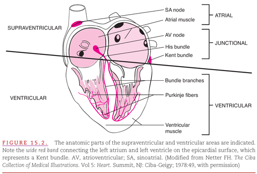
SVPBs may be either atrial premature beats (APBs) or junctional premature beats (JPBs). The term “junctional” is used instead of “nodal” because it is impossible to distinguish beats originating within the atrioventricular (AV) node from those originating in another structure located between the atria and the ventricles. Normally, the AV junction consists of only the AV node and the common bundle. Abnormally, however, an accessory AV conduction pathway (Kent bundle) is also a part of the AV junction as shown in Figure 15.2.
DIFFERENTIAL DIAGNOSIS OF WIDE PREMATURE BEATS
When SVPBs produce abnormally prolonged or wide QRS complexes (>0.12 second), identification of their supraventricular versus a ventricular origin may be facilitated by observing the effect on the regularity of the underlying sinus rhythm (Fig. 15.3). A VPB typically does not disturb the sinus rhythm, because it is not conducted retrogradely through the slowly conducting AV node into the SA node (see Fig. 15.3A). Although the SA node discharges on time, its impulse cannot be conducted antegradely into the ventricles because of the refractoriness following the VPB. The pause between the VPB and the following conducted beat is termed a compensatory pause because it compensates for the prematurity of the VPB. The interval from the sinus beat prior to the VPB to the sinus beat following the VPB is equal to two sinus cycles.
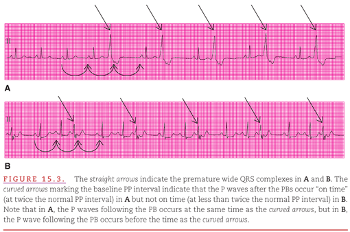
In contrast to a VPB (see Fig. 15.3B), an SVPB does disturb the sinus rhythm. Unlike the VPB, the SVPB can be conducted retrogradely into the SA node, discharging it ahead of schedule and causing the following cycle to also occur ahead of schedule. The pause between the SVPB and the following sinus beat is therefore less than compensatory. This is apparent because the interval from the sinus beat before the SVPB to the sinus beat after the SVPB is less than the duration of two sinus cycles. However, when the SVPB prematurely discharges the SA node, it occasionally suppresses SA nodal automaticity. This overdrive suppression may delay the formation of the next sinus impulse for so long that the resulting pause is compensatory, or even longer than compensatory. Thus, the compensatory pause must not be relied upon as the sole indicator of ventricular origin of a wide PB.
MECHANISMS OF PRODUCTION OF PREMATURE BEATS
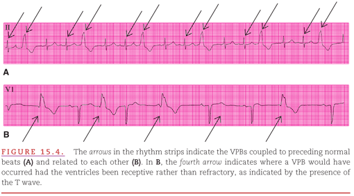
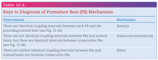
PBs may be caused by the two mechanisms indicated in Chapter 14: reentry or automaticity. It is usually difficult to determine the mechanism of PBs unless two or more occur in succession. Fortunately, the mechanism of a PB is usually not clinically important unless consecutive abnormal beats are present. When identification of the mechanism for a PB is considered clinically important, the following observations of the coupling intervals between beats may be helpful (Fig. 15.4): Reentry produces a constant relationship between normal and PBs (see Fig. 15.4A), whereas automaticity produces a varying relationship between normal and abnormal beats but a constant relationship between abnormal beats (see Fig. 15.4B) as in Table 15.2.
The usual APB has three features:
1. A premature and abnormal-appearing P wave.
2. A QRS complex similar to that of the normal sinus beats.
3. A following interval that is less than compensatory because of the retrograde activation of the SA node.
Usually, all of these characteristics are obvious, but “deceptions” occur (particularly when the APB is most premature) so that no one characteristic is completely reliable. In Figure 15.5A, the premature P waves appear normal; in Figure 15.5B, the premature QRS complexes are not always similar to those of the normal sinus beats. Some common ECG deceptions in recognizing APBs are:
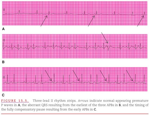
1. The P wave may be unrecognizable because it occurs during the previous T wave (see Fig. 15.5B, C).
2. The QRS complex may show aberrant ventricular conduction (see Fig. 15.5B).
3. The pause between the APB and the following P wave is compensatory, probably because of the extreme earliness of the APB (see Fig. 15.5C).
It is extremely rare to have all three of these deceptions appear at the same time. Therefore, with care, one usually has no difficulty in identifying an APB.
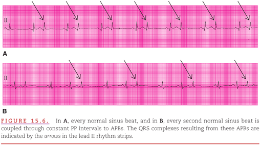
When an APB follows every sinus beat, the result is atrial bigeminy (Fig. 15.6A); when it follows every two consecutive sinus beats, the result is atrial trigeminy (see Fig. 15.6B).
When APBs occur very early (a short coupling interval), some parts of the heart may not have had time to complete their recovery from the preceding normal activation. This may result in failure of the premature atrial activation to cause any ventricular activation. Indeed, the most common cause of an unexpected atrial pause is a nonconducted APB (Fig. 15.7). It is better to refer to such beats as “nonconducted” rather than “blocked” because, by definition, “block” implies an abnormal condition. APBs fail to be conducted only because they occur so early in the cycle that the AV node is still in its normal refractory period. It is important to differentiate normal (physiologic) from abnormal (pathologic) nonconduction to avoid mistakenly initiating an antiarrhythmia treatment.

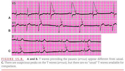
Nonconducted APBs that occur in a bigeminal pattern are particularly difficult to identify (Fig. 15.8). If the premature P waves are obscured by the T waves of the preceding normal beats, and if the earlier T waves during the regular sinus rhythm are not available for comparison, then the rhythm is often misdiagnosed as sinus bradycardia.
When APBs occur very early in the cardiac cycle of normal beats, they may have other effects on conduction to the ventricles (Fig. 15.9). In Figure 15.9A, there is prolonged AV conduction, whereas in Figure 15.9B there is both slightly prolonged AV conduction and also aberrant intraventricular conduction. In Figure 15.9B, there are varying coupling intervals (PP intervals) between normal sinus beats and APBs. When the PP interval is long, the premature PR interval is normal, but when the PP interval is short, the premature PR interval is prolonged. This inverse relationship occurs because of the uniquely long relative refractory period of the AV node: The longer the duration from its most recent activation, the better is the node able to conduct the following impulse and vice versa. This concept is vital to use of the ECG to differentiate a nodal versus Purkinje location of AV block.
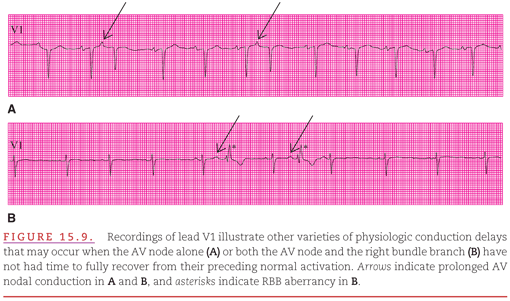
When an early APB traverses the AV junction but encounters persistent normal refractoriness in one of the bundle branches or fascicles, aberrant ventricular conduction occurs (see Fig. 15.9B). The morphology of the QRS complex is altered, and its duration may be so prolonged that it resembles a VPB. Detection of the preceding P wave and/or finding that the pause between the APB and the next sinus beat is less than compensatory usually establishes the diagnosis of an APB.
APBs may occur so early that even parts of the atria have not completed their refractory periods. During this time (the vulnerable period), the APB may initiate a reentrant atrial tachyarrhythmia (Fig. 15.10). In this instance, the APB becomes the first beat of atrial flutter/fibrillation (see Chapter 17). Killip and Gault1 developed the rule that when the PP interval is <50% of the previous PP interval, an APB is quite likely to initiate atrial flutter/fibrillation.

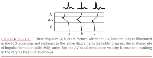
PBs arising in the AV junction may retrogradely activate the atria before, during, or after ventricular activation, and the retrograde P wave may therefore be seen preceding or following the QRS complex or may be lost within the QRS complex. These variations are illustrated in Figure 15.11.
The diagnosis of junctional origin of PBs is easiest when a premature normal QRS complex is closely accompanied by an inverted P wave (Fig. 15.12). As expected, the morphology of the P waves associated with JPBs is markedly different from that of the P waves of normal sinus rhythm. The polarity of the P waves associated with JPBs is approximately opposite that of the P waves of normal sinus rhythm, as is best seen in a lead with base-to-apex orientation, such as lead II. A P wave originating from the AV junction is also inverted in the other inferiorly oriented leads (e.g., aVF), is upright in superiorly oriented leads aVR and aVL, and is almost flat in leftward-oriented leads I and V5.

A JPB may be confused with an APB when a premature normal QRS complex is not closely accompanied by an abnormal P wave (Fig. 15.13
Stay updated, free articles. Join our Telegram channel

Full access? Get Clinical Tree


