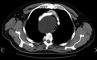
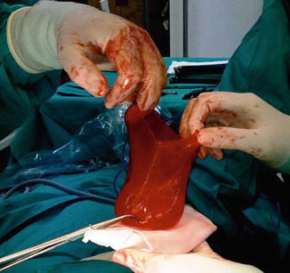
Fig. 15.1
Computed tomography of the cervicothoracic area showing a large anterior and right paratracheal cystic mass

Fig. 15.2
Computed tomography of the superior part of the chest showing the thoracic prolongation of the cystic mass into the middle mediastinum

Fig. 15.3
Unilocular mesothelial cyst of the mediastinum completely resected
15.3 Ectopic Mediastinal Parathyroid Adenoma
Abnormal ectopic parathyroid glands have been reported to occur in about 16 % of patients operated for hyperparathyroidism [19]. The most frequent locations of ectopic parathyroid glands are the thymus, the posterior mediastinum, the mediastinal fat of the anterosuperior mediastinum, the thyroid gland, and the fat of the aortopulmonary window [20–22]. In up to 25 % of cases, primary hyperparathyroidism is caused by an ectopic parathyroid adenoma that is mostly found in the anterosuperior mediastinum. Prior to the introduction of new metabolic and morphological imaging techniques for the assessment of the parathyroid glands, ectopic adenomas were responsible for more than 50 % of persistent or recurrent hyperparathyroidism after an initial surgical neck exploration [23–27].
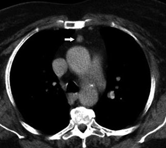
Different approaches can be used for the removal of ectopic mediastinal parathyroid adenomas according to the precise location of the mass and the experience of the surgeon. These include open or endoscopic transcervical approach, thoracoscopic or open transthoracic approach, sternotomy, and transmanubrial or parasternal mediastinotomy. However, most ectopic adenomas can be excised through a transcervical incision, and in less than 20 % of cases, an alternative approach will be required [28]. The transcervical approach is recommended for ectopic adenomas located in the anterosuperior mediastinum [21, 29] located cranial to the aortic arch (Fig. 15.4) [30].


Fig. 15.4
The thoracic computed tomography shows an ectopically placed parathyroid adenoma in the anterior mediastinum (white arrow). Parathyroid scintigraphy with 99mTc-MIBI showed focal activity
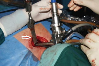
Different types of sternal retractors can be of great help to explore the mediastinum, especially if it is necessary to perform a transcervical closed thymectomy [26] or when the adenoma is located deeper in the mediastinum (Fig. 15.5) [31]. The dissection and visualization of the anterior mediastinum away from the innominate vein could be facilitated by introducing a 30º thoracoscope or a spreadable videomediastinoscope while open surgical instrument can be used. Some authors have described a combined thoracoscopic and transcervical approach, especially for adenomas located in the middle mediastinum [31]. Although it is possible to reach the aortopulmonary window through the neck, a detailed visualization and anatomical exposure of the structures is not easy, and probably an alternative approach should be planned in these cases (parasternal mediastinotomy, left videothoracoscopy, or a sternotomy). Liu et al. have described an original approach to the posterior and superior mediastinal adenomas, the so-called backdoor approach [21]. After performing an incision near the medial border of the sternocleidomastoid muscle, the authors reach the paraspinal space and the posterosuperior mediastinum by blunt dissection. Ectopic adenomas located in the posterior and superior mediastinum between the trachea and the esophagus could be completely excised through transcervical minimally invasive endoscopic surgical approach [32]. Also, transcervical endoscopic-assisted mediastinal parathyroidectomy could be done by carbon dioxide insufflation of the neck and mediastinum [33]. However, carbon dioxide insufflation has been associated with postoperative complications, such as massive subcutaneous emphysema, hypercapnia, cardiac arrhythmia, and acidosis [34].


Fig. 15.5
The image shows the bivalved spreadable videomediastinoscope and a sternal retractor (white arrow) during the excision of an ectopic mediastinal adenoma. The sternal retractor allows us the direct visualization of the anterior mediastinum. The videomediastinoscope can be of great help to explore the mediastinum away from the innominate vein
Some key points should be taken into account when the transcervical approach to excise an ectopic parathyroid adenoma is selected. Firstly, the incidence of double or multiple adenomas ranges from 1 to 10 % and not always all the functional foci of parathyroid tissue are accurately detected preoperatively. Intraoperative monitoring of serum levels of parathyroid hormone (PTH) is the best indicator of complete parathyroid removal. In addition to the frozen section study of the specimen that confirms the presence of parathyroid tissue, an intraoperative decrease of the PTH in the blood (less than 40 % of the initial range or to a normal level) should be demonstrated before awakening the patient. Secondly, in the case of ectopic adenomas located in the middle mediastinum, both the left and the right paratracheal recurrent laryngeal nerves should be monitored in order to avoid injury of them.
15.4 Descending Necrotizing Mediastinitis
Head and neck infections may extend to the mediastinum through the fascial spaces and the carotid sheaths. Descending necrotizing mediastinitis was described for the first time by Pearse in 1938, who identified the anatomical routes of the so-called secondary cervical suppuration [35]. Descending necrotizing mediastinitis is a life-threatening acute septic complication from an oropharyngeal or dental infection that spreads caudally along the fascial compartments of the neck (prevertebral fascia, retropharyngeal space, perivascular space, and pretracheal fascia) into the mediastinum. Gravity and the negative intrathoracic pressure facilitate the spread of cervical infections down to the mediastinum through the abovementioned cervicomediastinal fascial planes.
The diagnostic criteria to establish the diagnosis of this condition were reported by Estrera et al. in 1983 and still remain valid [36]. These include the following: (1) clinical manifestations of severe infection; (2) demonstration of characteristic radiographic features, such as mediastinal widening, mediastinal emphysema, and mediastinal fluid collection with bubbles or abscesses with air-fluid levels; (3) establishment of the relationship of oropharyngeal or cervical infection with the development of the necrotizing mediastinal process; and (4) documentation of necrotizing mediastinal infection at operation or postmortem examination or both.
Anaerobic bacteria play a central role in a septic process that is generally caused by a constellation of anaerobic and aerobic bacteria. Diagnosis is based on early clinical suspicion together with computed tomography scan of the chest and neck. Aggressive cervical and mediastinal debridement and drainage, effective pleural and pericardial drainage, and administration of broad-spectrum antibiotics, covering both aerobes and anaerobes, are essential to prevent involvement of the entire mediastinum leading to sepsis and multiorgan failure. Mortality rates still remain high and range between 0 and 40 % in most of the published series [37–44]. Although some authors have reported excellent results in the treatment of cervical necrotizing fasciitis with or without descending necrotizing mediastinitis with percutaneous catheter drainage [43, 44], the majority of the authors support the immediate transcervical debridement and drainage in combination (or not) with a transthoracic approach.
From a practical point of view, Endo et al. [45] classified descending necrotizing mediastinitis according to the extension of the infection on computed tomography scan, and the three proposed types are as follows: type I, the infection is localized to the upper mediastinum and above the tracheal bifurcation; type IIA, the infection extends to the lower anterior mediastinum; and type IIB, the infection extends to the lower anterior and posterior mediastinum. A management algorithm based on this classification has been proposed by many authors [39, 42, 45–47]. Type I descending necrotizing mediastinitis could be treated with a transcervical mediastinal debridement and drainage. Retropharyngeal, prevertebral, and pretracheal spaces should be exposed and debrided. Cervical and mediastinal drains should be left in place after surgery. Unilateral or bilateral chest drains could be necessary if there is evidence of pleural effusion. Types IIA and IIB should be treated by a combined cervical and transthoracic, transternal, or subxiphoid approaches depending on the need to have access to one or both pleural cavities [38, 48, 49].
Several important points should be mentioned in the management of patients with descending necrotizing mediastinitis. First, the need for a tracheostomy should be carefully evaluated. Second, CT scan should be repeated after 48–72 h postoperatively or in patients without clinical improvement in order to identify additional or persistent fluid or gas accumulation in the neck, chest, or superior abdomen requiring additional surgical procedures.
15.5 Miscellaneous
The rate of tracheobronchial injuries during a surgical exploration of the mediastinum is lower than 0.5 % [50–52]. Previous mediastinal radiation therapy or surgery may increase the risk. In some cases, repair via thoracotomy may be necessary [53, 54], but eventually tracheal or bronchial injuries may be repaired through the same videomediastinoscopic, open transcervical approach or a combination of both. Saumench-Perrramon et al. reported two cases of bronchial injuries during videomediastinoscopy [50]. In the first case, the injury was located in the anterior surface of the right main bronchus. Treatment consisted of the direct suture through a bivalve videomediastinoscope plus fibrin glue that was spread over the suture line. In the second case the only treatment consisted of covering the bronchial hole with a sealant patch.
Also, the transcervical approach has been used to remove foreign bodies lodged in the mediastinum [54, 55]. These foreign bodies could reach the mediastinum after a surgical procedure performed near it or accidentally. Surgical removal of foreign bodies located in the mediastinum should be performed as soon as possible in order to prevent infection and vascular or visceral injuries due to migration.
Although the most frequent approach to the cervicothoracic goiters is a combination of open transcervical and transternal (anterosuperior and middle mediastinal goiters) or transthoracic approach (middle and posterior mediastinal goiters) [56, 57], the videomediastinoscopic assistance to the standard cervical procedure has been described for resecting large mediastinal goiters or ectopic thyroid located anywhere in the mediastinum [58, 59]. The advantages of the transcervical endoscopic assistance are the direct visualization of the mediastinal structures, a better control of the mediastinal bleeding, and the possibility to avoid a partial or total sternotomy.
Kang et al. have described the videomediastinoscopic bilateral hilar-bronchial release to reduce anastomotic tension prior a transcervical resection of a long segment of trachea followed by a primary end-to-end anastomosis [60].
In addition to the lymphadenectomy of the central and lateral compartments of the neck, Khoo et al. [61] and Ducic et al. [62] have described the transcervical superior mediastinal lymphadenectomy down to the level of the brachiocephalic vein in cases of thyroid carcinoma. The incidence of lymph nodes metastases in this compartment is up to 35 % and has been related to the risk of recurrence after surgery.
Tracheal diverticulum is a rarely encountered entity, accounts for less than 2 % of cystic lesions of the mediastinum, and is usually full of air. Mediastinoscopic debridement of an infected tracheal diverticulum located in the paratracheal area has been reported [63]. The differential diagnosis could be difficult due to radiological features similar to lymph node metastasis.
15.6 Conclusion
Depending on the location of the mediastinal lesions and on the experience of the surgical team, the transcervical approach should be considered as first choice to avoid more invasive procedures and their morbidity.
References
1.
2.
3.
4.
Burjonrappa SC, Taddeucci R, Arcidi J. Mediastinoscopy in the treatment of mediastinal cysts. JSLS. 2005;9:142–8.PubMedCentralPubMed
Stay updated, free articles. Join our Telegram channel

Full access? Get Clinical Tree


