Masses, Tumors, and Source of Embolus


Masses, Tumors, and Source of Embolus


Normal Variants and Artifacts: Sources of False-Positive Findings

The echocardiographic evaluation of intracardiac masses is critically dependent on the ability to distinguish normal from abnormal findings. Ultrasound artifacts are common, even in high-quality studies, and may be mistaken for pathologic conditions. Near-field clutter and reverberations are examples of artifacts often confused with pathology (e.g., apical thrombi) on twodimensional echocardiography. Such artifacts, which are covered in Chapter 2, must be avoided whenever possible and correctly identified when present. Proper transducer selection and the use of multiple acoustic windows are among the strategies that can be used to avoid potential misinterpretations.

Anatomic variants are ubiquitous, may involve any chamber or valve structure, and are potentially confused with pathologic structures. A list of commonly encountered normal structures that often are interpreted as pathologic is provided in Table 23.1. The right atrium is the chamber that is most often a source of anatomic variants leading to inaccurate interpretation. The Chiari network, eustachian valve, and crista terminalis are examples of structures normally found in the right atrium that, due to individual variation, are frequently confused with pathologic entities. Fatty infiltration in the atrioventricular groove, especially around the tricuspid valve, is a common source of confusion. A benign condition, this fatty deposit is frequently mistaken for tumor or fluid. False tendons in the left ventricular apex are common and occasionally misinterpreted as thrombi (Fig. 23.1). In this example, the diagnosis of a false tendon is relatively straightforward. In some cases, the tendon can be mistaken for the surface of an apical thrombus. Color flow imaging or contrast echocardiography, by demonstrating flow on either side of the linear structure, can be helpful to make this distinction. Additional sources of confusion can be iatrogenic. For example, the suture line in the posterior atrial wall after cardiac transplantation and indwelling pacemaker leads or catheters are examples of “normal” structures that may be misinterpreted as pathologic. Figure 23.2 is an example of a right ventricular moderator band, another normal cardiac structure that can be confused with abnormal masses, such as thrombi.


Table 23.1 Normal Variants and Benign Conditions Often Misinterpreted as Pathologic


Right atrium


Chiari network


Eustachian valve


Crista terminalis


Catheters/pacemaker leads


Lipomatous hypertrophy of interatrial septum


Pectinate muscles


Fatty material (surrounding the tricuspid annulus)


Left atrium


Suture line following transplant


Fossa ovalis


Calcified mitral annulus


Coronary sinus


Ridge between LUPV and LAA


Lipomatous hypertrophy of interatrial septum


Pectinate muscles


Transverse sinus


Right ventricle


Moderator band


Muscle bundles/trabeculations


Catheters and pacemaker leads


Left ventricle


False chords


Papillary muscles


LV trabeculations


Aorta


Brachiocephalic vein


Innominate vein


Pleural effusion


LAA, left atrial appendage; LUPV, left upper pulmonary vein; LV, left ventricle.


Recognition of such normal variants depends on image quality and technique as well as experience. The use of multiple imaging windows and transducers of different frequency are additional strategies to ensure an accurate diagnosis. The availability of clinical information (such as whether the patient has a pacemaker) can be extremely valuable in avoiding errors.


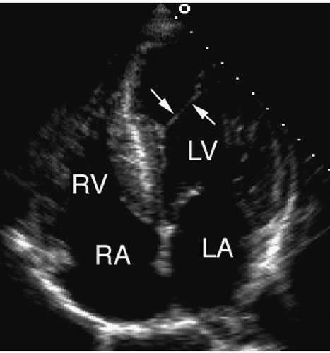
FIGURE 23.1. An apical four-chamber view demonstrates a false tendon (arrows) in the left ventricular apex.


FIGURE 23.2. A moderator band (arrow) is seen in the apex of the right ventricle.


Role of Echocardiography

Guidelines for the use of echocardiography in this setting have been published (Table 23.2). These include an evidence-based list of indications in which the value and utility of echocardiography have been demonstrated. Table 23.2 also contains the more recently developed appropriateness criteria that pertain to echo’s role in the evaluation of patients with known or suspected cardiac masses or source of embolus. Diagnostically, this application represents a broad category of conditions for which imaging is critical. Assessing cardiac anatomy and identifying abnormal structures are tasks well suited to echocardiography. For many patients, the ability to confidently exclude an intracardiac mass or potential source of embolus is often echocardiography’s most important contribution. When an anatomic abnormality is present, the imaging test must be able to detect it with high sensitivity; characterize its extent, location, and size; and distinguish it from artifact or normal variants. Through a careful anatomic assessment, echocardiography frequently provides important diagnostic information regarding the etiology of the mass and helps guide subsequent therapy. A limitation of echocardiography, however, is its inability to provide tissue or histologic diagnosis. Distinguishing a benign tumor from a malignancy, or a thrombus from a vegetation, is often impossible on the basis of ultrasound alone.


Table 23.2 Echocardiography in Patients with Cardiac Masses and Tumors


Indications


Class


1.


Evaluation of patients with clinical syndromes and events suggesting an underlying cardiac mass.


I


2.


Evaluation of patients with underlying cardiac disease known to predispose to mass formation for whom a therapeutic decision regarding surgery or anticoagulation will depend on the results of echocardiography.


I


3.


Follow-up or surveillance studies after surgical removal of masses known to have a high likelihood of recurrence (i.e., myxoma).


I


4.


Patients with known primary malignancies when echocardiographic surveillance for cardiac involvement is part of the disease staging process.


I


5.


Screening persons with disease states likely to result in mass formation but for whom no clinical evidence for the mass exists.


IIb


6.


Patients for whom the results of echocardiography will have no impact on diagnosis or clinical decision making.


III


Appropriateness Criteria


Appropriateness Score (1-9)


34.


Evaluation for cardiovascular source of embolic event (PFO/ASD, thrombus, neoplasm)


A (8)


35.


Evaluation of a cardiac mass (suspected tumor or thrombus)


A (9)


36.


Evaluation of pericardial conditions including but not limited to pericardial mass, effusion, constrictive pericarditis effusive-constrictive conditions, patients postcardiac surgery, or suspected pericardial tamponade


A (9)


Adapted from Cheitlin MD, Alpert JS, Armstrong WF, et al. ACC/AHA Guidelines for the Clinical Application of Echocardiography: a report of the American College of Cardiology/American Heart Association Task Force on Practice Guidelines (Committee on Clinical Application of Echocardiography) developed in collaboration with the American Society of Echocardiography. Circulation 1997;95:1686-1744 and Douglas PS, Khandheria B, Stainback RF, Weissman NJ. ACCF/ASE/ACEP/ASNC/SCAI/SCCT/SCMR 2007 appropriateness criteria for transthoracic and transesophageal echocardiography. J Am Coll Cardiol 2007;50:187-207, with permission. ASD, atrial septal defect; PFO, patent foramen ovale.


Cardiac Tumors


Primary Tumors

Echocardiography is useful to identify conditions in which masses may develop, is an accurate technique to detect and characterize masses once they occur, and provides a noninvasive means for surveillance after treatment or removal. Most tumors in the heart are the result of direct spread from adjacent malignancies or metastatic disease; primary cardiac tumors account for a small percentage of the total number. Primary tumors can be either benign or malignant and can occur in all age groups. The most common primary cardiac tumors are listed in Table 23.3. Of these, benign tumors outnumber malignant ones by a ratio of approximately 3 to 1.


Table 23.3 Relative Frequency of Primary Cardiac Tumors


Type


%


Benign


Myxoma


30


Lipoma


10


Papillary fibroelastoma


8


Rhabdomyoma


6


Fibroma


3


Hemangioma


2


Teratoma


1


Malignant


Angiosarcoma


8


Rhabdomyosarcoma


5


Fibrosarcoma


3


Mesothelioma


3


Lymphoma


2


Leiomyosarcoma


1


By far, the most common benign primary tumor of the heart is the myxoma, accounting for approximately 30% of all primary cardiac tumors. Myxomas are usually single and occur in the left atrium in 75% of cases where they most often arise from the area of the fossa ovalis (Fig. 23.3A). Their size, shape, and texture can be quite varied. Myxomas may be smooth surfaced but are more often irregularly shaped with filamentous fronds or have the appearance of a “cluster of grapes.” They are typically nonhomogeneous in texture with lucent centers or areas of calcification. Myxomas can be quite large, occupying most of the left atrium and resulting in obstruction to left ventricular filling. A large atrial myxoma is shown in Figure 23.3B. In this patient, the tumor nearly occludes the mitral orifice during diastole. The most important clue to the diagnosis is their location in the left atrium and origin from the midportion of the atrial septum. Given a typical presentation, echocardiography is virtually diagnostic of myxoma. Transthoracic imaging is usually sufficient, although small tumors or those that involve the right side of the heart may require transesophageal echocardiography for diagnosis. Three-dimensional echocardiography has also been used to more fully characterize atrial myxomas (Fig. 23.4). Myxomas sometimes involve the right atrium (15%) or the left or right ventricle (5% each) (Figs. 23.5 and 23.6). In the example shown in Figure 23.6, note the mobility of this right atrial myxoma and how it extends through the tricuspid valve in diastole resulting in obstruction to right ventricular inflow. In 5% of cases, myxomas are multiple. They are most often confused
with thrombi, although their characteristic location and attachment site is generally helpful in the differential diagnosis. After surgical excision, myxomas can recur. Therefore, surveillance echocardiograms should be obtained annually for several years to guard against this possibility.


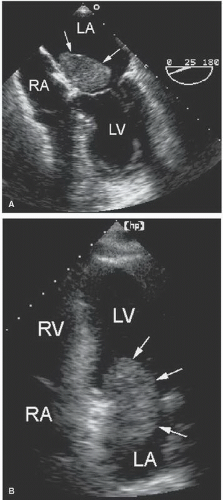
FIGURE 23.3. A: A myxoma (arrows) is seen in the left atrium on transesophageal imaging. The mass is attached to the fossa ovalis. B: A four-chamber view demonstrates a large myxoma within the left atrium partially obstructs the mitral orifice during diastole.


FIGURE 23.4. A large left atrial myxoma is demonstrated using three-dimensional imaging. The advantages of this modality are best appreciated when viewed in a cine loop format.


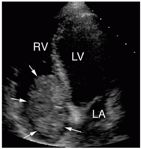
FIGURE 23.5. A large right atrial myxoma (arrows) is indicated by the arrows. The mass extends through the tricuspid valve into the right ventricle.


FIGURE 23.6. Upper left: A large mass is seen within the right atrium. Upper right: In diastole, note how the mobile mass protrudes through the tricuspid valve creating obstruction to right ventricular inflow. Lower panel: The degree of obstruction is demonstrated with pulsed Doppler, mean gradient = 9 mm Hg. The location, motion, and attachment site are consistent with right atrial myxoma.

Papillary fibroelastoma accounts for approximately 10% of all primary tumors. These are usually found in older patients and arise from either the aortic or mitral valve (Fig. 23.7). Because tumors arising from the heart valves are rare and often asymptomatic, establishing a diagnosis can be challenging and often relies on echocardiography. Among tumors that affect the valves, papillary fibroelastomas are by far the most common, accounting for more than 85% of valve-associated tumors. Myxomas and fibromas account for the remainder, whereas malignant tumors involving the valves are very rare.

Papillary fibroelastomas are small, generally 0.5 to 2.0 cm in diameter, and are often confused with vegetations. Making this distinction is difficult because of the similarity in the echocardiographic appearance. A correct diagnosis therefore depends on the clinical setting, that is, the presence or absence of signs of infection. These tumors usually attach to the downstream side of the valve by a small pedicle and are irregularly shaped with delicate frondlike surfaces (Figs. 23.8 and 23.9). Mobility is
common and generally considered a risk factor for embolization. Significant valvular regurgitation is rare. There is some confusion as to whether fibroelastomas are distinct from Lambl’s excrescences, which are smaller and frequently seen on otherwise normal valves in elderly patients (Fig. 23.10). Whether the two represent the distinct entities remains controversial. Fibroelastomas are also confused with blood cysts, which are unusual blood-containing cystic structures that develop within mitral leaflets (Fig. 23.11). Blood cysts have a broader base, are sessile, and are less mobile than fibroelastomas. Papillary fibroelastomas may be detected as an incidental finding on echocardiography. Because tumors can act as a nidus for the formation of fibrin-platelet aggregates, embolic events have been attributed to papillary fibroelastomas.


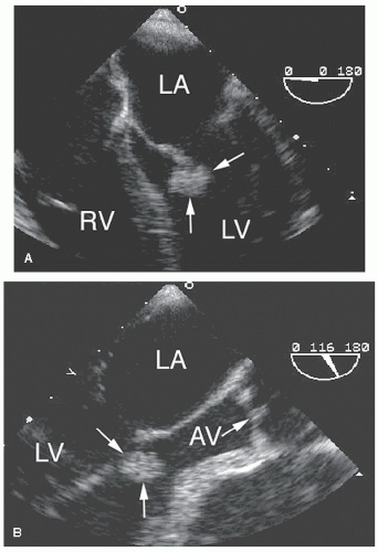
FIGURE 23.7. A transesophageal echocardiogram of the four-chamber (A) and long-axis (B) view show a papillary fibroelastoma of the mitral valve. The tumor was attached by a small pedicle to the anterior leaflet and was highly mobile. AV, aortic valve.


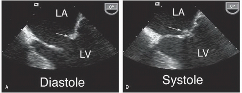
FIGURE 23.8. A small papillary fibroelastoma is seen in a patient who had a stroke. The mass (arrow) is seen on the posterior leaflet in diastole (A) and systole (B).


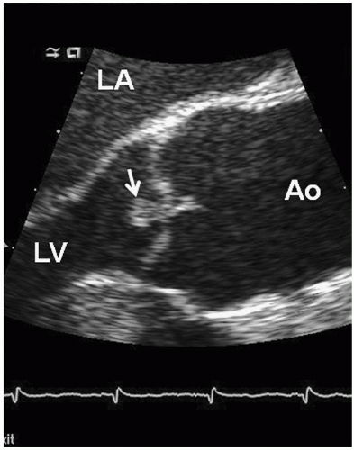
FIGURE 23.9. A transesophageal long-axis view of the aortic valve is shown from a patient who presented for evaluation of chest discomfort. The small, mobile mass attached to the aortic valve is a papillary fibroelastoma (arrow).

Lipomas are uncommon benign tumors involving the heart. Lipomatous hypertrophy of the atrial septum is one presentation. In this condition, the atrial septum is infiltrated by lipomatous material that results in dramatic thickening and increased echogenicity of its inferior and superior portions with sparing
of the fossa ovalis (Fig. 23.12). The fatty infiltrate is highly echogenic and results in a “dumbbell-shaped” appearance on two-dimensional echocardiography. The condition is thought to be benign and rarely associated with clinical manifestations.


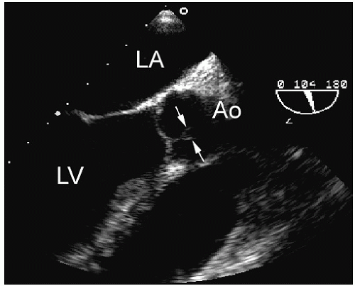
FIGURE 23.10. An example of Lambl’s excrescence of the aortic valve (arrows).

Rhabdomyomas are among the most common benign pediatric tumors (Fig. 23.13). They occur either within a cavity, sometimes as a pedunculated mass, or embedded within the myocardium. Such tumors can grow quite large and can obstruct blood flow within the heart. Fibromas are uncommon benign tumors, most often seen in children, and usually involve the left ventricular free wall. On echocardiography, they appear as distinct, highly echogenic, and well-demarcated masses that often extend into the cavity of the ventricle. Although benign, they occasionally result in obstruction to left ventricular filling and have been associated with ventricular arrhythmias. A rare condition that can be confused with a fibroma (or a thrombus) is endocardial fibroelastosis. This disease is usually seen in young children and is characterized by fibrous thickening of the left ventricular endothelium, probably as a nonspecific response to inflammation or infection. An example of endocardial fibroelastosis is provided in Figure 23.14. Unlike fibromas, the mass is endocardial rather than intramyocardial.

Malignant primary tumors of the heart are quite rare and include angiosarcoma, rhabdomyosarcoma, and fibrosarcoma.
Figure 23.15 is an example of a fibrosarcoma that occupies the right ventricular outflow tract. Its size and location combine to produce a significant outflow tract gradient, as evidenced by the Doppler recording. Such tumors tend to invade or replace myocardial tissue and thereby dramatically alter the appearance and/or function of the heart. A sarcoma involving the right and left atria is shown in Figure 23.16. The extension of the tumor through the atrial septum is suggestive of its malignant nature. As opposed to the well-circumscribed appearance of benign tumors, cardiac malignancies appear to infiltrate the tissues, disrupting normal anatomic planes, and invade or obliterate contiguous structures. The heart often appears tethered and relatively immobile, without its normal translational motion (Fig. 23.17). Contrast perfusion imaging may have a role in further characterizing intracardiac masses and distinguishing tumors from thrombi. Enhancement of the mass after contrast injection correlates with the degree of vascularity. Thus, malignant tumors and other vascular structures often demonstrate hyperenhancement while thrombi and other avascular masses, such as myxomas, show less contrast uptake.


FIGURE 23.11. A blood cyst (arrow) within the anterior mitral leaflet. The cyst is relatively immobile and the attachment is broad based. The mass is seen during diastole (A) and systole (B).


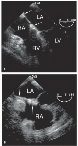
FIGURE 23.12. Lipomatous hypertrophy of the atrial septum. A: A mild degree of accumulation of lipomatous material is present (arrows). The fossa ovalis is characteristically spared. B: A more extreme form of lipomatous hypertrophy (arrows).


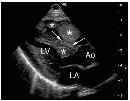
FIGURE 23.13. Rhabdomyoma is a common pediatric tumor. In this 12-year-old patient, multiple tumors are seen within the left and right ventricle (asterisks) and interventricular septum (arrows).

The echocardiographic assessment of these patients has several components. Because primary cardiac malignancy is so much less common than metastatic involvement, the echocardiographic demonstration of an invasive cardiac tumor should suggest the possibility of metastatic disease. In addition, the exact location and extent of a cardiac malignancy must be thoroughly assessed to determine whether resection might be possible. Some malignancies are likely to affect a given chamber or location within the heart. Angiosarcomas, for example, usually involve the right atrium, whereas rhabdomyosarcomas may occur anywhere. Associated pericardial effusion is common, sometimes leading to tamponade.


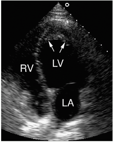
FIGURE 23.14. An example of endocardial fibroelastosis. Endocardial thickening in the left ventricular apex is present. Thrombus overlies the thickened endocardium (arrows).


Metastatic Tumors to the Heart

Echocardiography is often performed in patients with known or suspected malignancy. Among patients with cardiac symptoms, looking for evidence of metastatic spread has therapeutic and prognostic implications. Cardiac function helps determine whether a given patient may be a candidate for particular therapies, such as doxorubicin (Adriamycin). In patients who have
already received cancer therapy, echocardiography is useful to evaluate for side effects. Adriamycin, for example, can cause cardiomyopathy. Chest irradiation can result in constrictive pericarditis or scarring and fibrosis of the epicardial coronary arteries. In unstable or critically ill patients, the portability and noninvasive nature of ultrasound represent a significant advantage.


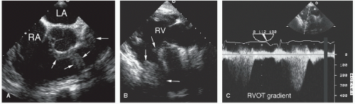
FIGURE 23.15. A primary fibrosarcoma is demonstrated in the right side of the heart. A: The tumor involves the right ventricular outflow tract and pulmonary artery. B: Narrowing of the right ventricular outflow tract is indicated by the arrows. C: Doppler imaging demonstrates a right ventricular outflow tract gradient of approximately 50 mm Hg.


FIGURE 23.16. A large sarcoma is shown involving the right atrium (black arrows) and left atrium (white arrowhead). Note how the invasive tumor restricts the normal motion of the heart on real-time imaging.

The heart is affected relatively less often by metastatic disease compared with other organs. Some investigators speculate that blood-borne malignant cells are destroyed by the contraction of the heart before they become established. Malignant tumors can spread to the heart through direct invasion from adjacent tumors, including lung and esophagus, from propagation through the venous system, or by hematogenous spread (Table 23.4). Melanoma, for example, has a high propensity for metastasizing to the pericardium and/or myocardium, involving the heart in more than 50% of cases. Intracardiac masses are frequently seen as a manifestation of malignant melanoma. Figure 23.18 is an example of a melanoma that has metastasized to the left ventricular apex. The presence of a mass is suggested on the transthoracic study but is best visualized after injection of a contrast agent. Although the appearance of the mass is similar to that of a thrombus, preserved apical contractility makes a thrombus unlikely and should suggest the possibility of alternative diagnoses. Figure 23.19 is taken from another patient with melanoma, metastatic to the right ventricular apex. Some leukemias also have a similarly high rate of cardiac spread. However, more common malignancies, such as breast or lung cancer, account for the greatest percentage of nonprimary cardiac tumors. There is also a high incidence of cardiac involvement among patients with lymphoma secondary to acquired immunodeficiency syndrome.


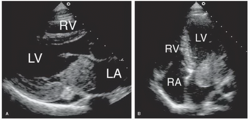
FIGURE 23.17. A, B: An example of angiosarcoma. The mass had infiltrated the lateral wall of the left atrium and left ventricle and invaded the mitral valve. Obstruction to mitral inflow was present. In real time, the heart appeared fixed due to infiltration by the malignancy. A pericardial effusion is also present.


Table 23.4 Metastatic Tumors to the Heart: Source and Cardiac Manifestations


Original Source


Cardiac Effect


Lung


Direct extension, often via pulmonary veins; effusion common


Breast


Hematogenous or lymphatic spread; effusion common


Lymphoma


Lymphatic spread, varied manifestations


Gastrointestinal


Variable manifestations


Melanoma


Intracardiac or myocardial involvement


Renal cell carcinoma


IVC to RA to RV; confused with thrombus


Carcinoid


Tricuspid and pulmonic valve thickening


IVC, inferior vena cava; RA, right atrium; RV, right ventricle.


FIGURE 23.18. Metastatic melanoma often involves the heart. A: Image quality prevents visualization of the apical mass. B: After contrast injection, the outline of the apical mass (arrows) is apparent.

The location of involvement of metastatic disease is frequently the pericardium, resulting in a pericardial effusion and epicardial involvement (Fig. 23.20). The usual signs and symptoms of pericarditis are often absent. In patients with known malignancies, the detection of a pericardial effusion should raise concern about cardiac metastases. However, it is almost impossible, based on echocardiographic findings alone, to establish the cause of a pericardial effusion. Patients with cancer may develop pericardial effusion for any of several reasons. For example, particular chemotherapies can cause pericardial effusion. In most cases, confirming that the effusion is malignant often has therapeutic implications. Pericardiocentesis, usually with biopsy, is generally appropriate but only diagnostic in approximately 50% of cases. When the pericardial involvement is due to metastatic disease, the prognosis is uniformly poor. Figure 23.21 is a case of metastatic disease involving the posterior left ventricular wall and pericardium. Over a period of several weeks, the tumor eroded through the myocardium, resulting in
formation of a pseudoaneurysm that gradually increased in size until the time of the patient’s death. Intramyocardial involvement is less common than pericardial metastases and usually occurs secondary to lymphoma or melanoma. Heart failure, obstruction to flow, and arrhythmias may develop as a result. Cardiac involvement is often established at autopsy as an incidental finding in patients with widely metastatic disease. Figure 23.22 is taken from a patient undergoing treatment of a B-cell lymphoma. The tumor had spread to the heart and can be seen filling the right atrium and extending into the left atrium. Figure 23.23 is an example of a pericardial mesothelioma. The mass is huge and grossly distorts the right side of the heart. Figure 23.24 shows a patient with lymphoma, before and after chemotherapy. The tumor involved the aortic root and posterior wall of the heart, including the area of the coronary sinus. After successful chemotherapy, normal anatomy is restored. In this case, serial echocardiography was critical to follow the progress of therapy and the reduction in tumor burden.

Only gold members can continue reading. Log In or Register to continue

Stay updated, free articles. Join our Telegram channel

Jun 22, 2016 | Posted by in CARDIOLOGY | Comments Off on Masses, Tumors, and Source of Embolus

Full access? Get Clinical Tree

Get Clinical Tree app for offline access