Fig. 1.1
Areas of the hand used to palpate localized pulsations, thrills, and heaves or lifts. The patient should be palpated in the supine position and the fingertips used to palpate localized pulsations. The flat of the hand should be used to feel thrills and the heel of the hand should be used to feel heaves or lifts
Thrills with the flat of the hand (Fig. 1.1).
Heaves or lifts with the heel of the hand (Fig. 1.1).
The apical impulse or point of maximal impact should be palpated first.
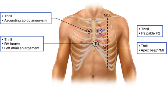
It should be located slightly medial and superior to the midclavicular line and the 5th intercostal space (Fig. 1.2).

Fig. 1.2
The aortic, pulmonary, mitral, and tricuspid areas should all be palpated as different pathologies can be felt at each location. MCL midcalvular line, A aortic, P pulmonary, M mitral, T tricuspid
It should be the lowest and most lateral impulse felt.
The left lateral decubitus position may make it easier to palpate the apical impulse.
In 50 % of normal patients, the apical impulse may not be palpable in the supine position.
The diameter of the impulse should be palpated and should be less than 2.5 cm in diameter, occupying only one intercostal space.
The amplitude should be small and the impulse should feel brisk and tapping.
If the apical impulse is displaced laterally or >3 cm in diameter, this indicates left ventricular enlargement.
Diastolic palpitation may be used to detect ventricular motion associated with a pathologic third or fourth heart sound.
A brief mid-diastolic impulse at the apex indicates an S3 gallop.
An impulse felt just before the systolic apical beat indicates an S4 gallop.
The right ventricle should not normally be palpable.
Right ventricle can be palpated at the left sternal border in the 3rd, 4th, and 5th intercostal spaces with the patient in the supine position (Fig. 1.2).
A palpable anterior systolic motion in the left parasternal area indicates right ventricular enlargement or hypertrophy.
In some patients, this may be easier to feel in the subxiphoid area.
It is important to distinguish between diffuse precordial action as seen with augmented stroke volume (which may be normal) and the sustained left parasternal outward thrust seen in right ventricular enlargement (always abnormal).
A prominent pulsation in the left second intercostal space indicates increased flow in the pulmonary artery and a palpable S2 indicates pulmonary hypertension (Fig. 1.2).
A palpable S2 at the right 2nd intercostal space (aortic area) suggests systemic hypertension and a pulsation here may indicate an aortic aneurysm (Fig. 1.2).
The pulmonic closure may be palpable in individuals with thin chest walls or in pulmonary hypertension.
Auscultation
Auscultation Technique
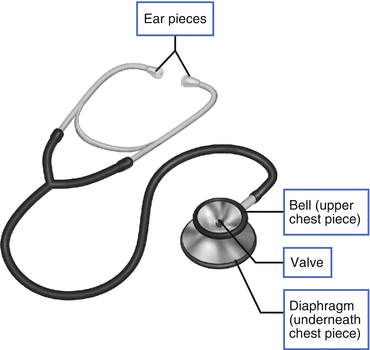
Auscultation is the act of listening to sounds arising from the internal organs for evaluation purposes. It is most often performed on fully exposed skin using a standard stethoscope (Fig. 1.3), though some physicians opt for more technologically advanced tools (e.g., electronic stethophones, fetoscope, Doppler stethoscope).

Fig. 1.3
Schematic of a standard stethoscope, identifying the bell (low frequency sounds) and diaphragm (high frequency sounds) components
Proper use of the stethoscope is crucial for accurate assessment of heart sounds and murmurs. In general, the diaphragm is used for high frequency sounds, while the bell is used for low frequency sounds; adjusting the bell pressure allows for a broader range of sounds to be heard.
Auscultation is classically performed parasternally at individual interspaces of the thoracic wall. In some patients, it is helpful to listen at routine non-precordial sites, including the axillae, back, lower right sternal border, and clavicles.
Proper auscultation technique is imperative for effective patient assessment. Once the stethoscope is placed on the patient, slowly inch the head along the body to listen for variation in sound. It is most productive to begin at the apex of the heart in the left lateral decubitis position and work toward the sternum.
Cardiac Auscultation
Normal Heart Sounds
Heart sounds heard on auscultation reflect turbulent blood flow through the heart as the heart valves snap shut. The “lub dub” beating of the heart is directly related to the normal heart sounds, S1 and S2. More specifically, S1 is produced upon closure of the atria-ventricular valves, and S2 is produced upon closure of the semilunar valves.
During inspiration, the S1 sound may be audibly split into two components. As the chest wall expands and intrathoracic pressure increases, venous return to the right atrium is also increased. This normal physiologic mechanism allows the pulmonic valve to remain open slightly longer than normal, thereby increasing the time between aortic and pulmonic valve closure.
Two additional heart sounds, S3 and S4, may be heard during routine auscultation. In children and trained athletes, S3 is associated with rapid ventricular filling during early diastole; it is benign. However, its re-emergence later in life may be an indication of left ventricular systolic dysfunction and congestive heart failure. Thus an S3 heart sound in older populations requires additional work-up. The presence of S4 in an adult is associated with turbulent blood flow being forced into a hypertrophic ventricle. S4 is sometimes referred to as a presystolic gallop; it is always an abnormal finding, indicative of reduced ventricular compliance.
Murmurs
Heart murmurs refer to abnormal heart sounds with an underlying physiologic pathology. They are typically caused by turbulent blood flow due to a diseased/abnormal valve or an abnormal blood flow pathway.
Stenotic valves are thickened, fused or calcified, narrowing the effective orifice for forward blood flow.
Regurgitant/incompetent valves do not close completely so blood can easily leak back into the previous chamber even when the valve is “closed”. This may occur secondary to structural valve changes caused by infection, inflammatory or degenerative changes, or nonvalvular abnormalities from chamber enlargement or wall motion abnormalities.
Abnormal blood flow resulting from an anatomic abnormality represents an error in development or persistence of fetal circulation. Some examples include atrial or ventricular septal defects, atria-ventricular canal, and patent ductus arteriosis.
Murmurs are characterized by:
Intensity – Intensity is graded on a 1–6 scale, related to the amount of blood flow.
Grade 1: Murmur is faintly heard with a stethoscope. It requires special effort to hear.
Grade 2: Murmur is soft but readily detectable.
Grade 3: Murmur is prominent by not loud.
Grade 4: Murmur is loud with a palpable thrill.
Grade 5: Murmur is very loud.
Grade 6: Murmur is extremely loud and even audible without use of the stethoscope.
Frequency – Frequency, or pitch, is related to the amount of blood flow.
Lower and slower flow relates to a lower pitch.
Higher and faster flow relates to a higher pitch.
Configuration – Configuration refers to the shape of the murmur with respect to its audibility. Murmurs may exhibit crescendo, decrescendo, flat, or crescendo-decrescendo configurations.
Duration – Described in terms of the length of systole or diastole in which the murmur occurs, for example mid-systolic, or holo-diastolic.< div class='tao-gold-member'>Only gold members can continue reading. Log In or Register to continue

Stay updated, free articles. Join our Telegram channel

Full access? Get Clinical Tree


