Echocardiography in Systemic Disease and Clinical Problem Solving


Echocardiography in Systemic Disease and Clinical Problem Solving


Echocardiography and Systemic Disease

There are many systemic diseases with cardiovascular manifestations for which echocardiography is an appropriate component of the clinical evaluation (Tables 24.1 and 24.2). Similarly, there are several clinical presentations for which echocardiography is a first-line investigative technique. This chapter discusses the integration of clinical and echocardiographic information for the management of patients with a variety of clinical presentations.


Hypertension

Clinically, echocardiography is used to detect end-organ cardiac damage due to hypertension, including left ventricular hypertrophy (Fig. 24.1), diastolic dysfunction, and later systolic dysfunction (Fig. 24.2). Numerous algorithms have been proposed for the determination of left ventricular mass and for quantifying left ventricular hypertrophy. The M-mode-derived Teichholz or cubed formula, which assumes spherical geometry of the left ventricle, was used in most early hypertension studies. Because the left ventricle does not adhere to spherical geometry, the absolute measurements are often inaccurate due to tangential imaging planes. Nevertheless, assuming no intervening event such as myocardial infarction, this methodology provides a relatively stable estimate of left ventricular mass over time in any given patient and has been used successfully for tracking left ventricular mass regression during therapeutic trials of antihypertensive agents.


Table 24.1 Appropriateness Criteria for the Application of Echocardiography in systemic Disease and Clinical decision Making


Indication


Appropriateness Score (1-9)


5.


Patients who have isolated APC or PVC without other evidence of heart disease


I (2)


6.


Patients who have sustained or nonsustained SVT or VT


A (8)


10.


Evaluation of known or suspected pulmonary hypertension including evaluation of right ventricular function and estimated pulmonary artery pressure


A (8)


15.


Initial evaluation of patient with suspected pulmonary embolism to establish diagnosis


I (3)


16.


Evaluation of patient with known or suspected acute pulmonary embolism to guide therapy (i.e., thrombectomy and thrombolytics).


A (8)


34.


Evaluation of cardiovascular source of embolic event (PFO/ASD, thrombus, neoplasm)


A (8)


37.


Known or suspected Marfan disease for evaluation of proximal aortic root and/or mitral valve


A (9)


38.


Initial evaluation of suspected hypertensive heart disease


A (8)


39.


Routine evaluation of patients with systemic hypertension without suspected hypertensive heart disease


I (3)


40.


Reevaluation of a patient with known hypertensive heart disease without a change in clinical status


I (3)


41.


Initial evaluation of known or suspected heart failure (systolic or diastolic)


A (9)


42.


Routine (yearly) evaluation of patients with heart failure (systolic or diastolic) in whom there is no change in clinical status


I (3)


43.


Reevaluation of known heart failure (systolic or diastolic) to guide therapy in a patient with a change in clinical status.


A (9)


51.


Baseline and serial re-evaluations in patients undergoing therapy with cardiotoxic agents


A (8)


58.


Evaluation (TEE) of patient with atrial fibrillation/flutter for left atrial thrombus or spontaneous contrast when a decision has been made to anticoagulate and not to perform cardioversion


I (3)


59.


Evaluation (TEE) for cardiovascular source of embolic event in a patient who has a normal TTE and normal ECG and no history of atrial fibrillation/flutter.


U (6)


Reprinted with permission of the ACCF from Douglas PS, Khandheria B, Stainback RF, et al. ACCF/ASE/ACEP/ASNC/SCAI/SCCT/SCMR 2007 appropriateness criteria for transthoracic and transesophageal echocardiography. J Am Coll Cardiol 2007;50(2):187—204.
A, appropriate; ASD, atrial septal defect; APC atrial premature contraction; ECG, electrocardiogram; I, inappropriate; LV, left ventricular; MRI, magnetic resonance imaging; PFO, patent foramen ovale; PVC ventricular premature contraction; SPECT, single-photon emission computed tomography; SVT, supraventricular tachycardia; TTE transthoracic echocardiography; TEE dtransesophageal echocardiography; U, uncertain; VT, ventricular tachycardia.


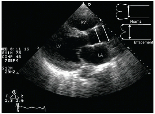
Other cardiac anomalies, which have a relatively greater prevalence in the hypertensive population, include left atrial dilation, calcification of the mitral annulus, and mild degrees of aortic valve insufficiency. With long-standing hypertension, there may be secondary dilation of the ascending aorta with effacement of the sinotubular junction. This has the effect of splaying the closure of the aortic cusps and resulting in secondary aortic insufficiency (Fig. 24.3). The degree to which aortic insufficiency is attributable to hypertension alone has been debated; however, there appears to be a fairly strong correlation between this type of functional aortic insufficiency and chronic hypertension. Additional abnormalities associated with long-standing hypertension include atherosclerosis of the aorta, which can be detected with transesophageal echocardiography, and peripheral vascular disease.


Table 24.2 Systemic diseases and Clinical Presentations in Which Echocardiography Plays a Valuable Role


Systemic disease conditions with cardiovascular manifestations


Hypertension


Diabetes mellitus


Pregnancy


Chronic renal insufficiency


Connective tissue disease


Systemic lupus erythematosus


Scleroderma


Marfan syndrome


Chronic hepatic disease


Pulmonary arterial hypertension


Miscellaneous Diseases


Thyroid disease


Sarcoidosis


Hemochromatosis


Muscular dystrophies


Friedreich ataxia


Carcinoid syndrome


Ergotamine toxicity


Clinical Presentations


Congestive heart failure


Dyspnea


Pulmonary embolus


Atrial fibrillation


Cardioembolic disease


Radiation therapy


Syncope


Athletic screening


Pregnancy


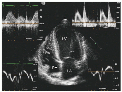
Diastolic dysfunction is one of the earliest manifestations of hypertensive heart disease. This is mild at first, but in advanced cases of severe untreated hypertension, it may progress to the point of being the predominant contributor to congestive heart failure symptoms. Methods by which diastolic dysfunction is evaluated in hypertensive patients are the same as for other diseases. Generally, in early hypertension, there is delayed relaxation of the myocardium because of hypertrophy and mild degrees of stiffening, which is manifested as a reduced E/A ratio of mitral valve inflow (Fig. 24.4). If left ventricular hypertrophy remains uncomplicated by concurrent systolic dysfunction, no other changes are anticipated. In severe long-standing hypertension, the left ventricle may develop systolic dysfunction as well. At this point, there may be evidence of more advanced diastolic dysfunction with a normal or high E/A ratio, representing pseudonormal filling or a restrictive physiology. Other echocardiographic modalities, including Doppler tissue imaging, have been employed in the hypertensive population. Generally, results of Doppler tissue imaging of the annulus parallel the abnormalities seen in the mitral valve inflow and consist of reduced early diastolic relaxation velocities (Fig. 24.4). Strain and strain rate, which provide a more detailed characterization of myocardial mechanics, may reveal subclinical abnormalities earlier in hypertensive cardiovascular disease than is apparent by detection of left ventricular hypertrophy or overt diastolic dysfunction. It should be emphasized that reduced strain and strain rate, while a sensitive marker for preclinical hypertensive cardiovascular disease, are nonspecific and have also been
reported in preclinical infiltrative and hypertrophic cardiomyopathies and are likely present in a broad range of othe disease states as well. As such, their utilization clearly needs to be put in context of the clinical situation.
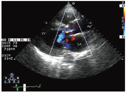


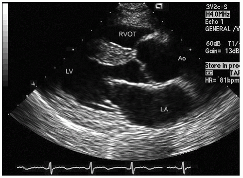
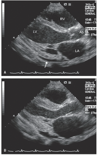
FIGURE 24.1. Parasternal long-axis view recorded in a 30-year-old patient with essential hypertension. In this diastolic frame, note the mild left ventricular hypertrophy but otherwise normal anatomy and preserved systolic function in the real-time image.


FIGURE 24.2. Parasternal long-axis image recorded in a patient with severe long-standing and poorly controlled hypertension. Note the left ventricular hypertrophy and the mild left atrial dilation. In the real-time image, note the global hypokinesis of the left ventricle. Also note the dilation of the ascending aorta with effacement at the sinotubular junction.


FIGURE 24.3. Parasternal long-axis echocardiogram with color Doppler flow imaging recorded from the same patient presented in Figure 24.2. Note the effacement of the sinotubular junction, which results in malcoaptation of the aortic cusps and a central aortic regurgitation jet.


FIGURE 24.4. Apical four-chamber view with mitral inflow, pulmonary vein flow, and Doppler tissue imaging of the annulus in a patient with essential hypertension. Note the reversal of the mitral E/A ratio, which is paralleled by reversal of annular velocities, all consistent with grade 1 diastolic dysfunction in this otherwise healthy 45-year-old patient. reported in preclinical infiltrative and hypertrophic cardiomyopathies and are likely present in a broad range of other disease states as well. As such, their utilization clearly needs to be put in context of the clinical situation.


Diabetes mellitus

Diabetes mellitus is associated with primary and secondary cardiovascular abnormalities. For patients with diabetes, the metabolic derangement results in premature coronary artery disease, sometimes in a very aggressive manner. For type 2 diabetes, especially if seen as part of a generalized “metabolic disorder,” there is an increased prevalence of lipid disorders and hypertension. The long-term effect of diabetes on the coronary vasculature is similar to that of coronary disease in those without diabetes; however, diabetes tends to result in more diffuse and premature atherosclerotic involvement. Detection of coronary disease in the population with diabetes is done in a manner identical to that of the population without diabetes, including the use of rest and stress echocardiography. From a clinical standpoint, it should be recognized that because of the autonomic neuropathy associated with diabetes, typical symptoms may not be present. As such, the indications for proceeding with provocative cardiovascular stress testing, and the end points for termination of a cardiovascular stress test, may not be the same as they are in the population of patients without diabetes.

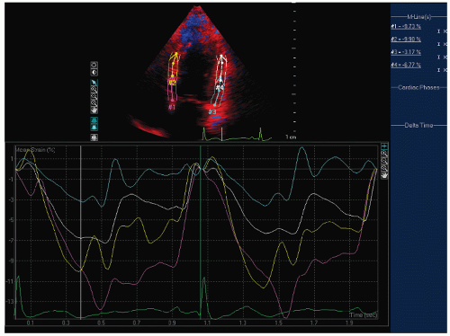
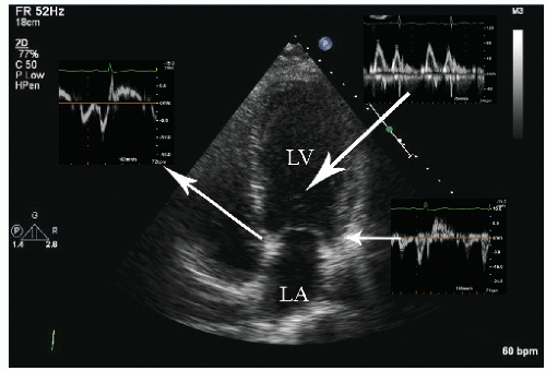
In addition to these secondary sequelae of diabetes that behave in a manner similar to that in patients without diabetes, there are subtle, less clinically obvious cardiovascular manifestations of diabetes. There is a well-recognized tendency to develop diastolic dysfunction even in the absence of “significant” hypertension or coronary artery disease. This is presumed to be due to accumulation of metabolic byproducts within the myocardial interstitium, which results in stiffening of the myocardium and delayed relaxation. In routine clinical practice, this is manifested as a reduced E/A ratio of mitral valve inflow. It is well recognized that the mitral valve E/A ratio diminishes with age; however, in the population with diabetes, the rate at which it diminishes exceeds that in the population without diabetes due to occult diastolic dysfunction (Fig. 24.5). Reductions in strain and strain rate have been demonstrated in preclinical diabetic heart disease as well (Fig. 24.6). The degree to which aggressive control of even borderline hypertension and scrupulous control of blood glucose will mitigate against these changes is yet to be determined.


FIGURE 24.5. Apical four-chamber view with multiple Doppler images in a 32-year-old female patient with diabetes but no evidence of hypertension or coronary artery disease. The geometry and size of the left ventricle are normal without evidence of overt left ventricular hypertrophy. Notice the pseudonormal mitral inflow with a mitral E/A ratio of approximately 1.2, but the reversed annular e′/a′ ratio of both the septal and lateral mitral annulus implying diastolic dysfunction.

Management of the patient with diabetes requires guidelines different from those for patients without diabetes. For a patient with diabetes requiring a major noncardiac surgical procedure, such as renal transplantation or vascular surgery, provocative stress testing, most often with dobutamine stress echocardiography, is typically warranted to identify occult coronary artery disease, even in the absence of classic symptoms, and at younger ages than typically recommended. Similarly, the frequency with which diagnostic testing should be repeated to ensure stability of the underlying substrate is greater than it is for the population without diabetes. After coronary artery bypass surgery, guidelines suggest routine postoperative stress testing only after 5 years. The likelihood of rapid progression is substantially greater in patients with diabetes, and many authorities have recommended earlier and more frequent provocative stress testing (including stress echocardiography) in diabetics.


Thyroid Disease

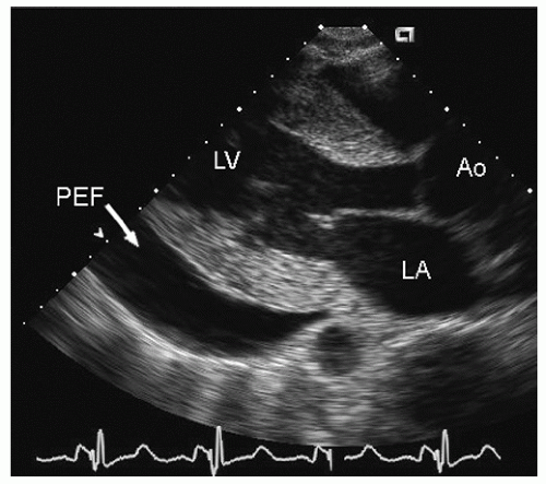
Both hyperthyroidism and hypothyroidism result in cardiovascular disease. Hyperthyroidism results in an increase in total blood volume as well as an increase in left ventricular contractility and a decrease in systemic vascular resistance. This results in a high-output state with an increased left ventricular stroke volume. In addition to these hemodynamic effects, hyperthyroidism results in sinus tachycardia and on occasion may trigger atrial fibrillation. In patients with underlying structural heart disease, the increase in heart rate and stroke volume may precipitate heart failure or unmask previously compensated heart failure or angina. Extreme hyperthyroidism may result in a high-output state sufficient to cause a picture identical to that of dilated cardiomyopathy (Fig. 24.7). The cardiomyopathy of hyperthyroidism typically reverses after successful treatment of the metabolic disorder. Hypothyroidism results in directionally opposite changes in left ventricular performance and cardiac output. Pericardial effusion occurs frequently, but even when severe, is an uncommon cause of hemodynamic compromise (Fig. 24.8).


FIGURE 24.6. Doppler tissue based strain imaging recorded from the same patient depicted in Figure 24.5. The strain images reveal reduced mean strain predominantly in the lateral wall with a lesser reduction in the two septal segments.


Chronic Renal Insufficiency

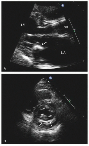
Chronic renal insufficiency results in a characteristic constellation of cardiac abnormalities. Patients with chronic renal insufficiency frequently have renal disease based on hypertension or diabetes, which, as discussed previously, result in premature coronary artery disease and other anatomic and/or physiologic cardiac abnormalities. In addition to the above secondary features, the metabolic derangement in chronic renal insufficiency, including hyperparathyroidism, results in ectopic calcification, predominantly of the fibrous skeleton of the heart. This is most often manifested as calcification of the mitral annulus (Fig. 24.9). The degree of annular calcification is related to the magnitude of hyperparathyroidism and can range from small focal deposits to extensive circumferential deposits of calcium in the annulus. In advanced cases, the calcification invades the proximal mitral valve leaflets and may cause functional mitral stenosis. Secondary features of chronic renal insufficiency
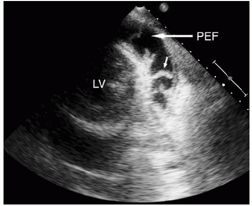
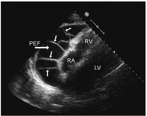
include left ventricular hypertrophy due to hypertension and an abnormal texture of the hypertrophied myocardium that mimics that seen in cardiac amyloid (Fig. 24.10). Other abnormalities seen in chronic renal insufficiency include pericardial effusion, which may range from small chronic effusions to presentation with cardiac tamponade. Uremia results in inflammatory and occasionally hemorrhagic pericarditis in which there is often evidence of “stranding” on the visceral pericardium (Figs. 24.11 and 24.12).


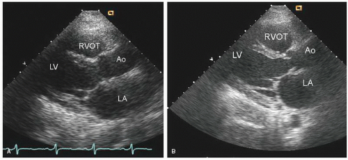
FIGURE 24.7. Parasternal long-axis echocardiograms recorded at end systole in a patient with severe thyrotoxicosis who presented with nonsustained ventricular tachycardia and congestive heart failure. A: Note the relatively preserved left ventricular internal dimension (52 mm) but the severe hypokinesis in systole in the real-time image. B: Recorded 6 months later, after successful therapy, and confirms substantial recovery of systolic function.


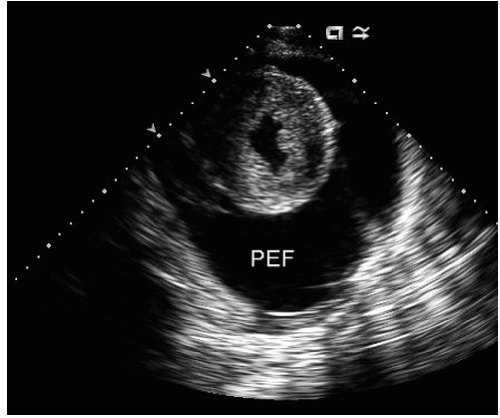
FIGURE 24.8. Echocardiogram recorded in a patient with profound hypothyroidism (TSH > 300). Note the large pericardial effusion (PEF) with a swinging heart in the real-time image. The patient had no clinical evidence of hemodynamic compromise. Incidental note is made of severe left ventricular hypertrophy, presumably related to long-standing hypertension.


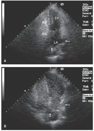
FIGURE 24.9. Parasternal long- and short-axis echocardiograms recorded in a patient with chronic renal insufficiency and calcification of the mitral annulus. A: In the parasternal long-axis view, notice the focal deposits in the posterior annulus (arrow), which have resulted in a side lobe artifact mimicking an associated mass. B: In the short-axis view, notice the crescent of calcium encompassing the posterior mitral annulus (arrows).


FIGURE 24.10. Parasternal long-axis echocardiogram recorded in a patient with end-stage renal disease. Left ventricular hypertrophy with abnormal myocardial texture, as well as a moderate pericardial effusion (PEF), is present.

On occasion, patients with chronic renal insufficiency develop systolic dysfunction, which cannot be related to uncontrolled hypertension, coronary artery disease, or other identifiable factors. The presumed etiology of the dysfunction is accumulation of metabolic byproducts, including metalloproteinases, in the myocardium. Numerous cases have been
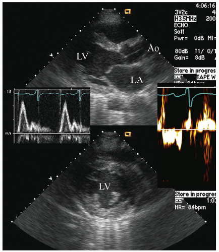
reported in which systolic function recovers after institution of more aggressive dialysis or renal transplantation. Figure 24.13 was recorded in a 34-year-old patient with end-stage renal disease related to glomerulonephritis. Note the significant systolic dysfunction in the real-time images and evidence of marked diastolic dysfunction. Figure 24.14 was recorded 6 months after renal transplantation and demonstrates marked reversal of both the systolic and diastolic dysfunction.
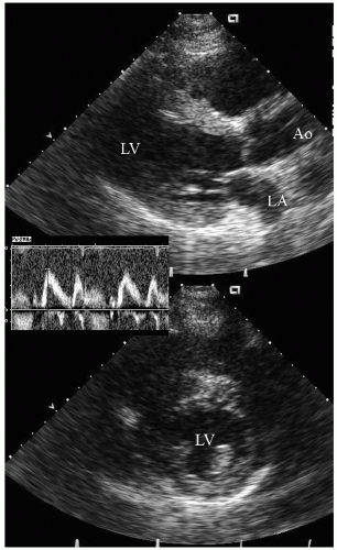


FIGURE 24.11. Parasternal short-axis view recorded in a patient with chronic renal insufficiency and uremic pericarditis. Note the moderate pericardial effusion (PEF) and the multiple strands connecting the visceral and parietal pericardium (arrow).


FIGURE 24.12. Subcostal echocardiogram recorded in a patient with chronic renal insufficiency and a large pericardial effusion (PEF) localized over the right atrium (RA) and right ventricle. Again, note the stranding between the visceral and parietal pericardium, implying a marked inflammatory response.


FIGURE 24.13. Parasternal long-and short-axis echocardiogram recorded in a patient with chronic renal insufficiency (known not to have coronary artery disease). In the real-time images, note the global hypokineses of the ventricle and the mildly abnormal myocardial texture. The Doppler insets demonstrate an elevated mitral E/A ratio with reduced annular e’/a’ ratio implying restrictive physiology.


FIGURE 24.14. Parasternal long- and short-axis echocardiogram recorded 6 months after renal transplantation in the same patient depicted in Figure 24.13. In the real-time images, note the almost complete recovery of systolic function. Also note the normalization of mitral inflow.


Connective Tissue/Autoimmune disease


Systemic Lupus Erythematosus

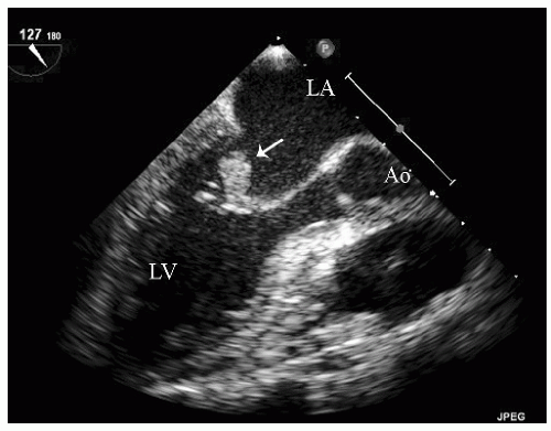
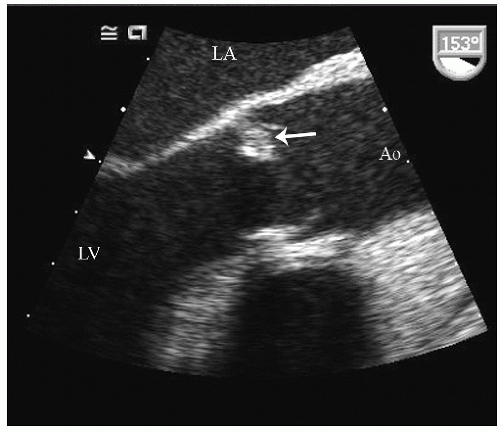
Systemic lupus erythematosus (SLE) may be associated with cardiovascular disease. There can be substantial crossover among many of the connective tissue diseases such as mixed connective tissue disease, SLE, Raynaud phenomenon, and scleroderma. A classic lesion encountered in patients with SLE is noninfectious endocarditis with the so-called Libman-Sacks vegetation (Figs. 24.15 and 24.16). These are most commonly encountered on the mitral valve and more frequently are on the atrial side of the leaflet. They tend to be less mobile than infectious vegetations. They may have an inflammatory component that can result in leaflet deformity and variable degrees of valvular regurgitation. When encountered on the aortic valve, they are usually on the arterial side. They may resolve with successful therapy of the underlying disease.

Other manifestations of SLE include coronary vasculitis, which can result in regional or global dysfunction and thereby mimic either an acute coronary syndrome or cardiomyopathy. A final manifestation of SLE may be acute pericarditis. There
are no characteristic features of the pericarditis or pericardial infusion seen in SLE. On rare occasion, SLE has been associated with pulmonary hypertension, although this association is far more common with scleroderma.


FIGURE 24.15. Transesophageal echocardiogram recorded in a patient with systemic lupus erythematosus and a neurologic event. Note the mobile mass on the atrial aspect of the mitral leaflet (arrow) representing a presumed Libman-Sacks vegetation in this patient without evidence of an infectious process.


Antiphospholipid Antibody Syndrome

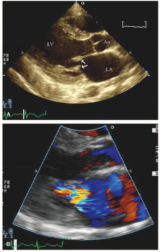
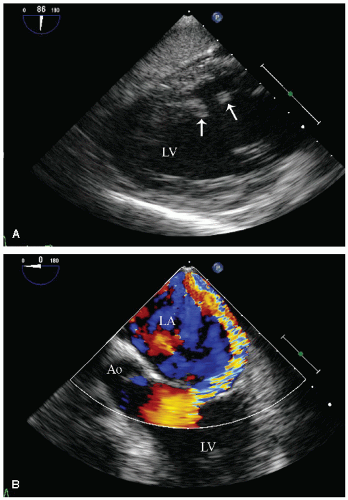
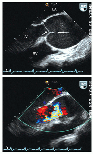
Antiphospholipid antibody syndrome is closely related to many connective tissue diseases and has been reported as an integral part of systemic lupus. This syndrome results in a variably hypercoagulable state with a tendency toward both venous and arterial thrombosis. In addition, patients with the antiphospholipid antibody syndrome develop sterile valvular vegetations similar to those seen in systemic lupus. Although not intrinsically destructive, they may result in valvular regurgitation (Fig. 24.17). They may resolve with successful treatment of the underlying systemic illness. In all likelihood, some individuals previously diagnosed with Libman-Sacks vegetative lesions may have had sterile vegetations related to the antiphospholipid antibody syndrome. On rare occasions, a catastrophic antiphospholipid antibody syndrome develops with acute severe multiorgan system failure related to microthrombosis of arterial and venous circuits. Myocardial necrosis may be a part of this syndrome. From an echocardiographic perspective, it will present as acute vegetative lesions and/or myocardial necrosis with instances of isolated papillary muscle rupture having been reported (Fig. 24.18).


FIGURE 24.16. Transesophageal echocardiogram recorded in a longitudinal view of the aorta revealing a mass on the ventricular aspect of the aortic cusp in a patient with systemic lupus erythematosus, representing a Libman-Sacks vegetation.


FIGURE 24.17. Parasternal long-axis view in a patient with connective tissue disease and documented antiphospholipid antibody syndrome. Note the small, immobile masses on the atrial aspect of both the anterior and posterior mitral valve leaflets (arrows) (A) and the moderate mitral regurgitation on color flow Doppler imaging (B).


Scleroderma/Raynaud Phenomenon

Many other connective tissue diseases can have cardiovascular manifestations. Diseases closely related to SLE such as mixed connective tissue disease represent a crossover category for which all the different manifestations of SLE may be seen. Patients with Raynaud phenomenon or with the full complex of scleroderma have a greater than usual prevalence of pulmonary arterial hypertension. In patients with scleroderma, pulmonary hypertension anatomically and physiologically is similar to
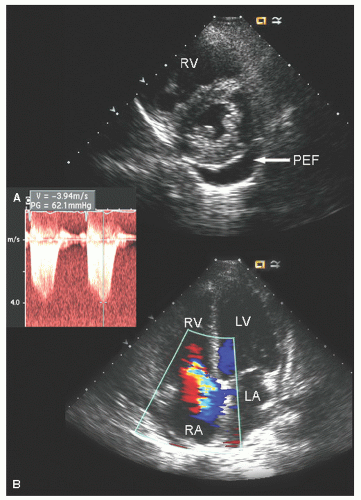
primary pulmonary hypertension with an increase in pulmonary vascular resistance at the arteriolar level (Fig. 24.19). Concurrent pericardial effusion may be more common in scleroderma than in pulmonary hypertension of other etiologies and is not necessarily an indicator of end-stage disease. The manifestations of pulmonary hypertension as a distinct entity are discussed further in this chapter, and the echocardiographic features of right ventricular pressure overload have been discussed in Chapters 8 and 13.


FIGURE 24.18. Transesophageal echocardiogram recorded in a 24-year-old patient with connective tissue disease and evidence of catastrophic antiphospholipid antibody syndrome. Note the rupture of the papillary muscle (arrows) (A) and the highly eccentric mitral regurgitation jet related to a flail mitral leaflet (B).


Marfan Syndrome

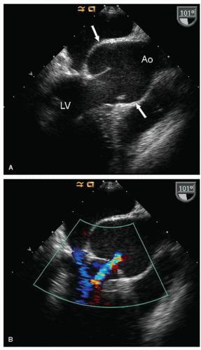
Marfan syndrome is a heritable disorder of connective tissue, which is associated with multiple cardiovascular abnormalities. Before the advent of corrective surgery, cardiovascular complications, especially aortic dissection and proximal aortic rupture, were the leading causes of mortality in patients with Marfan syndrome, resulting in death at an average age in the fourth decade. The cardiovascular manifestations of Marfan syndrome include cystic medial necrosis, which is a degeneration of the medial layer of the aorta. This results in dilation and weakening of virtually any portion of the aorta and as such should be considered a disease of the entire aorta. The most frequent area of dilation is in the proximal aorta and dilation may be confined to the aortic sinuses. Figure 24.20 was recorded in a patient with characteristic features of Marfan syndrome. Although the sinuses are the most common sites of dilation, it should be recognized that the underlying pathologic process extends throughout the entire aorta, and patients with Marfan syndrome are at risk of aneurysm formation, dissection, and rupture at any point along the course of the aorta. For most patients, initial screening can be undertaken with transthoracic echocardiography. Evaluation of the proximal aorta should be done systematically and measurements should be made at the level of the annulus, sinuses, sinotubular junction, and proximal ascending aorta (Fig. 24.21). Unfortunately, many laboratories often report only a single measurement of the aorta without specifying its location.


FIGURE 24.19. Transthoracic echocardiogram recorded in a patient with scleroderma and pulmonary hypertension. A: Note the small pericardial effusion (PEF) as well as the dilation of the right ventricle and the right ventricular overload pattern on the ventricular septum. B: In the apical four-chamber view, note the marked right heart dilation with tricuspid regurgitation. In the inset, note the elevated tricuspid regurgitation jet velocity consistent with significant pulmonary hypertension.

The anatomy of the normal aorta is well defined and consists of a relatively smaller annulus with dilation at the level of the sinuses, which measure approximately 6 mm/M2 more than the annulus. The aorta then narrows to within 2 to 3 mm of the annular dimension at the sinotubular junction and tapers very slightly throughout its course distally. Failure to narrow at the level of the sinotubular junction is referred to as effacement. The aortic cusps insert at the level of the sinotubular junction, and effacement or frank dilation of the sinotubular junction results in malcoaptation and subsequent aortic regurgitation (Figs. 24.20 and 24.22). In patients with Marfan syndrome, this is the most common mechanism of aortic regurgitation. Much of the older literature referred to dilation of the aortic annulus
as a cause of aortic insufficiency. Dilation of the actual aortic annulus is uncommon, and in most patients, aortic insufficiency is the result of effacement of the sinotubular junction and not an abnormality of the annulus.
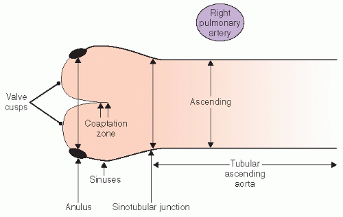


FIGURE 24.20. Transesophageal longitudinal view of the ascending aorta in a patient with Marfan syndrome. A: Note the dilation of the proximal aorta, confined to the sinus of Valsalva with relatively normal dimensions of the sinotubular junction and ascending Ao. B: Color flow Doppler demonstrates mild aortic regurgitation, which is a result of malcoaptation of the aortic cusps.


FIGURE 24.21. Schematic representation of normal aortic anatomy and the different components of the proximal aorta as well as recommended sites for making measurements.


FIGURE 24.22. Transesophageal echocardiogram recorded in a patient with Marfan syndrome and marked proximal aortic dilation. There is significant effacement of the sinotubular junction resulting in malcoaptation of the aortic cusps. Note the relatively normal position in diastole of the right aortic cusp (horizontalarrow (A) and the abnormal closure position of the noncoronary cusp, which fails to contact the opposing cusp, resulting in a highly eccentric aortic regurgitation jet (B).

Management of patients with Marfan syndrome involves serial imaging to evaluate aortic size and monitor progression of dilation. Most authorities believe that, at the time of detection,
a patient should undergo an evaluation of the entire extent of at least the thoracic aorta, which can be performed with transesophageal echocardiography, computed tomography, or magnetic resonance imaging. If there is no evidence of distal aortic dilation, follow-up usually can be performed with transthoracic echocardiography because the proximal ascending aorta is the single most likely site to be involved in subsequent dilation. It should be emphasized that follow-up should include serial measurements as noted previously for comparison. A maximum aortic dimension of 55 mm is considered an indication for elective surgical intervention. However, a threshold of 50 mm has been recommended in the presence of a bicuspid aortic valve or in the Marfan syndrome and is also used as a general indication in high-volume centers. In addition, an interval increase in size of 5 mm over a period of 12 months or less is considered an indication for prophylactic aortic replacement. The need to index aortic size to body size is not firmly established; however, the implications of dilation less than 55 mm in a smallstatured individual are obvious. Aortic dilation associated with clinically relevant aortic insufficiency has been considered an indication for surgery as well. After surgical repair, continued surveillance is crucial because this is a systemic process involving all portions of the aorta. However, after replacement of the ascending aorta in a patient with Marfan syndrome, follow-up may require transesophageal echocardiography, computed tomography, or magnetic resonance imaging because additional disease will typically not be in the field of view of transthoracic echocardiography.

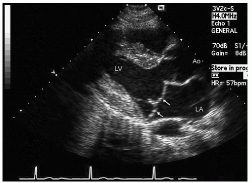
The full spectrum of cardiovascular abnormalities in Marfan syndrome includes not only disease of the aorta but also an increased prevalence of myxomatous degeneration of the mitral valve with mitral valve prolapse (Fig. 24.23). When present, it has the same appearance and clinical implications of myxomatous degeneration and prolapse occurring in the non-Marfan patient. Typically, the leaflets are diffusely thickened and redundant and have characteristic buckling or prolapse behind the plane of the mitral annulus. Echocardiographic imaging in clinical management of mitral valve disease is discussed in Chapter 12.

In many cases, mitral valve prolapse with mitral regurgitation and aortic insufficiency may both be noted. However, if aortic regurgitation is the predominant lesion, the left ventricle may dilate, resulting in reduction of the anatomic appearance of mitral valve prolapse and occasionally in a reduction in the amount of visualized mitral regurgitation. After aortic valve replacement, ventricular size diminishes, at which point mitral valve prolapse again becomes apparent and mitral regurgitation of a clinically relevant degree may again be appreciated. For patients undergoing aortic valve replacement, who have mitral valve anatomy that is suspicious for myxomatous change, or in whom this lesion complex is suspected, intraoperative evaluation of mitral valve prolapse and regurgitation should be undertaken after aortic valve replacement so that a combined aortic and mitral valve procedure can be performed if necessary.


FIGURE 24.23. Parasternal long-axis echocardiogram recorded in a young patient with Marfan syndrome and only mild dilation of the ascending aorta. This patient also has classic mitral valve prolapse (arrows).

Patients with Marfan syndrome are at an increased risk to develop an acute coronary syndrome secondary to spontaneous dissection of a proximal coronary artery. Spontaneous coronary dissection may occur in association with pregnancy or in the postpartum period and these patients will present with typical features of acute myocardial infarction. Identification of a regional wall motion abnormality in a patient with Marfan syndrome or a closely related connective tissue disease, who is otherwise not at risk of atherosclerotic coronary artery disease, should heighten the awareness of spontaneous coronary dissection as a possible etiology.

In addition to the Marfan syndrome, there are other heritable disorders of connective tissue as well as genetic syndromes, which can present with similar aortic pathology. These include connective tissue diseases such as the Ehlos-Danlos syndrome and genetic syndromes including Turner syndrome (karyotype XO). The aortopathy of Turner syndrome has become increasingly appreciated and the syndrome also may be associated with an increased prevalence of bicuspid aortic valve. The combination of bicuspid aortic valve and aortic dilation in Turner syndrome may confer substantial risk of dissection, and patients with Turner syndrome and aortic disease probably warrant surveillance and follow-up similar to that provided for patients with the Marfan syndrome.


Chronic Liver Disease and Cirrhosis

There are several clinical situations in which cardiac disease results in hepatic dysfunction and several hepatic diseases that secondarily result in cardiac disease (Table 24.3). Clinical liver disease can occur as a result of cardiovascular disease when either poor cardiac output with malperfusion occurs, or there is long-standing right ventricular dysfunction with elevated systemic venous pressure. Poor perfusion due to low cardiac output may result in multisystem organ dysfunction, and typically the liver is only one of several organs involved. In this instance, there usually will be biochemical evidence of both synthetic dysfunction and reduced clearance of metabolites. In rare occasions, either poor hepatic perfusion or elevated venous
pressures with hepatic congestion result in an obstructive biochemical pattern.


Table 24.3 Heart and Liver Disease


Cardiac disease with an impact on hepatic function


Malperfusion (hypotension/low-output state)


Passive venous congestion


Pericardial constriction


Pulmonary hypertension


Severe tricuspid regurgitation


Cardiovascular sequelae of chronic liver disease


Lowered systemic vascular resistance


Fluid retention


High-output state


Pulmonary hypertension


Pulmonary arteriovenous malformations


In patients with chronic right heart failure, systemic venous pressures are chronically elevated, which results in passive hepatic venous congestion. Chronically, this results in the syndrome of “cardiac cirrhosis,” which has distinct histologic features. This syndrome should be suspected when there is evidence of chronic hepatic dysfunction and cardiac disease, likely to cause elevation of hepatic venous pressure, is also present. Cardiovascular diseases that may result in this syndrome are constrictive pericarditis, restrictive cardiomyopathy, primary pulmonary hypertension, and mitral stenosis or dilated cardiomyopathy with secondary pulmonary hypertension. Cardiac cirrhosis occasionally develops in patients with severe tricuspid regurgitation without pressure elevation, such as after tricuspid valve resection.

There are also secondary effects of liver disease on the cardiovascular system. Advanced cirrhosis of any etiology is frequently associated with pathologically low systemic vascular resistance. This results in a chronic high-output state in which the resting cardiac output may exceed 10 L/min. In this situation, the normal heart has hyperdynamic left ventricular function with a resting ejection fraction exceeding 65% (Fig. 24.24). For patients with chronic liver disease, the echocardiographer should be cognizant of the anticipated supernormal left ventricular function and the relatively high ejection fraction. A normal or below normal ejection fraction in the presence of chronic liver disease should raise suspicion of an occult cardiomyopathy or concurrent coronary disease.


FIGURE 24.24. Parasternal long-axis viewrecorded in diastole(A) and systole (B) in a patient with end-stage liver disease and a high-output state. Resting cardiac output was 16 L/min in the catheterization laboratory. Note the mild dilation of the left atrium and left ventricle and the hyperdynamic motion of the left ventricle at rest. Incidental note is made of a small pericardial effusion (arrow).


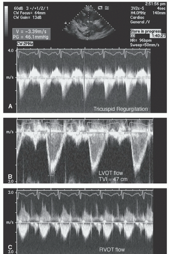
FIGURE 24.25. Spectral Doppler imaging recorded in the patient presented in Figure 24.24. Note the peak tricuspid regurgitation velocity of 3.4 m/sec and the greater than usual time velocity integral (TVI)of both left ventricular outflow tract and right ventricular outflow tract.

In addition, because of the elevated flow, pulmonary artery systolic pressures of 35 to 60 mm Hg may be seen with normal pulmonary vascular resistance (Fig. 24.25). This is analogous to the elevation in pulmonary artery systolic pressure seen in a left-to-right shunt, such as atrial septal defect, or in the high-output state of pregnancy. Mild elevation of pulmonary artery systolic pressure in chronic liver disease is not necessarily an indication of intrinsic abnormalities of the pulmonary vasculature. Pulmonary hypertension with elevated pulmonary vascular resistance (not as a result of high flow) also has been associated with chronic liver disease. There may be greater prevalence of this syndrome in chronic liver disease due to hepatitis C, suggesting a common autoimmune pathophysiology.

Other anomalies that can be seen in patients with chronic liver disease include pulmonary arteriovenous malformations (AVMs). These can be detected with contrast echocardiography and result in a delayed right-to-left shunt compared with an early phasic shunt seen with an atrial septal defect (Figs. 24.26 and 4.38). Additional features of a pulmonary AVM include a gradual increase over time in the contrast appearing in the left heart and identification of saline contrast in the pulmonary veins. In the presence of a large pulmonary AVM, contrast intensity in the left heart progressively increases over time and may, after a delay, exceed the intensity in the right heart. For patients with chronic liver disease presenting with hypoxia, contrast echocardiography should be performed to identify any
pathologic right-to-left shunt due to pulmonary AVMs. If the magnitude of shunting is significant, percutaneous closure of the pulmonary AVM may be beneficial. Identification of such a shunt also assists in clinical management because it may provide an explanation for otherwise unexplained arterial desaturation.


FIGURE 24.26. Apical four-chamber view with intravenous saline contrast recorded in a patient with end-stage liver disease and pulmonary arterial venous malformations. A: Contrast is present in the right atrium and right ventricle but has not yet appeared in the left atrium or left ventricle. The two pulmonary veins are free of contrast (arrows). B: Recorded 27 seconds after image A and shows opacification of the left atrium and left ventricle. Note also that the contrast can be clearly seen in the pulmonary veins (arrows), documenting that the level of shunt is not directly at the atrial level but rather due to a pulmonary arteriovenous malformation.

Patients with chronic liver disease may have abdominal distention due to either an enlarged liver or ascites. The effect of this is to elevate the diaphragm and compress the heart from below, occasionally resulting in the need for atypical imaging windows. Because the posterior wall is frequently compressed, “pseudodyskinesis” of the posterior wall may be noted. The genesis of this phenomenon is illustrated in Figure 24.27. In this situation, the posterior wall is compressed anteriorly by the diaphragm and hence assumes abnormal short-axis geometry in diastole. With active myocardial contraction, the ventricle reassumes circular geometry and normal thickening and contraction then ensue. The genesis of this phenomenon is analogous to the right ventricular volume overload pattern with paradoxical septal motion. Focusing on myocardial thickening rather than endocardial excursion can help avoid confusing this phenomenon for myocardial ischemia.

Occasionally, when performing transesophageal echocardiography in a patient with end-stage liver disease, one encounters large cystic vascular structures adjacent to the esophagus (Fig. 24.28). These represent dilated venous collaterals due to portal hypertension, analogous to true esophageal varices.

Only gold members can continue reading. Log In or Register to continue

Stay updated, free articles. Join our Telegram channel

Jun 22, 2016 | Posted by in CARDIOLOGY | Comments Off on Echocardiography in Systemic Disease and Clinical Problem Solving

Full access? Get Clinical Tree
Get Clinical Tree app for offline access