Fig. 2.1
Normal splitting of S2 during inspiration. P2 sounds later during inspiration due to increased venous return and blood flow through the pulmonic valve (Used with permission of Texas Heart Institute)
Accordingly, systolic murmurs that increase in intensity during deep inspiration are indicative of being right sided in origin.
Pooled data from Rothman and Goldberger suggest that the sensitivity of the deep inspiration in diagnosis of tricuspid regurgitation is 61 % [1].
The Valsalva Maneuver
The Valsalva maneuver is performed by forcibly exhaling against closed glottis.
Physiological response (Fig. 2.2): Venous return is reduced due to increased intrathoracic pressure and stroke volume is reduced due to increased mean arterial pressure during the strain phase of the Valsalva maneuver.
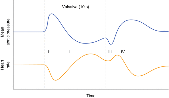
Fig. 2.2
Physiological tracing of mean aortic pressure and heart rate during a valsalva maneuver
The Valsalva maneuver diminishes nearly all systolic murmurs (Fig. 2.3), with the exception hypertrophic cardiomyopathy (HOCM) [2]. In a patient with HOCM, decreased left ventricular volume during the maneuver increases subaortic muscular obstruction increasing murmur intensity of HOCM.
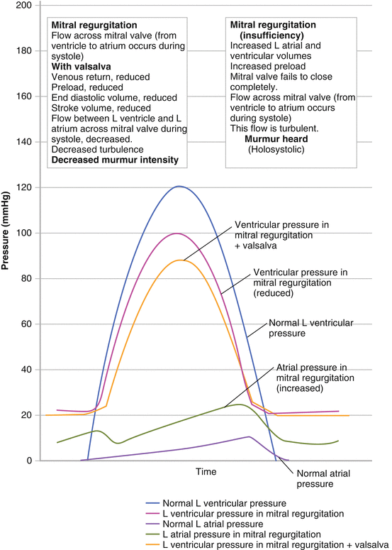
Fig. 2.3
Atrial and Ventricular pressure tracings of a normal heart; resting heart with the common systolic murmur, mitral regurgitation (MR); and a heart with MR during a Valsalva maneuver. Note how the Valsalva maneuver reduces pressure in the ventricle due to decreasing venous return to the heart. The effect is a diminished intensity of the common systolic MR murmur (Used with permission from Salazar et al. [2])
Diagnostic Value of Valsalva Maneuver for HOCM: Sensitivity: 65 %, Specificity: 96 %, Positive Predictive Value: 81 %, Negative Predictive Value: 92 % [3].
The Valsalva maneuver can also help differentiate right vs. left heart murmurs. After release of the Valsalva maneuver, right-sided murmurs have been shown to return to baseline after two cardiac cycles while left-sided murmurs require 4–11 cycles to return to baseline intensity [4].
Squatting and Standing
To perform the maneuver: From a standing position, have patient squat down bringing knees to the axilla region. Auscultate as the patient remains squatting for approximately 30 s. This is most easily done while the physician sits beside the patient, so as to reduce movement and allow for better concentration. These maneuvers are best used to distinguish HOCM and Mitral valve prolapse.
Physiological response: Squatting increases the venous return to the heart and additionally increases total peripheral resistance intensifying most systolic murmurs. The increased venous return allows for increased blood flow through a stenotic pulmonary and aortic valve, accentuating associated murmurs. Meanwhile, elevated peripheral resistance brought on by the maneuver increases murmurs of mitral and tricuspid regurgitant valves. See Fig. 2.4 [2].
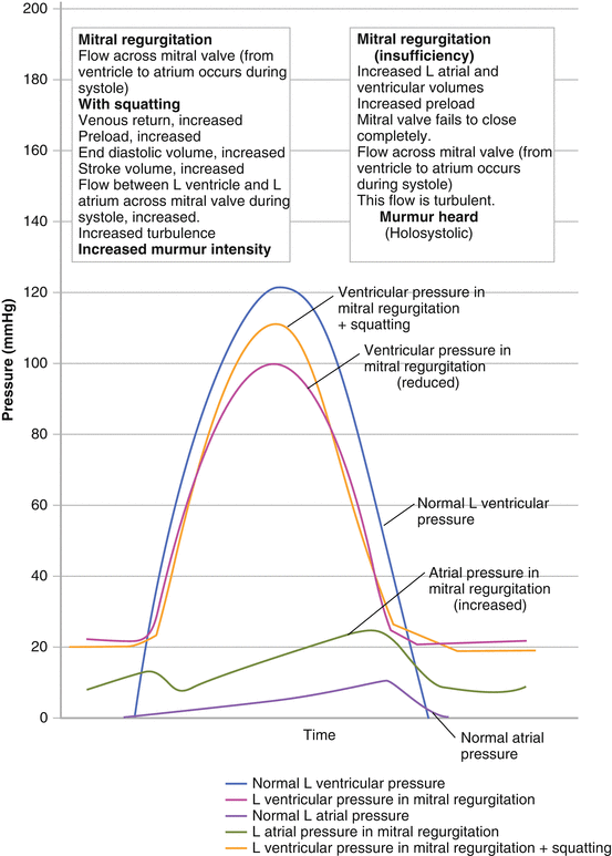
Fig. 2.4
Atrial and Ventricular pressure tracings of a normal heart; resting heart with the common systolic murmur, mitral regurgitation (MR); and a heart with MR during a squatting maneuver. Note how the squatting maneuver increases pressure in the ventricle. This is due to increased venous return to the heart. Therefore, the effect of the maneuver is to increase the intensity of the common systolic MR murmur (Used with permission from Salazar et al. [2])
Importantly, squatting diminishes the HOCM murmur. During a squat, increased preload stretches the LV, reducing sub-aortic muscular obstruction.
The decreased murmur intensity associated with HOCM during the squatting maneuver has been shown to have 95 % Sensitivity, 85 % Specificity, 61 % Positive Predictive Value, and a 99 % Negative Predictive Value [3].
Click here to listen to an example of the effect of squatting on HOCM as described by Dr. W. Proctor Harvey (Video 2.1).
Squatting helps distinguish the origin of the systolic click of mitral valve prolapse. The Mitral Valve Prolapse Systolic “Click” moves away from S1 during the maneuver (Fig. 2.5).
< div class='tao-gold-member'>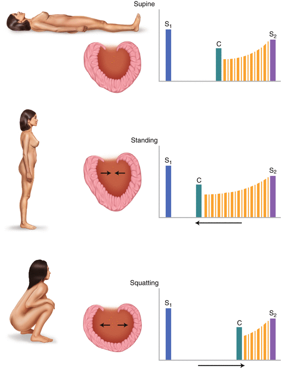 Only gold members can continue reading. Log In or Register to continue
Stay updated, free articles. Join our Telegram channel

Full access? Get Clinical Tree


