Coronary Artery Bypass Grafting using Cardiopulmonary Bypass


Coronary Artery Bypass Grafting using Cardiopulmonary Bypass


Ahmet Kilic

Chittor Sai-Sudakar

Juan Crestanello

Robert S.D. Higgins


INTRODUCTION

Atherosclerotic heart disease continues to be the leading cause of death in the United States. While coronary artery bypass surgery has enjoyed a storied evolution since the 1960s to be among the most spectacular advances in medicine, the field of cardiothoracic surgery is undergoing a significant evolution with the advent of more effective medical therapies, alternative percutaneous and hybrid revascularization techniques, and percutaneous valve procedures. Ironically as these alternative strategies evolve, the need for effective surgical myocardial revascularization techniques using cardiopulmonary bypass is becoming even more critical in patients with advanced atherosclerotic heart disease and left ventricular dysfunction. Central to this expanding need is the refinement of indications for coronary artery bypass procedures in critically ill patients. Older patients with advanced disease, left ventricular dysfunction, and multiple comorbidities create even greater challenges for the practicing cardiac surgeon.

In this chapter, we will review the evolving assessments of coronary artery anatomy using innovation imaging techniques. A full understanding of assessments of myocardial viability and suitability for revascularization continues to be of critical importance in this discussion. We will also review the impact of left ventricular dysfunction on surgical revascularization outcomes in patients with low ejection fraction. The importance of surgical risk reduction in complicated coronary revascularization particularly in patients on platelet inhibitor therapies, that is, clopidogrel as well as evolving perioperative management techniques, has significantly reduced the morbidity and perioperative complications of coronary revascularization. The indications for hybrid and multivessel disease management as well as left main coronary revascularization will also be reviewed.

No discussion of the management of coronary artery disease (CAD) by conventional revascularization techniques can ignore the importance of the development of appropriateness criteria for the management of multivessel disease and left main coronary disease. Evidence highlighting the benefits of coronary artery bypass grafting (CABG) continues to demonstrate significant survival advantage in patients with multivessel disease compared with percutaneous coronary intervention (PCI). In these recent contributions to the literature, critical evidence from randomized clinical trials and case-matched patient studies, CABG was associated with lower risk of death than stenting underscoring the comparative effectiveness of CABG in these patients. As we appreciate the comparative benefits of CABG versus PCI, we also have to be mindful of the challenges in the current reimbursement environment to manage preventable complications, so as to reduce the financial impact of readmissions in the current healthcare economy. Only if the surgical profession can minimize the risk in appropriately selected patients, enhance patient outcomes, reduce perioperative complications, and readmission rates after CABG, will we be able to continue to demonstrate the comparative benefits of CABG.


ASSESSMENT OF CORONARY ANATOMY


Coronary Angiography

Coronary angiography remains the gold standard in defining coronary anatomy and graft patency. It is an invasive procedure and is associated with procedural risks including stroke, native vessel dissection, acute myocardial infarction (MI), ventricular arrhythmias, and puncture site morbidities such as hematomas and pseudoaneurysm formation. In addition, its deficiency in three-dimensional definition has led to the investigation of other noninvasive imaging techniques to define the overall coronary pathoanatomy. Initial attempts to utilize electron beam tomography and helical scan computed tomography (CT) were not encouraging due to motion artifacts, the need for prolonged breath holding, and image distortion due to metallic clips. Newer imaging techniques such as multidetector computed tomography (MDCT) have begun to revolutionize the diagnostic field.


Multidetector Computed Tomography

MDCT allows for fast data acquisition in a single breath hold of 25 to 30 seconds with varying slice thickness collimation, and Gantry rotation speeds following intravenous administration of 80 to 100 ml of nonionic contrast agent and a predetermined delay period to allow the contrast to reach the arterial circulation. Preprocedure beta-blockade allows for a heart rate of <60 beats per minute since faster hearts along with atrial fibrillation and
ectopic beats impair the quality of images obtained. Image reconstruction and manipulation is facilitated by electrocardiographic gated reconstruction algorithms.


Evaluation of Native Coronary Anatomy

Martuscelli and coworkers investigated the accuracy of MDCT in detection of significant (>50%) stenosis using a scanner equipped for 16 × 0.625 mm collimation and compared it with coronary angiography in 64 patients with suspected CAD. Eighty-four percent of the angiographic segments were evaluable. Severe calcification, cardiac motion artifacts, poor opacification, and blending of the segments with veins hindered the evaluation of the remaining segments. In the segments that were evaluable, namely, vessels >1.5 mm in diameter, MDCT had a sensitivity, specificity, positive predictive value, and negative predictive value of 89%, 98%, 90%, and 98%, respectively. MDCT had a 100% specificity and sensitivity in detecting total occlusion. Overall, MDCT detected 78% of stenoses detected by angiography. Cury et al. demonstrated an excellent correlation between 16-slice MDCT and angiography in quantifying the degree of stenosis. However, MDCT tended to overestimate the stenosis. The evaluation of the coronary anatomy and the results were significantly better than those obtained with the 4-slice MDCT. With the advent of 64-slice MDCT, Rubinshtein et al. analyzed its efficacy in the emergency room in evaluating patients with chest pain. They observed that emergency room MDCT had a high positive predictive value for diagnosis of acute coronary syndrome and a negative study predicted a low rate of major adverse cardiovascular event. In this study, only 4.6% of the coronary segments were of low image quality testifying to improvements in MDCT technology and also aided by the low calcium burden. These diagnostic tests have also been helpful in planning for minimally invasive revascularization procedures. During the procedures of minimally invasive direct coronary artery bypass (MIDCAB) or totally endoscopic coronary artery bypass (TECAB), the visualization of the target vessels is often limited and it is critical to identify the target vessel correctly and bypass it at the correct site. A common source of error is the bypassing of a large diagonal instead of the left anterior descending (LAD) artery during minimally invasive surgery. In addition, the target vessels could be buried under epicardial fat, covered by a myocardial bridge, run an intramural course, or heavily calcified. Newer generation MDCT scanners have been shown to identify these variations in coronary anatomy better than coronary angiography. These findings can dictate the type of surgery offered to the patients. Coronary calcification is obscured by the dye used in angiography. In an open procedure, such areas can be palpated and anastomosis can be performed in the noncalcified segments of the coronary vessels. However, tactile feedback is absent in the minimally invasive procedures. Regions of coronary vessels burdened by calcification as evidenced by MDCT can be avoided for anastomosis.


Evaluation of Graft Patency

Reoperative cardiac surgery is associated with increased morbidity and mortality and the potential to inadvertently injure the bypass grafts. MDCT allows for the accurate three-dimensional visualization of the course of the left internal mammary artery (LIMA) graft in relation to the midline, location of other conduits, and the degree of adherence of the right ventricle to the posterior table of the sternum. Unusual location of the grafts and their proximal anastomoses pose difficulties in evaluation by conventional angiography, especially in the absence of radio-opaque markers for the proximal anastomoses. Faster data acquisition allows a manageable breath hold for the patient allowing for visualization of the most proximal portions of the internal mammary artery grafts not seen in the early generation scanners. Martuscelli et al. analyzed 285 conduits in 96 patients with 16-slice MDCT and compared the results with those obtained by conventional angiography. The sensitivity of MDCT in diagnosing significant stenosis was 96%. Evaluation of graft patency and occlusion improved with the advent of 64-slice MDCT. The accuracy of 64-slice multislice CT for graft occlusion and stenosis is given in Table 51.1. A recent analysis suggests that the sensitivity and specificity for graft occlusion approach 100% across the studies, with only two cases of missed occlusions in bypass grafts, which were not visualized on MDCT. There are currently no reports of false-positive occlusion. When diagnosing bypass graft stenosis, sensitivity ranged from 75% to 100%. Missed stenoses at distal anastomoses due partly to calcified wall plaque and membranous-like stenoses accounted for falsenegative results. The specificity for diagnosis of stenosis of bypass grafts ranges from 89.3% to 100%. Poor opacification combined with small vessel size, vessel kinking, and surgical clip artifact account for false-positive results. The results from these eight studies of 1,169 coronary artery bypass grafts are similar to those obtained by coronary angiography. Several studies have reported that grafts not visualized on coronary angiography were visualized on MDCT, thus, providing important additional clinical information.


Table 51.1 Diagnostic Accuracy of 64-Slice MSCT in Coronary Artery Bypass Grafting Assessment (Assessable Grafts Only)


Occlusion


Stenosis


Author


Grafts


Art


Ven


Sens (%)


Spec (%)


Sens (%)


Spec (%)


Dikkers


69


52


17


100


100


100


98.7


Feuchtner


70


46


24


100


100


75


95


Jabara


147


47


100


93.3


100


100


100


Malagutti


109


45


64


100


98.3


Meyer


397


144


253


100


100


97a


97a


Onuma


146


72


74


100


100


100


98.1


Pache


93


22


71


97.8


89.3


Ropers


138


37


101


100


100


100


94


Art, arterial grafts; Sens, sensitivity; Spec, specificity; Ven, venous grafts.


a Combined results for stenosis and occlusion.


From: Jones CM, et al. Coronary artery bypass graft imaging with 64-slice multislice computed tomography: literature review. Semin Ultrasound CT MR 2008;29(3):204-213.


However, assessment of native coronary arteries is impaired in the presence of atherosclerosis, calcification, and small diameter vessels. In addition, MDCT does not provide the flow characteristics of either the grafts or the distal run off. Arrhythmias, metallic clips on the side branches of the grafts, and higher heart rates continue to hinder adequate evaluation of the bypass grafts. Improved temporal and spatial resolution of the 64-slice CT scanner overcomes the artifacts caused by metal clips. Evaluation of in-stent stenosis by MDCT is difficult. Coronary stents are usually 2.5 to 4 mm in diameter. Blooming artifacts are produced by the metal in both the bare metal and drug-eluting stents. Cardiac motion compounds the problem. Partial volume effects lead to decreased visibility of the stent lumen, underestimates the luminal diameter, and overestimates
the outer diameter. Inside the lumen of the stent, there is an increase in the attenuation that can be minimized by utilizing submillimeter collimation and a sharp reconstruction kernel. With improving technological areas, the larger diameter stents in the left main coronary artery and the proximal coronary vasculature can be analyzed for evidence of decreased luminal diameter due to intimal hyperplasia. The native coronary vasculature in the immediate vicinity of the stent is difficult to evaluate for stenosis due to beaming artifacts. In addition, beam hardening produces dark areas adjacent to the clips stimulating stenosis especially at the anastomotic points.

MDCT can be effectively used as a one-stop test to evaluate the different causes of chest pain following CABG. Dual source and 256-slice scanning allow for fast data acquisition, may further simplify the test, improve diagnostic accuracy, and eliminate the current limitations imposed by rapid hearts rates. MR coronary angiography is being evaluated to analyze its potential as a noninvasive diagnostic tool. The summary statement released by the American Heart Association in collaboration with other councils has recommended that neither coronary artery CTA nor MRA should be used to screen for CAD in patients who have no signs or symptoms of CAD (Class III, level of evidence C). The potential benefit of noninvasive coronary angiography is likely to be greatest and is reasonable for symptomatic patients who are at intermediate risk for CAD after initial risk stratification including patients with equivocal stress-test results (Class IIa, level of evidence B). Concerns regarding radiation dose limit the use of coronary CTA in hig-hrisk patients who have a very low pretest likelihood of coronary stenoses; they are likely to require intervention and invasive catheter angiography for definitive evaluation; thus, CTA is not recommended for those individuals (Class III, level of evidence C). Pronounced coronary calcification may negatively impact interpretability and accuracy of coronary CTA and thus, the usefulness of CTA is uncertain in these individuals (Class IIb, level of evidence B).”
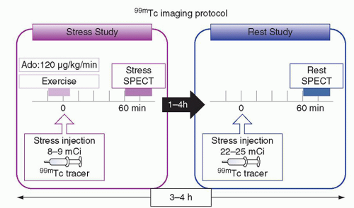
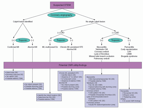
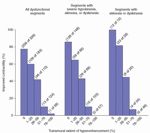
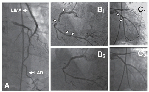


Table 51.2 Common Myocardial Blood Flow Tracers


Tracer


PET/SPECT


Tracer production


Half-life


Scan time (min)


Positron energy (MeV)


Radiation dosage (m)*Sv


Technetium-99m sestamibi/tetrofosmin


SPECT


Generator


6 h


12–15 (dual head – camera)


1-d: stress-rest. Sestamibi/tetrofosmin 12/10.6 (40 mCi)


2-d: stress-rest. Sestamibi 17.5 (30 mCi + 30 mCi)


Thallium-201


SPECT


Cyclotron


72.9 h


12–15 (dual head camera)


25.1 (4 mCi)


Rubidium-82


PET


Generator


76 s


6–10


3.15


16 (90 mCi)


Oxygen-15 water


PET


Cyclotron


110 s


6–10


1.72


2.5 (60 mCi)


Nitrogen-13 ammonia


PET


Cyclotron


9.97 min


2–4


1.19


2.4 (30 mCi)


F-18 FBnTP


PET


Cyclotron


110 min


12–15 (dual head camera)


0.63


Not available


From: Yoshinaga K, Manabe O, Tamaki N. Physiological assessment of myocardial perfusion using nuclear cardiology would enhance coronary artery disease patient care: which imaging modality is best for evaluation of myocardial ischemia? (SPECT-side). Circ J 2011;75(3):713-722.


ASSESSMENT OF MYOCARDIAL ISCHEMIA AND VIABILITY

Total or sub-total occlusion of any portion of the coronary vasculature leads to varying spectra of myocardial viability including infarction, myocardial stunning, and hibernating myocardium. Reestablishment of the coronary perfusion by fibrinolytic therapy or percutaneous interventions is the cornerstone of the ST elevation myocardial infarction (STEMI) protocols adopted by nearly all the hospitals across the country. In less emergent cases or patients presenting in outpatient settings with symptoms of angina or congestive heart failure and documented evidence of critical CAD and depressed systolic function, there is considerable debate about the utility of further assessment for myocardial viability prior to any intervention.


Nuclear Imaging

Nuclear imaging utilizes technetium-labeled radioisotopes or positron emission tomography (PET) tracers under rest and stress conditions to detect the extent and severity of myocardial ischemia (Table 51.2), and single-photon-emission computed tomography (SPECT) myocardial perfusion imaging is considered a useful tool for risk assessment for CAD. Following the injection of the thallium or technetium-labeled tracers (sestamibi or tetrofosmin), their uptake by the myocyte is dependent on the regional myocardial blood flow. The subsequent washout of these radiopharmaceuticals is faster from normal myocardium compared with the ischemic myocardium. The images obtained at this time point are usually considered the resting images. Subsequently, stress images are obtained using exercise stress protocols (Fig. 51.1). In patients who are unable to exercise or who have other pathologies such as an abnormal EKG, left bundle-branch block, paced rhythm, chronic obstructive pulmonary disease, or asthma pharmacological stress is induced utilizing several agents (vasodilator therapy, adenosine, dipyridamole, or dobutamine. The images obtained at rest and stress are compared and myocardial perfusion defects are graded as myocardial ischemia, reversible or fixed perfusion defects. Myocardial perfusion imaging utilizing SPECT has been validated in several studies as a useful screening tool for CAD in patients with risk factors and in addition for the detection of extent and degree of myocardial ischemia. Glucose metabolism substitutes free fatty acid metabolism as the main energy source in ischemic but viable myocardium. 18F fluorodeoxyglucose (FDG), a glucose analog combined with a tracer (Rubidium-82), is used to evaluate the glucose metabolism in the myocyte. Following the detection of a fixed perfusion defect on perfusion imaging, preservation of 18F-FDG uptake in that region by a PET scan is suggestive of hibernating myocardium and indicative of the beneficial effect of revascularization in patients with ventricular dysfunction.


Disturbances in fatty acid metabolism in the myocyte persist for up to 30 hours following the cessation of symptoms from a transient coronary event. This pathway has been investigated as a potential diagnostic tool for a recent coronary event by using radioiodine-labeled branched chain fatty acid β-methyl-p-[1231]-iodophenyl pentadeconic acid (BMIPP) SPECT scans to detect acute coronary syndromes in the emergency room with a sensitivity, specificity, and negative predictive value of 81%, 61%, and 83%, respectively.


Fig. 51.1. Exercise/pharmacological stress 99mTc myocardial perfusion imaging protocol. (From Yoshinaga K, Manabe O, Tamaki N. Physiological assessment of myocardial perfusion using nuclear cardiology would enhance coronary artery disease patient care: which imaging modality is best for evaluation of myocardial ischemia? (SPECT-side). Circ J 2011;75(3):713-722.)


Fig. 51.2. Algorithm for potential usage of cardiovascular magnetic resonance imaging in patients with suspected ST elevation myocardial infarction. From Kim HW, Farzaneh-Far A, Kim RJ. Cardiovascular magnetic resonance in patients with myocardial infarction: current and emerging applications. J Am Coll Cardiol 2009;55(1)1-16, with permission.


Cardiovascular Magnetic Resonance

Over the last decade, cardiovascular magnetic resonance (CMR) has gained in popularity as a reliable diagnostic modality for cardiac structure and function and myocardial ischemia to the extent that clinical utility pathways are being developed to utilize this technology for patients presenting with acute coronary syndromes (Fig. 51.2).


Cardiovascular Magnetic Resonance Techniques

The powerful magnetic field exerted by CMR scanner with a field strength of 1.5 to T (30,000 times stronger than the earth’s magnetic field) aligns the hydrogen atoms in the human body. Subsequently, radiofrequency pulses delivered by the scanner excite the hydrogen nuclei and receiver coils detect the signals as the hydrogen nuclei relax. The strength of the signal is dependant on the density of the protons in any given tissue and accounts for the contrast in the MR images. In addition, the images are also dependant on two distinct relaxation patterns of the protons. T1 is the longitudinal relaxation time and is the time required for a substance to be magnetized after being placed in a magnetic field, whereas T2, the transverse relaxation time is a measure of how long the resonating protons remain in phase following a radiofrequency pulse.


  • Spin Echo Imaging: Blood is black and tissues are bright.


  • Gradient Echo Imaging: Blood is bright and cardiac tissue is dark.


  • Cine CMR: Provides important information regarding segmental and global ventricular function, wall thickness at different time points of the cardiac cycle, and structural heart defects including aneurysms, septal defects, and valve pathologies.


  • Perfusion Imaging: For the assessment of myocardial perfusion and used in conjunction with vasodilators. Vasodilation with adenosine or dipyridamole following a bolus contrast injection causes a three- to five-fold increase in blood flow in myocardial regions supplied by normal coronary arteries resulting in bright areas in contrast to the areas supplied by stenotic coronary arteries, which appear as dark regions on imaging.


  • Delayed Contrast Imaging: For the assessment of the ability of myocardium to clear the contrast (gadolinium diethylenetriamine penta-acetic acid). Normal myocardium clears the contrast quickly and appears dark. Ischemic or scar tissue appears bright due to delayed clearance of contrast.


ASSESSMENT OF MYOCARDIAL VIABILITY

In canine models of acute and chronic MI, contrast MRI was used to distinguish between reversible and irreversible ischemic injury independent of wall motion and infarct age. Kim et al. observed that contrast MRI in combination with cine MRI could identify acute MI (hyperenhanced with contractile dysfunction), injured but viable myocardium (not hyperenhanced but with contractile dysfunction and normal myocardium (not hyperenhanced and with normal function). Subsequently, the same group investigated the use of contrast-enhanced MRI to predict improvements following revascularization in regions of abnormal ventricular contraction. Gadolinium-enhanced MRI was performed in 50 patients with ventricular dysfunction before either surgical or percutaneous revascularization to assess the transmural extent of the hyperenhanced myocardium postulated to represent nonviable myocardium. In addition, cine MRI was performed to assess the regional contractility at the same locations before and after revascularization. Following analysis of the data, the authors demonstrated that the likelihood of improvement in regional contractility after revascularization decreased progressively as the transmural extent of the hyperenhancement before revascularization increased. The amount of left ventricular mass that was dysfunctional and not hyperenhanced before revascularization strongly related to the degree of improvement in the wall motion score and ejection fraction after revascularization. The investigators clearly demonstrated that reversible myocardial dysfunction can be identified by contrast-enhanced MRI before coronary revascularization (Fig. 51.3). Further confirmatory studies established the value of delayed enhancement MRI as a powerful predictor of myocardial viability after surgery.


HYBRID CORONARY REVASCULARIZATION

While conventional revascularization is routine in patients with triple-vessel CAD, hybrid coronary revascularization (HCR) has evolved to combine (a) surgical revascularization to the LAD coronary artery with the LIMA through a minimally invasive approach and (b) percutaneous coronary revascularization (PCI) to the circumflex and right coronary artery (RCA) territories. HCR is an alternative revascularization strategy for patients with triple-vessel CAD. HCR achieves complete revascularization and combines the advantages of bypass surgery and PCI (Fig. 51.4).


Fig. 51.3. Relationship between the transmural extent of hyperenhancement before revascularization and the likelihood of increased contractility after revascularization. (From Kim RJ, et al. The use of contrast-enhanced magnetic resonance imaging to identify reversible myocardial dysfunction. N Engl J Med 2000;343(20):1445-1453.)


Fig. 51.4. Hybrid coronary revascularization in a patient with triple-vessel coronary artery disease. (A) Completion angiogram of the left internal mammary artery to left anterior descending. (B1) Right coronary artery (RCA) lesions that were treated by percutaneous coronary intervention (PCI). (B2) RCA after PCI. Lesions in the circumflex coronary artery before (C1) and after (C2) PCI.

Although traditional CABG is highly effective in improving symptoms and increasing survival, it is associated with morbidity and mortality mostly related to the invasiveness of the procedure. Revascularization of the LAD coronary artery with the LIMA provides the major clinical advantage of CABG and provides the majority of the survival advantage provided by CABG with the best long-term patency rate. Compared with PCI, LIMA bypass for isolated lesions of the LAD results in better angina relief, and lower incidence of major adverse cardiovascular and cerebrovascular events and need for repeat revascularization. Long-term patency of saphenous vein bypass grafts (SVG) is inferior to that of arterial grafts. It is unknown how the patency of SVG compares to the patency of DES in the same position.

Table 51.3 summarizes the potential advantages of HCR and compares them with PCI and CABG. HCR achieves complete revascularization potentially combining the advantages of both revascularization strategies: low procedural morbidity and mortality, low restenosis rate, and low repeated revascularization rates.

HCR builds on the concept of “heart team” approach for the management of coronary revascularization. The heart team, integrated by cardiac surgeons, interventional cardiologists, clinical cardiologists, and primary physicians, evaluate the patient’s clinical and angiographic characteristics and offer the patients a revascularization strategy based on best evidence and clinical guidelines. For HCR, interventional cardiologist and surgeons should agree that the patients are candidates for both (a) PCI to the non-LAD territories and (b) minimally invasive bypass to the LAD using the LIMA.


Only gold members can continue reading. Log In or Register to continue

Stay updated, free articles. Join our Telegram channel

Jun 15, 2016 | Posted by in CARDIAC SURGERY | Comments Off on Coronary Artery Bypass Grafting using Cardiopulmonary Bypass

Full access? Get Clinical Tree

Get Clinical Tree app for offline access

Table 51.3 Comparison of Potential Benefits of Percutaneous Coronary Intervention (PCI), Conventional Coronary Artery Bypass Grafting (CABG), Minimally Invasive CABG, and Hybrid Coronary Revascularization (HCR)