Transhiatal Esophagectomy


Transhiatal Esophagectomy


Mark B. Orringer


INTRODUCTION

The technique of transhiatal esophagectomy (THE) without thoracotomy and a cervical esophagogastric anastomosis (CEGA) was rediscovered in the mid-1970s. Since then, the operation has been progressively refined and has emerged as a recognized alternative to traditional transthoracic esophagectomy for both benign and malignant disease requiring resection and reconstruction. Since the earliest reported series of transthoracic esophagectomy and an intrathoracic esophagogastric anastomosis, the leading causes of postoperative morbidity and mortality have been (1) respiratory insufficiency resulting from a combined thoracoabdominal operation and (2) mediastinitis from an intrathoracic anastomotic leak. THE addressed both issues. By eliminating a thoracic incision, postoperative pulmonary complications were less. By placing the esophagogastric anastomosis in the neck, a leak meant a salivary fistula which drained externally and was not generally associated with mediastinitis. Detractors of THE have argued that the procedure is done “blindly” and has the potential for excessive bleeding, and failure to permit an extensive mediastinal lymph node dissection makes it less attractive from an oncologic standpoint. These concerns have not been substantiated in now more than 30 years of experience with THE, which the author therefore regards as the approach of choice in most patients with benign and malignant disease requiring esophageal resection and reconstruction. This chapter focuses upon the technique of THE, particularly the nuances of gastric mobilization, transhiatal mobilization of the esophagus, preparation of the gastric conduit and positioning it in the mediastinum and neck, and construction of the CEGA.


PATIENT SELECTION AND PREOPERATIVE PREPARATION

Critical to a successful THE are careful patient selection and preoperative assessment and conditioning. Physiologic age is of far more important than chronologic age, and there is no absolute age limit to performing this operation. Baseline pulmonary function tests in those with a history of cigarette smoking and cardiology assessment of risk in those with heart disease are routine. The presence of cirrhosis and associated ascites and/or portal hypertension absolutely preclude an esophagectomy by jeopardizing venous drainage of the esophageal replacement and therefore a safe esophageal reconstruction. In those who have undergone multiple prior gastric operations that jeopardize use of the stomach as an esophageal replacement, or those with carcinoma of the cardia that may require a major gastric resection to achieve an adequate margin, the suitability of the colon as an esophageal substitute must be determined in advance. A barium enema examination to exclude severe diverticular disease and a bowel prep preoperatively are undertaken. It has not been the author’s
practice to utilize preoperative mesenteric angiography to evaluate the blood supply of the colon unless there is a history of mesenteric vascular disease or possible interruption of the inferior mesenteric artery in a prior abdominal aortic aneurysm repair. Abstinence from cigarette smoking for a minimum of 3 weeks before the operation is an absolute requirement of the author, even in those with cancer. Those suspected of continued smoking have a urine cotinine level determined, and if recent tobacco use is confirmed, the operation is cancelled. Patients are issued an incentive spirometer at the initial consultation and are requested to use it on a set schedule throughout the day until they are admitted for their operation. They are instructed to walk 3 miles a day to condition themselves for early postoperative ambulation. The importance of this “surgeon-patient contract” is emphasized. With severe esophageal obstruction and inability to obtain adequate fluid and caloric intake by mouth, a nasogastric feeding tube is placed into the stomach, if necessary with fluoroscopic control, and tube feedings for home use are initiated. Every effort is made to avoid a hospitalization and the need for intravenous hyperalimentation before surgery, and patients are typically admitted for the esophagectomy on the day of the scheduled procedure. A mechanical cleansing of the large bowel is undertaken in those in whom there is concern about the suitability of the stomach as an adequate esophageal replacement.


Table 17.1 Indications for Transhiatal Esophagectomy (2007 Patients)


Group I 1,063 Pts


Number (%) Group II 944 Pts


Total 2,007 Pts


1976-1998


1998-2006


1976-2006


Benign conditions


278


(26%)


204


(22%)


482


(24%)


Neuromotor dysfunction


92


(33%)


47


(23%)


139


(29%)


Achalasia


69


44


113


Spasm/dysmotility


21


3


24


Scleroderma


2


0


2


Stricture


74


(27%)


21


(10%)


95


(20%)


Gastroesophageal reflux


40


7


47


Caustic ingestion


18


6


24


Radiation


4


2


6


Other


12


6


18


Barrett mucosa with high-grade dysplasia


53


(19%)


90


(44%)


143


(30%)


Recurrent gastroesophageal reflux


21


(8%)


6


(3%)


27


(6%)


Recurrent hiatus hernia


14


(5%)


14


(7%)


28


(6%)


Acute perforation


15


(5%)


9


(5%)


24


(5%)


Acute caustic injury


5


(2%)


1


(1%)


6


(1%)


Other


4


(1%)


16


(8%)


20


(4%)


Carcinoma of the intrathoracic esophagus


Site


785


(74%)


740


(78%)


1,525


(76%)


Upper third


35


(4%)


16


(2%)


51


(3%)


Middle third


164


(21%)


63


(9%)


227


(15%)


Lower third and/or cardiaa


586


(75%)


661


(89%)


1,247


(82%)


a Includes pathologic gastric carcinomas involving the cardia and lower esophagus.


Source: Reproduced with permission from Orringer MB, Marshall B, Chang AC, Lee J, Pickens A, Lau CL. Two thousand transhiatal esophagectomies: Changing trends, lessons learned. Ann Surg 2007;246:363-374.


SURGICAL TECHNIQUE

The four phases of THE—abdominal, cervical, mediastinal (transhiatal), and anastomotic —are performed in a sequential and reproducible fashion from patient to patient. Placement of an epidural catheter preoperatively for both intraoperative and postoperative pain control is an important advance. Flexible esophagoscopy is performed after induction of general anesthesia to insure the availability of an adequate proximal esophageal margin as well as a healthy gastric conduit for esophageal replacement. Excessive air insufflation should be avoided. After the endoscopy, a 16F nasogastric tube is placed. A radial artery catheter for intraoperative monitoring of blood pressure during the mediastinal dissection is routine. This catheter is well-secured to avoid its dislodgment during this operation in which the arms will be at the sides, not secured to arm boards. The patient is positioned supine on the operating table with the head turned toward the right and supported on a soft head ring, and the neck extended by a small shoulder roll under the scapulae. The wrists, elbows, and arms are carefully padded with foam, placed at the sides, and secured in place with the drape sheet. This positioning allows the surgeon and his/her assistant unobstructed access to the patient’s neck, chest, and abdomen. Table-mounted brackets for securing an upper-hand retractor during the abdominal phase are positioned at the nipple line. “Bumping up” one side or another for an anterolateral thoracotomy in case a transthoracic exposure is required is not done. If conversion to a transthoracic esophagectomy is required, the abdomen is quickly closed temporarily with 4 or 5 through-and-through full-thickness #2 nylon sutures, the wound covered with a plastic surgical drape, and the patient turned and repositioned in the lateral decubitus position for a true posterolateral thoracotomy. After completion of the transthoracic esophagectomy, the chest is closed and the patient turned supine once again and positioned as before for completion of the procedure. The operative field extends from the mandible to the pubis and from one midaxillary line to the other. Two suction catheters, one at the head of the table and one from below, are used routinely. This operation is best performed from the left side of the operating table, and the following operative description of THE is how the procedure would be viewed by the surgeon so positioned.


Abdominal Phase

The sequence of this phase of the operation is gastric mobilization, distal esophageal transhiatal mobilization, Kocher maneuver,
pyloromyotomy, and finally feeding jejunostomy. Through an upper midline supraumbilical incision (Fig. 17.1, inset), the abdomen is explored, the triangular ligament of the liver divided with electrocautery, and the left lobe of the liver retracted to the right with a liver blade retractor secured to the upper-hand supporting bar. A standard upper-hand abdominal wall retractor secured to the bar is used to retract the left side of the abdominal wall. The high greater curvature of the stomach is localized, and the adjacent greater omentum retracted to the left. A “clear space” between the omentum and the gastric wall is developed until the lesser peritoneal sac is entered. An index finger passed into this opening behind the greater omentum elevates the omentum away from the stomach and is used to define the high short gastric vessels and left gastroepiploic arcade. These vessels are sequentially clamped with 13-inch long right-angle clamps, divided, and tied with 2-0 silk, taking care to stay well away from the stomach to avoid ischemic necrosis. The gastric wall must be handled gently throughout this mobilization and unnecessary traction on the stomach avoided. Care must also be taken to avoid excessive downward traction on the high greater omentum that may be adherent to the spleen and result in a splenic capsular tear. Before dividing all of the high short gastric vessels, the direction of the dissection is changed, and the remaining left gastroepiploic vessels sequentially identified, clamped, divided, and ligated moving along the greater curvature toward the pylorus. The right gastroepiploic artery is identified by palpation and vision as it either terminates in the gastric wall or communicates through fine vessels with the left gastroepiploic arcade. The greater omentum is divided 1.5 to 2 cm inferior to the right gastroepiploic artery, carefully preserving it and repeatedly monitoring for the presence of a pulse before dividing and ligating each branch. The greater omentum is separated from the stomach to the level of the pylorus. Once this portion of the distal stomach has been mobilized, it can be better retracted to the right exposing the highest short gastric vessels still to be divided. The peritoneum overlying the hiatus is incised and the esophagogastric junction identified. The author no longer encircles the esophagogastric junction with a Penrose drain in order to minimize the chance of a traction injury to the upper stomach. The stomach is elevated and any adhesions between the retroperitoneum and posterior gastric wall divided with electrocautery.


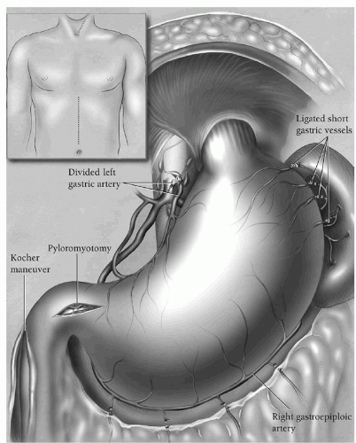
Fig. 17.1. The cervical and supraumbilical incisions used for THE (inset), and the key components of the abdominal phase: ligation of the high short gastric and left gastroepiploic vessels of the stomach; preservation of the right gastroepiploic vascular arcade, the primary blood supply of the mobilized stomach; division of the left gastric artery and vein; a generous Kocher maneuver; and a pyloromyotomy. (Reproduced from Orringer MB. Transhiatal esophagectomy without thoracotomy. Oper Tech Thorac Cardiovasc Surg 2005;10(1):63-83.)

Attention now shifts to the lesser curvature of the stomach. One hand is passed behind the mobilized greater curvature of the stomach into the lesser peritoneal sac until the fingers can be seen through the filmy gastrohepatic omentum on the lesser curvature side of the stomach. The gastrohepatic omentum is incised with electrocautery progressing superiorly toward the diaphragmatic hiatus, carefully palpating for a pulse in an aberrant left hepatic artery arising from the left gastric artery. This aberrant vessel is preserved if present by dividing the left gastric artery distal to its origin. By retracting the stomach to the left, the lesser curvature soft tissues are tensed, facilitating identification, mobilization, clamping, dividing, and ligating of the left gastric vein and then the left gastric artery with 2-0 silk ties. The artery is doubly ligated at its origin from the celiac trunk, and all lymph nodes in this area dissected and swept to the left attached to the lesser curvature. Similarly, all soft tissue and lymph nodes along the lesser curvature of the stomach anterior to the right crus of the diaphragmatic hiatus are swept to the left with the stomach. With mobilization of the greater curvature and high lesser curvature of the stomach completed, dissection of the esophagogastric junction and lower esophagus is begun.

The mobilized stomach is gently retracted downward as the phrenoesophageal tissue at the level of the hiatus is incised and the posterior mediastinum entered anterior to the esophagus. This space is enlarged bluntly with a sweeping
motion of the fingers so that a narrow heart-shaped retractor can be inserted and upward traction on it exerted. This exposes the distal esophagus in the posterior mediastinum. If tumor at the cardia is adherent to the diaphragm, a rim of diaphragm may need to the resected with the specimen to achieve an adequate margin. The posterior aspect of the esophagus is gently mobilized away from the spine by two, three, and then four fingers inserted into the hiatus behind the esophagus. Using a long 13-inch right-angle clamp and a long electrocautery tip, the posterior mediastinal paraesophageal soft tissues are elevated and divided. Visible paraesophageal lymph nodes are resected. The dissection alternates from one side to the other of the esophagus moving progressively toward the carina. If either lung is seen in the field, the mediastinal pleura on that side has been violated, and a chest tube will be required later. Inserting a hand into the low mediastinum and gently “rocking” the esophagus from side to side allows assessment of the degree of fixation between the esophagus and contiguous structures, particularly the spine, prevertebral fascia, and descending thoracic aorta, which could complicate transhiatal esophageal mobilization. During this low esophageal dissection, blood pressure as recorded through the radial artery catheter is carefully watched to avoid prolonged hypotension resulting from cardiac displacement by the hand inserted into the mediastinum. After mobilizing approximately 10 cm of the distal esophagus, the low posterior mediastinum is packed with a large abdominal pad to facilitate hemostasis.

The duodenum is mobilized with a generous Kocher maneuver to facilitate the eventual upward reach of the stomach through the posterior mediastinum. After an adequate Kocher maneuver, the pylorus should be able to be grasped and moved from its usual position in the right upper quadrant to the level of the xiphoid process. A pyloromyotomy is now performed. Two 3-0 silk figure-of-eight traction sutures are placed, one through the superior aspect and the other through the inferior aspect of the pylorus. The anterior pylorus is thus elevated as the pyloromyotomy is begun. Using a low cutting current from a needle-tipped electrocautery, the gastric serosa and superficial muscle layers are incised beginning approximately 1.5 to 2 cm on the gastric side of the pylorus. A fine right-angle clamp is then used to elevate the remainder of the muscle away from the submucosa as the muscle is progressively divided moving toward the pylorus. The dissection then becomes more superficial as the serosa and a few muscle fibers overlying the pylorus are cut, and the serosa and muscle of the duodenum divided for 0.5 to 1 cm. The duodenal submucosa is distinctly different from the gastric submucosa, the former being more fatty, more yellow in color, and having fine veins coursing over it, while the gastric submucosa is more pink and has relatively fewer veins on its surface. With the gastric and duodenal submucosa exposed on either side of the pylorus, the pyloric muscle is readily identified, dissected, and elevated with the right-angle clamp, and progressively divided with the needle-tipped electrocautery. Freely bulging submucosa between the two cut ends of the pylorus signifies a complete myotomy. At the conclusion of the pyloromyotomy, a silver hemoclip is placed near the pylorus at the base of either traction suture to mark the level of the pylorus for future radiographic evaluation. Should the integrity of the pyloroduodenal mucosa be violated, the pyloromyotomy is not converted to a pyloroplasty. Rather, the hole is closed with several interrupted 5-0 polypropylene sutures. This avoids a pyloroplasty suture line at right angles to the vertical axis of the stomach when the stomach is pulled upward through the posterior mediastinum. If repair of a pyloroduodenal mucosal tear is required, once the stomach has been brought through the mediastinum and the fundus delivered to the neck, the pyloromyotomy site, which typically comes to rest approximately 3 to 4 cm below the level of the hiatus, is buttressed with adjacent omentum or greater curvature fat loosely “tacked” in place with several interrupted 4-0 sutures. A feeding jejunostomy is performed using a 14F rubber catheter with side holes cut at its tip inserted into the bowel 18 to 20 cm distal to the ligament of Treitz. The tube is secured in place with a 4-cm long Witzel maneuver. The jejunostomy tube is not yet brought through the abdominal wall but rather covered with a towel clamped on either side to the drapes with hemostats as it emerges from the lower end of the abdominal incision to prevent its dislodgment during the remainder of the operation. Attention is now turned toward the neck.


Cervical Phase

Palpation of the cricoid cartilage identifies the level of the cricopharyngeus sphincter, the esophageal introitus. A 5 to 7 cm long oblique left cervical incision that parallels the anterior border of the sternocleidomastoid muscle is made (Fig. 17.2). The incision extends no more than 2 cm superior to the cricoid cartilage; as there is no esophagus above this point, a long cervical incision toward the angle of the mandible contributes nothing to exposure of the esophagus. The incision is deepened through the platysma muscle and then the fascia along the anterior edge of the sternocleidomastoid muscle. The sternocleidomastoid muscle is separated from the underlying cervical muscle using blunt finger dissection in the direction of the muscle. The sternocleidomastoid muscle is retracted laterally, and the underlying omohyoid muscle can be seen coursing obliquely in the opposite direction of the sternocleidomastoid muscle. The tendinous midpoint of the muscle is elevated with a fine right-angle clamp and divided with electrocautery (Fig. 17.3). Traction on the cut medial side of the omohyoid muscle tenses the omohyoid fascial layer, which is incised with electrocautery in the direction of the cervical incision. This omohyoid fascial layer leads to the underlying carotid sheath and its contents. Throughout the entire cervical portions of this operation, no metal retractors are placed against the tracheoesophageal groove in order to avoid injury

Only gold members can continue reading. Log In or Register to continue

Stay updated, free articles. Join our Telegram channel

Jun 15, 2016 | Posted by in CARDIAC SURGERY | Comments Off on Transhiatal Esophagectomy

Full access? Get Clinical Tree

Get Clinical Tree app for offline access