(1)
Department of Medicine, Johns Hopkins University School of Medicine, Baltimore, MD, USA
Keywords
HypertrophyStrainLeft atrial abnormalityP pulmonaleP mitraleAny of the four chambers of the heart may become enlarged. Unfortunately, the electrocardiogram (EKG) is only slightly useful in detecting chamber enlargement, and that may be a generous characterization. The electrocardiographic criteria often cited to suggest the presence of chamber enlargement are of only limited value and have improved little in the past half century. This chapter is intended primarily to familiarize the reader with the terminology related to EKG associations with chamber enlargement, not to suggest that EKG criteria are reliable in establishing chamber enlargement or hypertrophy.
Left Ventricular Hypertrophy
There are several methods of determining left ventricular hypertrophy (LVH). One that has been used for years is called the Estes system [1]. The Estes system is a point system that assigns certain weight to various features on the EKG (Table 6.1). The point assignments for the Estes system are as follows: (1) increased QRS amplitude, specifically an R or S wave in any limb lead of 20 mm, an S wave in V1 or V2 of 30 mm, or an R wave in V5 or V6 of 30 mm = 3 points; (2) ST–T changes, specifically ST depression and inverted or biphasic T waves = 1 point if the patient is taking digitalis, 3 points if the patient is not taking digitalis; (3) “left atrial involvement,” in which the terminal portion of the P wave in V1 is greater than 0.04 s in duration and greater than 1 mm deep = 3 points; (4) left axis deviation of at least −30° = 2 points; (5) QRS duration of 0.09 s or more = 1 point; and (6) increased intrinsicoid deflection in V5–6, which means the time between the beginning of the QRS and the peak of the R wave is 0.05 s or more = 1 point. This system gives a potential total of 13 points. Five points or more is considered definite LVH, while four points is “probable” LVH.
Table 6.1
Estes scoring system for left ventricular hypertrophy
Criterion | Points |
|---|---|
Voltage | 3 points |
“Strain” | 3 points (1 point if patient takes digitalis) |
Left atrial involvement | 3 points |
Left axis deviation | 2 points |
Prolonged QRS | 1 point |
Increased intrinsicoid | 1 point |
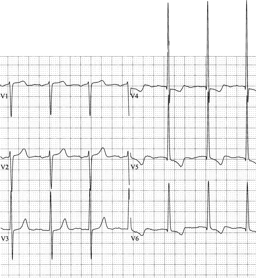
Some reflections are reasonable at this juncture. The first and most important criterion for LVH in any system is voltage. In my opinion, if there is not adequate voltage for LVH, one cannot make the EKG diagnosis, regardless of what other criteria may be present, since the nonvoltage findings may be present for many reasons other than LVH. Undoubtedly, requiring that the voltage criterion is always met will lead to some false negative readings of no LVH when it really is present. Nevertheless, insisting on the voltage criterion will provide increased specificity in exchange for less sensitivity. There are a variety of different findings that may satisfy the voltage criterion in addition to those mentioned above. The one that I use most is the amplitude of the R wave in lead V5 plus the amplitude of the S wave in lead V1. If the sum of these amplitudes is greater than or equal to 35 mm, that is adequate voltage for LVH. There are other variations in the criteria for voltage. Some authors accept an R wave amplitude of 11 mm or greater in aVL, but I believe that criterion is too dependent upon the QRS axis. The ST–T wave change associated with LVH is called “left ventricular strain” (Fig. 6.1). It looks like ischemia, but it is not acute ischemia because it is a persistent finding (not reversible within minutes) and is unassociated with symptoms of myocardial ischemia. One usually sees strain in leads I, aVL, V5, and V6. There may be variable degrees of ST and T wave abnormality, but just about any nonspecific ST–T change associated with voltage qualifies for strain. The reduced score for ST–T changes in the presence of digitalis is appropriate since the drug itself can cause ST segment abnormalities. Some authors object to the term “strain” because it implies a physiological statement related to hemodynamic work [2]. I find it a short, tidy EKG term that substitutes for “nonspecific ST–T changes associated with voltage criteria for LVH.”


Fig. 6.1
Left ventricular hypertrophy with strain. The R wave in V5 is 30–31 mm, the S wave in V1 is 15 mm, and ST depression and T wave inversion are present in V4–V6
A prolonged intrinsicoid deflection is intuitively compatible with LVH since the increased left ventricular mass would take longer to completely depolarize. As the volume of the left ventricular wall gets larger, then the intrinsicoid area would get bigger. The QRS duration and the intrinsicoid deflection criteria may not be especially useful because conduction disturbances independent of LVH (but not severe enough to cause bundle branch block) may produce similar changes.
Right Ventricular Hypertrophy
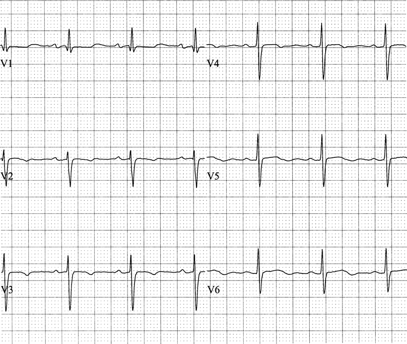
Right ventricular hypertrophy (RVH) is suggested by right axis deviation (RAD) beyond 90°, early R wave development in V1–3, and persisting S waves in V5–6. One finds an unusually tall R wave in the early precordial leads and a persisting S wave in the lateral precordial leads, so the QRS complexes can look nearly the same across the precordium (Fig. 6.2). The R wave in the right precordial leads is large as a reflection of the increased size or thickness of the right ventricle. The persisting S wave in the lateral precordial leads correlates with right ventricular depolarization after the left ventricle is depolarized. So both of these changes reflect the abnormally increased mass of the right ventricle. Some criteria require a certain amplitude for the R wave in V1, while others emphasize the importance of the relative sizes of R to S waves compared to the normal precordial R wave progression. Regardless of the criteria, none are felt to be especially reliable [2, 3]. Nevertheless, in the presence of RAD and early precordial R wave prominence, one may consider RVH.


