(1)
Department of Medicine, Johns Hopkins University School of Medicine, Baltimore, MD, USA
Keywords
BradycardiaTachycardiaFibrillationFlutterReentryArrhythmiaIn the initial approach to an arrhythmia, three key questions must be answered. If the following questions are answered correctly, one can diagnose just about any arrhythmia:
1.
Is it too fast or too slow?
2.
Is it ventricular or supraventricular in origin?
3.
Is it regular or irregular?
The first question can be answered easily. “Too fast” is a rate over 100 beats per minute; “too slow” is a rate less than 60 beats per minute. The answer to the second question is provided primarily with inspection of the duration of the QRS complex, and only secondarily with the presence or absence of P waves. If the QRS duration is normal (i.e., <0.12 s) then the rhythm must be supraventricular. If the QRS is prolonged (i.e., ≥0.12 s) one cannot immediately be sure if the rhythm is supraventricular in origin with aberrant conduction (e.g., bundle branch block) or is ventricular in origin. In cases of prolonged QRS complexes, the presence of P waves (or other evidences of supraventricular activity, such as flutter waves) and their constant relationship to QRS complexes can be helpful in distinguishing supraventricular from ventricular arrhythmias. In some cases, however, the distinction cannot be made with complete certainty from the surface 12-lead electrocardiogram (EKG). In such cases, esophageal or intracardiac leads may be required to absolutely determine the origin of the rhythm. The third question refers to the regularity of ventricular depolarization, or, in other words, the constancy of the RR interval. One should keep in mind that a minimal variation in RR interval is normal.
Once the determination is made of whether a rhythm is too fast or slow, regular or irregular, and ventricular or supraventricular, then one has taken the most important steps in distinguishing arrhythmias (Figs. 7.1 and 7.2). Each category of arrhythmias will be discussed separately.
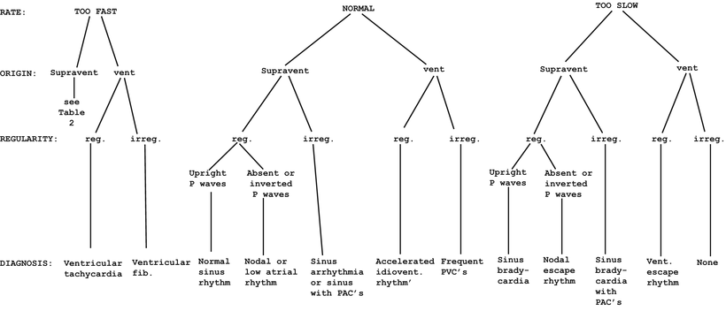
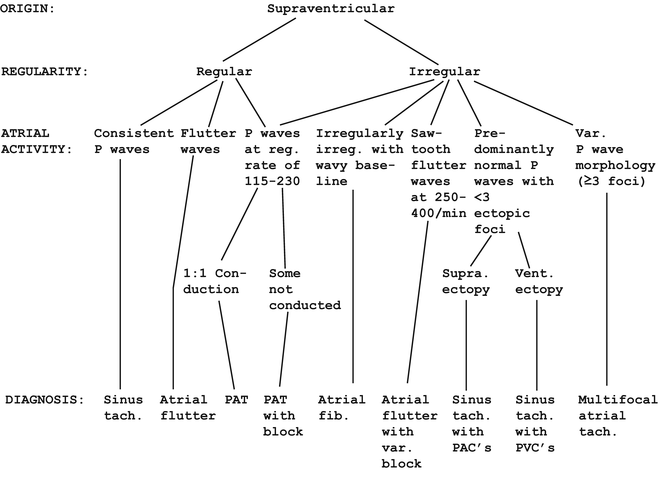
Fig. 7.1
Systematic approach to rhythm
Fig. 7.2
Flow sheet for supraventricular tachycardia
Supraventricular Tachycardias
There are five primary supraventricular tachycardias (Fig. 7.2 and Table 7.1):
Table 7.1
Characteristics of the supraventricular tachyarrhythmias
SVT | Too fast? | Supraventricular? | Regular? | Atrial ratea | Ventricular ratea | Response to CSMb |
|---|---|---|---|---|---|---|
Sinus tachycardia | + | + | +c | 101–190 | 101–190 | Transient atrial and ventricular slowing |
Atrial fibrillation | + | + | −d | 400–800 | 101–190 | Transient ventricular slowing |
Atrial flutter | + | + | +e | 250–400 [1] (mean = 299) | 101–190 (usually 2:1 block) | Transient ventricular slowing |
Paroxysmal atrial tachycardia (PAT) | + | + | + | 115–230 [1] | 115–230 [1] | No response, or conversion to sinus rhythm |
Multifocal atrial tachycardia (MAT) | + | + | − | 101–190 | 101–190 | Transient atrial and ventricular slowing |
1.
Sinus tachycardia
2.
Atrial fibrillation
3.
Atrial flutter
4.
Paroxysmal (or reentrant) atrial tachycardia (PAT)
5.
Multifocal atrial tachycardia (MAT)
The general category specified for these arrhythmias answers two of the three rhythm questions, namely that they are too fast and they are supraventricular. Now we need to answer the other question, namely whether these rhythms are regular or not (see Table 7.1). The (usually) regular supraventricular tachycardias are sinus tachycardia, atrial flutter, and PAT, while atrial fibrillation and MAT are irregular. After the question of regularity is determined, the atrial and ventricular rates may suggest one of the supraventricular tachycardias as opposed to another.
Sinus Tachycardia
In sinus tachycardia, the rhythm is regular. As already mentioned, there can be some minor variations in the RR interval with any sinus rhythm, but in general the RR interval is quite constant in sinus tachycardia. Each QRS is preceded by a P wave, which is usually but not always obvious in each lead (Fig. 7.3). The atrial rate with sinus tachycardia is 101 to about 190. It is impossible to make a normal heart go as fast as 250 beats per minute with sinus tachycardia. The ventricular rate in sinus tachycardia is the same as the atrial rate. The atrial rate limitation can be exceeded by atrial pacing, but at rates greater than about 200 beats per minute the normal atrioventricular (AV) node fails to conduct all of the supraventricular beats, and a pattern of AV block appears, usually Mobitz I (see Chap. 4). Thus, the AV node imposes a limit to the ventricular rate even with supraphysiological atrial rates.
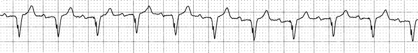
Fig. 7.3
Sinus tachycardia
Atrial Fibrillation
Atrial fibrillation is characterized by an irregularly irregular ventricular rhythm and a “wavy” baseline as a result of the chaotic, disorganized atrial activity. There is generally no pattern whatever to the sequence of ventricular (QRS) complexes in atrial fibrillation (Fig. 7.4). Sometimes there can be brief periods of regular or nearly regular ventricular depolarizations, but such regularity is short lived and may not be quite as regular as it may initially appear on casual inspection after when one takes a moment to carefully measure the RR intervals (Fig. 7.5).
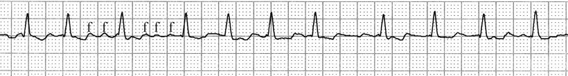
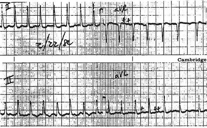
Fig. 7.4
Atrial fibrillation. Note the irregularly irregular fluctuation of the ventricular (QRS) complexes. The letter “f’ is marked above typical fibrillatory waves
Fig. 7.5
Atrial fibrillation. The ventricular complexes are fairly regular at first glance, but with careful measurement of the RR intervals, especially near the end of the strip, the irregularity can be more easily appreciated. The letter “f” is marked above typical fibrillatory waves, which give the baseline a “wavy” appearance
Rarely, in patients with a history of atrial fibrillation who have received excessive digitalis (“digitalis intoxication”), one can observe a regular rhythm. That regularization of rhythm as a manifestation of digitalis toxicity will be further described later in this chapter since it actually represents a different arrhythmia. However, these conditions of regularity associated with atrial fibrillation are rare, and atrial fibrillation is almost always irregularly irregular. In fact, any irregularly irregular rhythm should raise the likely possibility of atrial fibrillation until further evaluation proves otherwise. The presence of the small fibrillatory waves (“f” waves) serves to confirm the diagnosis. When the fibrillatory waves are large, the term “coarse” atrial fibrillation is sometimes used, as opposed to “fine” atrial fibrillation where the fibrillatory waves are small and nearly inapparent.
The atrial rate with atrial fibrillation is very fast, reaching 400–800 beats per minute. The ventricular rate is limited to about 200, just as with atrial pacing, since the normal AV node cannot conduct more rapidly than that. In the uncommon circumstance of an accessory pathway that bypasses the AV node, as with Wolff–Parkinson–White (WPW) or other preexcitation syndromes, the conduction can be much faster and may greatly exceed 200 beats per minute (Fig. 7.6). Therefore, in patients with preexcitation syndromes, the development of atrial fibrillation can be a catastrophic event since cardiac output falls greatly with rapid heart rates because ventricular filling time is severely shortened. Occasionally atrial fibrillation is present with a normal or slower-than-normal ventricular heart rate (Fig. 7.7). This usually occurs in patients on a medication that reduces AV conduction (e.g., verapamil) or with intrinsic AV node disease.
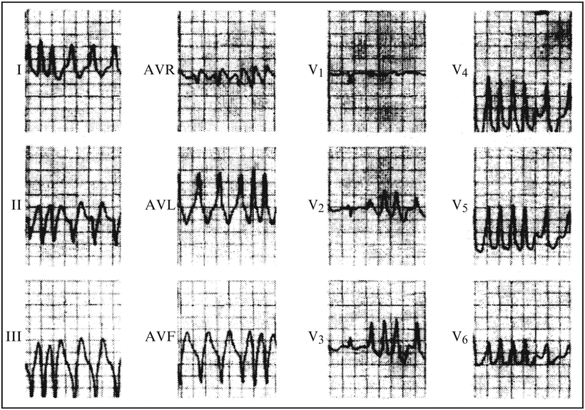
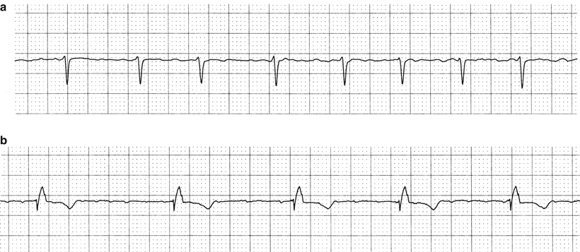
Fig. 7.6
Atrial fibrillation in a patient with Wolff–Parkinson–White syndrome. Note that the ventricular rate is frequently over 300 beats per minute. Reprinted from The American Journal of Medicine, 76(6), German LD, Gallagher JJ, Functional properties of accessory atrioventricular pathways in Wolff–Parkinson–White syndrome: Clinical implications, 1079–1086, Copyright 1984, with permission from Elsevier
Fig. 7.7
Atrial fibrillation. (a) Normal ventricular rate (92 beats per minute). (b) Slow ventricular rate (51 beats per minute with pause of 1.35 s between the first and second beats)
Atrial Flutter
It is interesting that the atrial rate with atrial flutter is usually within the fairly narrow range of about 300 ± 30 beats per minute, even though the rate can range between 250 and 400 beats per minute. The ventricular rate is usually about 150, since there is generally a fixed 2 to 1 block of conduction at the AV node when the atrial rate is so fast. The distinguishing flutter (F) waves in the classical “saw-tooth pattern” are often but not always recognizable on a rhythm strip, but generally are demonstrable on a 12-lead tracing (Fig. 7.8). Sometimes there is a greater degree of AV block, but this is not common and usually occurs, as with low rates in atrial fibrillation, if the patient is on a medication that increases AV block (e.g., verapamil) or the patient has intrinsic AV node disease (Fig. 7.9). Occasionally, the AV block with atrial flutter is variable, and this leads to an irregular ventricular rate. Any supraventricular tachycardia with a regular ventricular rate of 150 beats per minute should raise the possibility of atrial flutter and this should be the working diagnosis until further examination of the tracing confirms some other arrhythmia [1]. At least some cases of atrial flutter involve a large reentrant circuit involving the right atrium [2].
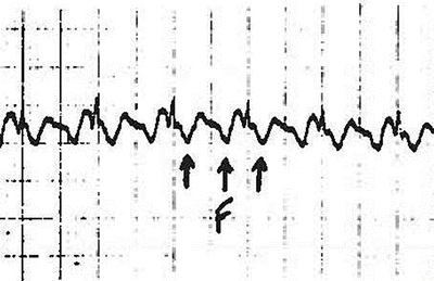
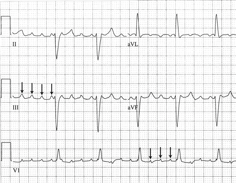
Fig. 7.8
Atrial flutter with 2:1 A:V block. Arrows indicate flutter (“F”) waves. Note atrial rate is 300 beats per minute (FF interval = 0.2 s), while ventricular rate is 150 beats per minute (RR interval = 0.4 s)
Fig. 7.9
Atrial flutter with 4:1 A:V block. Flutter waves, marked with arrows, appear at a rate of approximately 285 beats per minute
Paroxysmal Atrial Tachycardia
The atrial rate for PAT is usually 115–240 with an equal ventricular rate (1:1 AV conduction). If both the sino-atrial (SA) node and the AV node have rate limits of about 200 beats per minute, how is it possible that PAT achieves a rate over 200 beats per minute? First, PAT usually does not involve the SA node, and so the rate limitation imposed by the SA node is avoided. That also explains why the atrial depolarization with PAT usually causes abnormal P waves or no apparent P waves at all (hidden by QRS and/or T waves). Second, PAT is usually due to a reentry mechanism that involves the AV node. When electrophysiological testing is done and confirms that the reentry involves the AV node, it is properly called AV nodal reentry tachycardia, or AVNRT. When electrophysiologic studies show that the reentry does not include the AV node, it is called AV reciprocating tachycardia, or AVRT. Because at least part of the AV node (the usual bottleneck of conduction into the ventricle) is bypassed, the ventricular rate can reach 240 beats per minute. Whenever there is a supraventricular tachycardia with a ventricular rate greater than 200, the first thing that should come to mind is PAT. In fact, it is not likely to be anything else. Rarely, one might see a brief burst of atrial fibrillation that will go 200 beats per minute or so, but with rates around 240 the rhythm is usually PAT. The only exception to this dictum is the situation where there is an accessory pathway bypassing the AV node, in which case atrial flutter or fibrillation may achieve rates as high or higher than PAT (see Fig. 7.6).
At this point it is appropriate to elaborate on WPW syndrome, the classic preexcitation syndrome. Accessory pathways between the atria and the ventricles are typical for preexcitation syndromes, and in WPW an accessory pathway is responsible for the short PR interval (because the impulse gets into the ventricle faster than going through the AV node) and the pathognomonic “delta wave” (from slightly aberrant initial depolarization of the ventricle) (Fig. 7.10). WPW syndrome is not an arrhythmia—it is a syndrome that predisposes to PAT because the accessory pathway can become part of a conduction circuit. WPW can be Type A (tall R wave in V1) or Type B (negative QRS in V1).
Fig. 7.10
Wolff–Parkinson–White syndrome. Arrows indicate diagnostic delta waves, the slurred upstroke of the QRS, which is distinctly different than the usual steep, linear upstroke
In the presence of digitalis toxicity, PAT may develop with a 2:1 or greater block, and the rhythm may still be regular (Fig. 7.11). When digitalis toxicity occurs in a patient previously in atrial fibrillation, the irregular rhythm of atrial fibrillation may be replaced by a regular PAT in fixed block. Without an EKG, this situation could be mistaken on physical examination for normal sinus rhythm. Therefore, when a patient previously in atrial fibrillation treated with digitalis presents with a regular rhythm, one must obtain a tracing to determine if the rhythm is really sinus rhythm or is PAT or atrial flutter with fixed block. On the other hand, PAT may rarely occur with variable block, and without an EKG it would be indistinguishable from atrial fibrillation. While PAT with block has classically been observed in digitalis intoxication, it may also occur in other settings (Fig. 7.12).
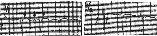
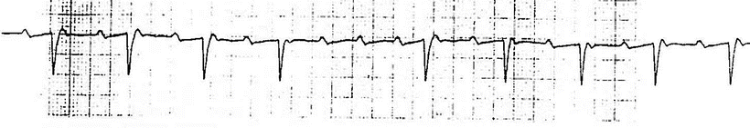
Fig. 7.11
PAT with 2:1 A:V block. Arrows show atrial depolarization
Fig. 7.12
PAT with block. Most of the strip is 2:1 block, but there is a cycle of 4:1 block in the middle of the strip
Multifocal Atrial Tachycardia
MAT is an uncommon supraventricular tachycardia that is usually irregular. The diagnosis of MAT requires that the patient has a supraventricular tachycardia with at least three different configurations of P waves and varying PR intervals (Fig. 7.13). This means that there are at least three separate foci of supraventricular activation. It is called MAT when the rate is over 100 beats per minute. When the rate is normal and there are three or more supraventricular foci of activation, the rhythm is termed “wandering atrial pacemaker.” Wandering atrial pacemaker, in a sense, is slow MAT, or MAT is wandering atrial pacemaker with a rapid rate.
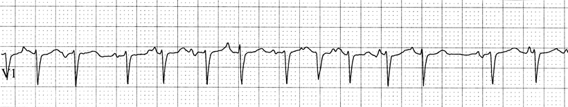
Fig. 7.13
Multifocal atrial tachycardia. Note the varying P wave configuration and PR intervals
Differentiating Supraventricular Tachycardias
It is clear that a patient with a supraventricular tachycardia and a ventricular heart rate of about 155 beats per minute could possibly have any of the five arrhythmias discussed above. If the rhythm is irregularly irregular, it is most likely atrial fibrillation, though atrial flutter or PAT with variable block, MAT, or sinus tachycardia with frequent premature contractions can give an irregularly irregular rhythm (Table 7.2).
Table 7.2
Differential diagnosis in a patient with a rapid, irregularly irregular pulse
Atrial fibrillation |
Atrial flutter with variable block |
PAT with variable block |
Sinus tachycardia with frequent PACs |
Sinus tachycardia with frequent PVCs |
Multifocal atrial tachycardia |
When the rhythm is regular, there is sometimes difficulty in distinguishing sinus tachycardia, atrial flutter with 2:1 AV block, and PAT. When P waves or F waves are obvious and recognizable, their presence can allow one to distinguish the rhythm. Sometimes, however, P waves and F waves are fairly flat in some leads and not well seen, or they may be “hidden” in T waves or QRS complexes. In such cases, where the distinction between these regular supraventricular tachycardias is not clearly obvious, there is a simple and cheap diagnostic maneuver that one can consider using. It is carotid sinus massage, which increases vagal tone and thereby increases the degree of block at both the SA and AV nodes. Other maneuvers may be used to increase vagal tone, but seem to be either more barbaric [e.g., eyeball pressure or putting the patient’s face into cold water (“diver’s reflex”)] or may interfere with the quality of the EKG record (Valsalva maneuver, cough). The mechanics of carotid sinus massage are fairly simple (Table 7.3). It should be done with the patient supine, the EKG limb leads attached, and the machine running. It is desirable to have an intravenous line in place.
Table 7.3


Appropriate precautions with carotid sinus massage
Stay updated, free articles. Join our Telegram channel
Full access? Get Clinical Tree
