(1)
Department of Medicine, Johns Hopkins University School of Medicine, Baltimore, MD, USA
Keywords
Bundle branchFascicleBlockTrifascicular blockAxisQRSThe conduction system of the heart is shown in Fig. 5.1. Under normal circumstances, the electrical activity of the heart arises from the sino-atrial (SA) node, whose intrinsic rate of electrical depolarization is normally faster than any other portion of the heart. The electrical impulse leaves the SA node, spreads across the atria, goes through the atrioventricular (AV) node, and enters the His bundle system at the inferior aspect of the AV node. The bundle of His bifurcates into the right and left bundle branches, and the left bundle branch divides again into two fascicles, the left anterior fascicle and the left posterior fascicle. Thus, by the time the electrical impulse reaches the end of the bundle branch conduction system it is running in three fascicles: (1) the left anterior fascicle, (2) the left posterior fascicle, and (3) the right bundle branch. Either of the bundle branches can have some process which interferes with conduction, thus leading to a bundle branch block, and either fascicle of the left bundle branch can have an impairment to conduction leading to a “hemiblock.” It is important to emphasize that hemiblocks and bundle branch blocks may represent simply a delay in electrical conduction, rather than a total absence of conduction down the fascicle in question.

Fig. 5.1
Conduction system of the heart
Bundle Branch Block
Two criteria must be met to diagnose a bundle branch block: (1) the QRS duration must be abnormally prolonged (0.12 s or greater) and (2) there must be a supraventricular origin of electrical activity. The other common situation in which one observes prolonged QRS complexes is ventricular rhythms; less common causes of prolonged QRS complexes are hyperkalemia and Wolff–Parkinson–White syndrome, so these must be ruled out in order to be sure that the QRS prolongation is from a bundle branch block. A supraventricular rhythm is documented if there are P waves with a consistent PR interval before each QRS complex (sinus rhythm) or if there are other evidences of supraventricular rhythms (see Chap. 7). Only on rare occasions is it difficult to distinguish a ventricular rhythm from a supraventricular rhythm with a bundle branch block.
If the QRS complex is wide and there is a supraventricular focus of activation, then the most likely diagnosis is a bundle branch block. The issue then is whether it is a right bundle branch block or a left bundle branch block. The distinction is made by examining the QRS configuration in three leads: I, V1, and V6 (Fig. 5.2). With a left bundle branch block, there is a tall, broad R wave in I and V6 and a QS or rS in lead V1. With a right bundle branch block, the QRS configuration is markedly different, with a broad terminal S wave in leads I and V6 and an rsR′ or a tall broad R wave in V1.
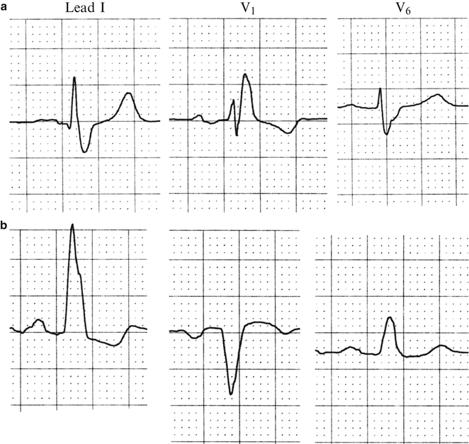
Fig. 5.2
Bundle branch blocks. (a) Right. (b) Left. For description, see text
The electrophysiology that creates these patterns may help you remember them. The key element of this electrophysiology is what is called the “terminal forces,” or the last part of ventricular depolarization. With a right bundle branch block, the impulses go through the entire conduction system normally until the bifurcation of the bundle of His. At that point, the impulse continues normally (and quickly) down the left bundle, but it is delayed as it tries to traverse the right bundle. When the depolarization is complete on the left side, the wave of depolarization then sweeps towards the undepolarized tissue on the right, and the depolarization down the right bundle that had been delayed also may be able to finally get through. For either or both reasons, the terminal forces, those at the end of ventricular depolarization, are to the right (Fig. 5.3). Because leads I and V6 have their positive directions to the left, the terminal forces in these leads are negative, leading to the typical “broad, terminal S waves” characteristic of right bundle branch block in these leads.
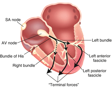
Fig. 5.3
Electrophysiology underlying the QRS configuration in right bundle branch block. The normal wave of depolarization originates in the SA node, goes through the atria, and then through the AV node into the bundle of His. It continues unimpeded down the left bundle branch (arrow), but is delayed going down the right bundle branch (dotted arrow). When the depolarization finishes through the left side of the heart, it then sweeps to the right side as that tissue is yet undepolarized because of the delay in the right bundle branch. When the delay in the right bundle branch is finally penetrated, the depolarization continues to the right. In both cases, the “terminal forces” (large arrows), which represent the last part of ventricular depolarization, sweep to the right, leading to the large “terminal S waves” in leads 1 and V6 and the R′ in V1
The changes in lead V1 are likewise interesting in a patient with right bundle branch block. The normal QRS configuration in V1 is a small r wave followed by a deep, narrow S wave as shown in (Fig. 5.4). Keeping in mind that the “right” side of the heart is not only to the right of the patient’s body but also anterior, the terminal forces with a right bundle block are coming almost directly at V1. Therefore, the normal rS pattern is altered by the substantial positive forces at the end of ventricular depolarization, causing the classic rsR′ in V1 (and often in V2). Sometimes the terminal forces totally obscure the normal S wave and one may see just a tall, broad R wave in V1, which is just as good as an rsR′ in suggesting right bundle branch block. ‘Incomplete right bundle branch block’ is when the QRS duration is normal and there is a small r’ in V1; this finding is of little clinical consequence except that patients with incomplete right bundle branch block have a greater chance of developing complete right bundle branch block than other patients.
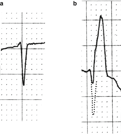
Fig. 5.4
QRS configuration in V1. (a) Normal, with rS. (b) With right bundle branch block, where terminal forces anteriorly interrupt S wave and cause tall R′. Dotted lines indicate S wave appearance without interruption by terminal forces
The electrophysiology of a left bundle branch block is, as one would expect, somewhat “opposite” of what is found with a right bundle branch block. The initial ventricular depolarization goes quickly down the unimpaired right bundle, with the terminal forces sweeping to the left. Because the left ventricle is so much greater in thickness and muscle mass than the right ventricle, almost all of the QRS complexes reflect the terminal forces. Therefore, one sees a tall, broad R wave in I and V6 (as the terminal forces sweep towards the positive sides of these leads), and a QS or rS in V1 (as the terminal forces sweep directly away from the lead). Especially in the QRS configurations in V1, one can easily see the opposite appearance of the right vs. left bundle branch block.
Stay updated, free articles. Join our Telegram channel

Full access? Get Clinical Tree


