(1)
Division of Thoracic Surgery, CHUM Endoscopic Tracheobronchial and Oesophageal Center (CETOC), University of Montreal, 1560 rue Sherbrooke Est, 8e CD – Pavillon Lachapelle, Bureau D-8051, Montréal, QC, H2L 4 M1, Canada
7.1 Introduction
7.2 Historical Note
7.3 Indications
7.5 Technique
7.6 Results
7.7 Complications
7.8 Conclusions
7.1 Introduction
Minimally invasive approaches in thoracic surgery took off with the advent of video-assisted thoracoscopic surgery (VATS) in the early 1990s [1–4]. Many procedures are now possible using VATS with significant benefits compared to the traditional approach by thoracotomy. Minimally invasive surgery permits reduced scar, decreased postoperative pain, diminished blood transfusion requirements, diminished morbidity and mortality, earlier return to baseline, decreased inflammation, and decreased hospital length of stay [5, 6].
Cervical video-assisted thoracoscopic surgery (C-VATS) is a minimally invasive technique combining mediastinoscopy, VATS, and flexible endoscopy. It requires no intercostal incision and is therefore less invasive than the traditional VATS approach. The lack of intercostal incisions prevents both initial postoperative pain with breathing and chronic post-thoracotomy/post-thoracoscopy pain. C-VATS permits unilateral and bilateral thoracoscopy using a single cervical incision with minimal immediate postoperative and long-term pain.
7.2 Historical Note
Mediastinoscopy was first described by Carlens in 1959 and has become a common procedure in thoracic surgery, especially for lung cancer staging [7]. Endobronchial ultrasound (EBUS) and endoscopic ultrasound (EUS) are quickly replacing cervical mediastinoscopy in lung cancer staging; however, mediastinoscopy still has a role [8]. C-VATS borrows from the techniques of both cervical mediastinoscopy and thoracoscopy giving it a new place in the surgeon’s thoracic cavity access armamentarium.
Deslauriers described the first pleuroscopy performed through a cervical approach in 1976 [9]. However, the rigid nature of the mediastinoscope limited pleural cavity exploration and procedural options. This technique did not become part of standard practice in thoracic surgery. Today, with new and improving instruments, we are now able to perform thoracoscopy using flexible endoscopes through a cervical incision, overcoming the previous limitations associated with the use of rigid instruments.
7.3 Indications
C-VATS is a minimally invasive technique allowing for pleural investigation and procedures through a single cervical incision using a flexible endoscope. Unilateral or bilateral pleural interventions can be carried out within the same operative setting.
Patients presenting with a pleural effusion of unknown etiology can benefit from C-VATS. Standard VATS is now recognized as the gold-standard procedure for the investigation of pleural effusions of unknown etiology [10]. Similar to VATS, C-VATS permits pleural fluid aspiration and analysis, complete thoracoscopy, pleural biopsies, and peripheral lung biopsies for diagnostic purposes. Talc pleurodesis for therapeutic purposes can also be achieved. It offers the advantage over bilateral VATS especially when bilateral interventions are necessary.
C-VATS may contribute to diagnosis and staging of malignant pleural effusions in primary lung and metastatic cancers [9, 11, 12]. Biopsies allow for the identification of molecular markers when indicated [12]. C-VATS can also be adapted for staging and pleurodesis of malignant pleural mesothelioma [13].
7.4 Contraindications
Contraindications include the inability to achieve full neck extension or cervical spine instability. This would preclude proper positioning for the procedure. C-VATS is contraindicated in patients with previous neck or mediastinal surgery because it compromises dissection to reach the parietal pleura by passing through the mediastinum. Similarly, previous mediastinal irradiation and mediastinits prevent C-VATS. Active cervical cutaneous or deep cervical infections are also contraindications to C-VATS.
Patients with significant peri-mediastinal tumor burden as assessed by preoperative CT scan possess a relative contraindication to the procedure. Adequate space between the peri-tracheal space and the mediastinal pleural envelope is required in order for the surgeon to find his/her way to the pleural cavity using the mediastinoscope. Suspected important pleural adhesions are a relative contraindication to the procedure. They compromise the safe entering into the pleural cavity with the mediastinoscope and the endoscope. Because it is not always possible to predict important pleural adhesions, clinical judgment should be used. C-VATS can be attempted and aborted if entering the pleural cavity through the cervical route is unsafe or impossible. In such cases, another approach, such as VATS, should be chosen.
C-VATS is a technique combining mediastinoscopy, VATS, and flexible endoscopy. Therefore, skills in all three techniques are required for safety and facility with the technique.
7.5 Technique
Equipment for a standard mediastinoscopy, including a standard or video mediastinoscope and a suction-cautery device, are necessary. A standard adult video gastroscope is used for the procedure. This gastroscope must be gas sterilized following the manufacturer’s guidelines. The sterile gastroscope should be opened onto the sterile field. For safety, standard VATS equipment as well as equipment for open thoracotomy or sternotomy should be prepared and opened in a sterile manner.
The patient is placed under general anesthesia with a left-sided double-lumen endotracheal tube. Positioning should be supine with shoulders elevated, neck extended, and a 30° Fowler position. Skin preparation and draping should include the anterior portion of the neck, the entire anterior thorax, and hemithorax in which the procedure will be carried out. This is for complications that may require conversion to a standard VATS technique, a thoracotomy, or a sternotomy.
A transverse 1.5- to 2.0-cm incision is performed 0.5–1.0 cm above the suprasternal notch. As in standard mediastinoscopy, the platysma is divided transversely, and the strap muscles are divided in the midline and retracted to expose the pretracheal fascia. The fascia is opened with a sharp incision, and blunt finger dissection is performed in the pretracheal and bilateral paratracheal spaces. The mediastinoscope is inserted anterior to the trachea. The suction-cautery device is used through the mediastinoscope for dissection. Lymph nodes may be biopsied if needed at this step of the procedure depending on the clinical diagnosis. The parietal pleura is then dissected out on the side where C-VATS is to be performed. On the right side, the parietal pleura is exposed about 1 cm above the azygos vein. On the left side, the parietal pleura is exposed by passing between the common carotid artery and the left subclavian artery [9]. Special care has to be taken on the left to identify and protect the left recurrent laryngeal nerve. If there is doubt concerning the position of a vascular structure, the curvilinear EBUS scope can be used with Doppler through the mediastinoscope.
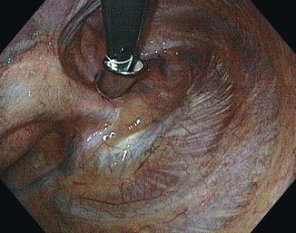
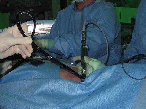
The anesthetist then isolates the lung on the side of the procedure, and the parietal pleura is opened with the suction-cautery device. The mediastinoscope is passed through this opening and advanced about 2.0-cm into the pleural cavity (Fig. 7.1). The mediastinoscope is left in place for the following steps of the procedure. The sterile gastroscope is then passed through the mediastinoscope into the pleural cavity (Fig. 7.2). The required procedures can now be performed depending on the patient’s clinical condition. Pleural fluid aspiration is done directly with the gastroscope, and fluid can be collected and sent for analysis. Complete thoracoscopy is possible due to the flexible nature of the instrument. The following structures can be examined: parietal pleura, diaphragm, lung lobes, hilum, pericardium, sympathetic chain, superior vena cava, ribs, and phrenic nerve. If indicated, biopsies on the parietal pleura or lung lobes are accomplished with endoscopic biopsy forceps. Finally, sterile talc is insufflated with a rigid talc poudrage insufflator passed through the mediastinoscope for pleurodesis when necessary. This step is done under direct endoscopic vision by keeping the gastroscope in the pleural cavity.


Fig. 7.1
Mediastinoscope inserted in the right pleural cavity

Fig. 7.2
Gastroscope inserted into the right pleural cavity through the stabilized mediastinoscope
A separate stab incision is made in the subclavicular area on the ipsilateral side of the procedure. A soft silicone drain is then passed through this opening into the mediastinal tract and then directed into the pleural cavity under direct vision. The lung is reinflated under endoscopic vision by returning to double-lung ventilation. The gastroscope and the mediastinoscope are the retracted.
Stay updated, free articles. Join our Telegram channel

Full access? Get Clinical Tree


