(1)
Department of Medicine, Johns Hopkins University School of Medicine, Baltimore, MD, USA
Keywords
First degreeSecond degreeThird degreeAV blockWenckebachMobitz IMobitz IIEscapeDissociationThere are three types of atrioventricular (AV) node block: first-degree, second-degree, and third-degree. These are sometimes abbreviated 1°, 2°, and 3°, respectively (Table 4.1). These abbreviations should not be confused with “primary, secondary, and tertiary,” which can carry the same annotation. While AV block may be a transient phenomenon (e.g., associated with ischemia, infarction, or drug intoxication), the block may be permanent.
Table 4.1
Atrioventricular block
First degree (“1°”): PR > 0.20 s |
Second degree (“2°”) |
Mobitz I (Wenckebach): Gradual prolongation of PR interval until beat dropped |
Mobitz II: Proportional conduction usually at a constant ratio and equal (usually normal) PR intervals for conducted beats |
Third degree (“3°”): Also known as “complete heart block” No relationship between P waves and QRS complexes, each with independent rate |
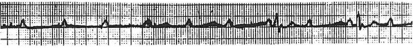
First-degree AV block is simply a prolongation of the PR interval above the normal range, i.e., >0.20 s (Fig. 4.1). At slow heart rates the normal PR interval may extend up to 0.21 s, but for simplicity it is reasonable to read any prolongation of the PR interval over 0.20 s as first-degree AV block, and simply keep in mind that slight prolongations at slow heart rates are of little clinical consequence. In fact, first-degree AV block is essentially a benign condition and is very unlikely to be associated with progression to a higher degree of AV block [1].
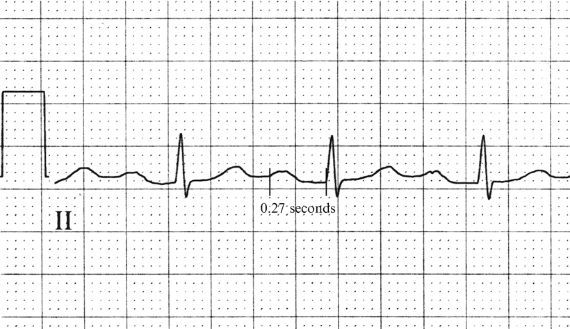
Fig. 4.1
First-degree AV block. The PR interval is 0.27 s in duration
Second-degree AV block is of two subtypes: Mobitz I and Mobitz II. Mobitz I is also commonly known as Wenckebach block. In Mobitz I (Wenckebach), there is gradual prolongation of the PR interval duration until finally one P wave is not conducted through the AV node to the ventricles (Fig. 4.2). As a consequence, a P wave is not followed by a QRS complex. Following the “dropped beat” (the missing QRS after a P wave), the PR interval is once again relatively short and then gradually prolongs again until another beat is dropped, and the cycle recurs. The length of the cycle (i.e., the number of conducted beats before the nonconducted beat) may vary. The extent of block is given in a ratio with the number of atrial beats observed in the cycle followed by a colon and then the number of ventricular beats in the cycle. For Mobitz I, the number of atrial beats in the cycle is always just one greater than the number of ventricular beats (e.g., 3:2, 4:3). This repetitive prolongation of PR intervals until a P wave is not conducted continues as long as the factor responsible for the block persists without improvement to a lower degree of block (first degree) or deterioration into a higher degree of block.
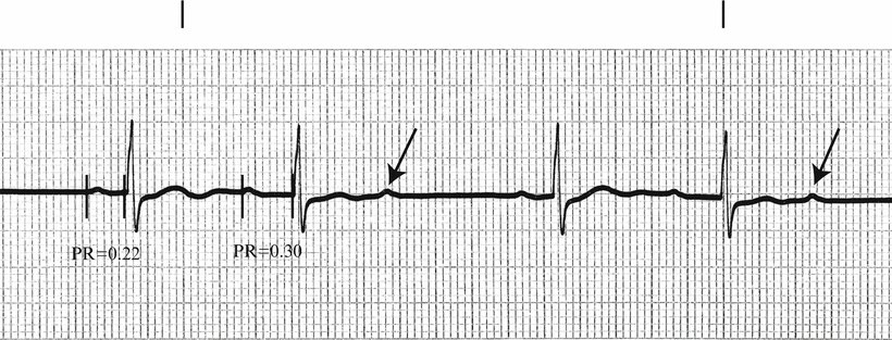
Fig. 4.2
Mobitz I (Wenckebach). The first and fourth P waves are conducted with a PR interval of 0.22 s. The second and fifth P waves are conducted with a PR interval of 0.30 s, and the third and sixth P waves are not conducted. This is a 3:2 block. Arrows indicate nonconducted P waves
While the PR interval gradually increases in Mobitz I (Wenckebach), in classic cases the R–R interval actually shortens. This seems paradoxical, if not contradictory, at first glance. Yet the apparent paradox is possible because the increment of change in successive PR intervals gradually decreases, causing the R–R interval to shorten. This phenomenon is diagrammatically demonstrated in Fig. 4.3.
Fig. 4.3
Mobitz I (Wenckebach). The diagram shows how the R–R interval may decrease while the PR interval gradually increases. The P–P interval is constant at 0.8 s (heart rate of 75 beats per minute). The PR gradually increases until the sixth P wave is not conducted into the ventricles (arrow). The RP interval is simply the P–P interval minus the PR interval. The R–R interval is determined by adding the RP and PR intervals of consecutive beats. The first PR interval is 0.16 s, and the second is 0.24 s, or an increment of 0.08 s. The third PR interval is 0.30 s, so the increment of increase in PR between the second and third P wave is 0.06 s. In this example, the increment of increase between consecutive PR intervals is 0.08, 0.06, 0.04, and 0.02 s. The R–R interval decreases, then, because the increment of change in consecutive PR intervals decreases
Even though in classic cases of Mobitz I the consecutive R–R intervals decrease, it is clear that this is not always true (Fig. 4.4).
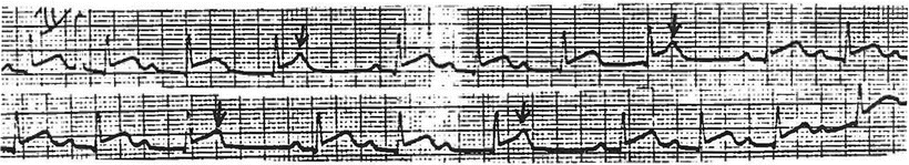
Fig. 4.4
Mobitz I (Wenckebach). The PR interval gradually increases until a beat is dropped. The dropped beats actually fall in the middle of ventricular repolarization, and so they distort the normal configuration of the T waves (arrows). In this example, the R–R intervals do not shorten
Mobitz II is a higher degree of AV block than Mobitz I wherein a relatively constant fraction of P waves are conducted through the AV node to cause ventricular depolarization (Fig. 4.5). The PR intervals of the conducted beats are quite constant, rather than varying as in Mobitz I. The common ratios of conduction in Mobitz II are 3:1, 2:1, 4:1. While the ratio of P waves to QRS complexes is often constant, the ratio also may vary somewhat.
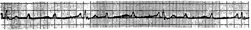
Fig. 4.5
Mobitz II. In this example the block is 3:1, with three P waves present for each QRS complex. Note that the PR intervals of the conducted beats are constant at 0.18 s
It should be noted that Mobitz II with a 2:1 block cannot be distinguished from Mobitz I block with a cycle length of only 2. In cases of 2:1 block, it is appropriate to simply call the phenomenon “second-degree AV block with 2:1 block” and not specify Mobitz I or II, unless other cycles clearly demonstrate the presence of one or the other (Fig. 4.6).
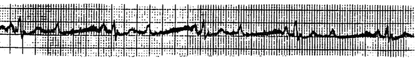
Fig. 4.6
Second-degree AV block with 2:1 conduction ratio. Without other cycles with typical features, this cannot be labelled either Mobitz I or Mobitz II with certainty
Mobitz I is generally considered to be a benign problem, primarily because it is usually transient in the setting of inferior myocardial infarction, and because in most cases only one beat out of every three or four fails to conduct through the AV node, so heart rate and cardiac output is not seriously affected. In the case of chronic Mobitz I, however, the prognosis appears to be just as poor as with chronic Mobitz II, with a 5-year survival of only about 60% [2].
Third-degree AV block is also known as complete heart block. It means that none of the P waves are being conducted through the AV node to the ventricles. As a consequence, the heart would stop depolarizing if “subsidiary pacemakers” below the point of block in the AV node did not take over the initiation of electrical activity (Fig. 4.7). These subsidiary pacemakers are in the node below the point of block, or in the conduction bundles, or in the ventricles themselves. When complete heart block is present and a subsidiary pacemaker takes over the initiation of electrical activity, the rhythm is called an “escape” rhythm, and the location of the subsidiary pacemaker identified, i.e., “nodal escape” or “ventricular escape.” Obviously, escape rhythms occur as a consequence of the nature of all cardiac cells to automatically depolarize. The rate at which this automatic depolarization occurs is fastest in the sino-atrial (SA) node, which therefore acts as the normal pacemaker for the heart. The automatic depolarization rate is slower in the node (about 45–55 beats per minute) and even slower in the ventricles (about 35–45 beats per minute). The rate of the escape rhythm, in addition to the QRS complex duration, can therefore give a clue as to where the escape rhythm originates. If the QRS complex is normal in duration (<0.12 s), the escape focus (point where electrical activity originates) must be in the node or high in the His bundle (before the bifurcation into the right and left bundle branches) with a normal or near normal conduction pattern. If the QRS complex is prolonged (≥0.12 s), the activation focus is either in the ventricles or in the bundle system below the bifurcation of the bundle of His, and the QRS complexes have the configuration of a bundle branch block. In these instances where the QRS is prolonged, a slower rate (35–45) favors a ventricular focus, while a relatively faster rate (45–55) favors a nodal or bundle focus. Occasionally there is what might be called an “iatrogenic escape rhythm,” otherwise known as a transvenous pacemaker (Fig. 4.8).