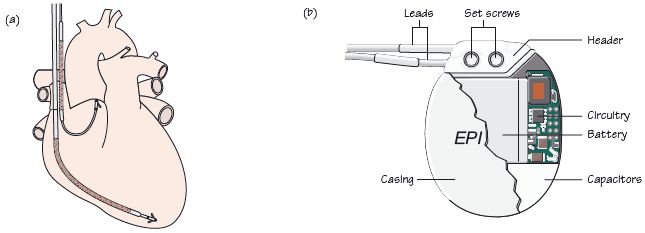
Fig. 60.2 (a) Electrodes from an implantable cardioverter defibrillator (ICD), one positioned to pace/sense the right atrium, one to pace/sense the right ventricle. The shocking circuit is between the two heavily hatched areas on the two electrodes. (b) Components of an ICD.

Fig. 60.3 Implantable cardioverter defibrillator (ICD) – overdrive pacing terminating ventricular tachycardia (VT). Top = atrial electrogram, middle = ventricular electrogram, bottom = timings between P and R waves. Initially VT is present (P waves less frequent than R waves, no relationship between them). Short burst of ventricular pacing (partly seen by the atrial electrode but best seen in the ventricular electrode trace), terminating the arrhythmia. Sinus rhythm is then restored, with each P wave followed by an R wave.

Fig. 60.4 Anti-atrial fibrillation (AF) pacing. Two paced ventricular beats, then a non-paced QRS beat, then a large number of pacing spikes through which non-paced QRS beats can be seen. Following this, AF is seen. This is a system with algorithms for anti-AF pacing. On this occasion, the device failed to terminate AF.

Anti-tachycardia and heart failure devices are commonly implanted; 2700 implantable cardioverter defibrillators (ICDs) were implanted in the UK in 2004, and 1500 cardiac resynchronization therapy (CRT) devices (50% combined ICDs, 50% only pacing). These numbers are increasingly rapidly, by 10% per year (ICDs), 30–50% per year (CRT).
Anti-bradycardia pacing to suppress tachycardias
Occasionally ‘standard’ anti-bradycardia devices are implanted specifically to suppress tachycardias: (i) AAI pacing to prevent atrial fibrillation (AF) in sick sinus syndrome-induced bradycardia; (ii) long QT dependent torsade-de-pointes (TDP) type ventricular tachycardia (VT). Pacing (DDD) increases heart rate, shortens QT interval and so prevents TDP, especially acutely (e.g. drug toxicity + heart disease + low K+
Stay updated, free articles. Join our Telegram channel

Full access? Get Clinical Tree


