Antepartum Fetal Monitoring


Antepartum Fetal Monitoring


Because more than two-thirds of fetal deaths occur before the onset of labor (1), it would be natural to extend the principles of intrapartum fetal heart rate (FHR) monitoring to the antepartum period in an effort to prevent these fetal deaths. A substantial number of antepartum deaths occur in women who have risk factors for uteroplacental insufficiency (UPI) (2). Other causes include hydrops fetalis, intrauterine infections, cord accidents, congenital anomalies, and a number of unknowns. An ideal test for assessing the antepartum fetus would allow intervention before fetal death or asphyxial damage. Before the availability of such tests, the only method for attacking this problem was to prematurely deliver such fetuses based on empirical risk data, as in the method proposed by Priscilla White for managing diabetics (3). The problem with such an approach is twofold: The majority of such prematurely delivered fetuses were not in jeopardy, and the morbidity and mortality from premature intervention often exceed those of the original risk factor. It would be preferable to treat the disease process and allow the fetus to go to term; however, we have made few advances in treating UPI.

Several biochemical tests have been proposed to evaluate the antepartum fetus. Historically, these include maternal estriol, human placental lactogen, diamine oxidase, and heatstable alkaline phosphatase. Since these biochemical tests are no longer used, they will not be discussed here. Currently available methods for antepartum fetal assessment include


  • Fetal movement (FM) counting


  • Assessment of uterine growth


  • Antepartum fetal heart rate (AFHR) testing


  • Biophysical profile (BPP) testing


  • Doppler velocimetry


PHYSIOLOGY AND PATHOPHYSIOLOGY

UPI implies inadequate delivery of nutritive and/or respiratory substances to appropriate fetal tissues. The term UPI may be applied specifically to inadequate exchange within the placenta due to decreased blood flow, decreased surface area, or increased membrane thickness. The term may also be applied more generally to problems of inadequate maternal delivery of nutrients or oxygen to the placenta, as in starvation or cyanotic cardiac disease, or to problems of inadequate fetal uptake (e.g., fetal anemia). Kubli et al. (4) suggested that UPI be divided into nutritive and respiratory components: nutritive deficiency leading to intrauterine growth retardation (IUGR) and respiratory insufficiency leading to asphyxial damage and subsequent fetal death. Parer (5) suggested that fetal nutritive function generally precedes fetal respiratory compromise (except in diabetics). Figure 11.1 is a theoretical scheme of the stages through which a fetus with declining placental function might pass. The rapidity with which this occurs may vary, being gradual in such cases as chronic hypertension or happening very suddenly as in abruption. Other conditions, perhaps including diabetes, might bypass the stage of nutritive insufficiency completely.


RISK IDENTIFICATION

To apply this knowledge to patient treatment, one must first identify the patients at risk who need evaluation. This risk identification must include data from the patient’s history, physical examination, ongoing patient assessment (including uterine growth and blood pressure), and laboratory data. Those conditions that place the patient at risk for UPI are listed in Table 11.1. In addition, some obstetric/fetal conditions apparently unrelated to maternal disease may also be associated with UPI. The most common reasons for AFHR testing are postdate pregnancy, hypertension, diabetes, clinical IUGR, and the history of a stillbirth. However, distribution or indications vary depending upon the reported testing protocol (Table 11.2) (6,7 and 8). Similarly, the rate of abnormal test result varies depending on the primary testing modality used (Table 11.3).


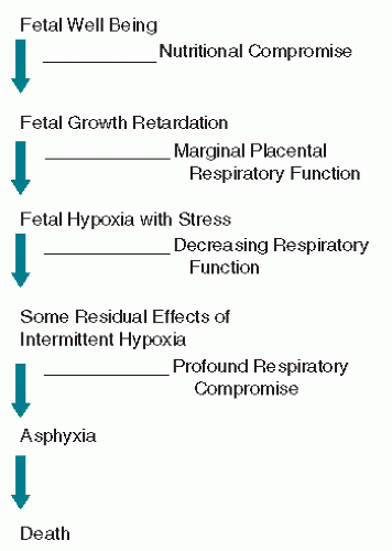
Figure 11.1. Theoretical scheme of fetal deterioration with progressive uteroplacental insufficiency.


TABLE 11.1 Conditions placing the fetus at risk for UPI


Preeclampsia


Chronic hypertension


Collagen vascular disease


Diabetes mellitus


Renal disease


Fetal or maternal anemia


Blood group sensitization


Hyperthyroidism


Thrombophilia


Cyanotic heart disease


Postdate pregnancy


Fetal growth restriction


UPI, uteroplacental insufficiency.


FETAL MOVEMENT COUNTING

Long before electronic monitoring devices were available, clinicians recognized that maternal perception of a decrease in FMs may be a sign of impending fetal death. In a prospectively randomized antepartum fetal surveillance study from Denmark, Neldam (9) had over 1,000 patients followed by a FM counting protocol. In those women randomized to the protocol of daily monitoring of FM, there was a significant reduction in the relative risk of stillbirth to 0.25 (0.07 to 0.88) and of avoidable stillbirth to 0.27 (0.08 to 0.93). This method of fetal surveillance costs nothing and, when done in a systematic fashion, especially in low-risk populations, may contribute significantly to the detection of otherwise unsuspected fetal jeopardy.


TABLE 11.2 Indications for antepartum testing by primary surveillance test


Indication


CST (%)


MBPP (%)


BPP (%)


Postdate


39


44


12


Hypertension


31


8


18


Diabetes


Gestational


5


6


7


Insulin dependent


10


2


Intrauterine


9


24


21


growth


restriction


Previous stillbirth


4


2


4


Other


5


6


38


Totals are >100% because some patients had more than one testing indication.


BPP, biophysical profile; CST, contraction stress test; MBPP, modified biophysical profile.


From Freeman R, Anderson G, Dorchester W: A prospective multiinstitutional study monitoring. I. Risk of perinatal mortality according to antepartum fetal heart rate test results. Am J Obstet Gynecol 143:771, 1982; Manning F, Morrison I, Lange I, et al.: Fetal assessment based on fetal biophysical profile scoring: experience in 12,620 referred high-risk pregnancies. Am J Obstet Gynecol 151:343, 1985; Nageotte M, Towers C, Asrat T, et al.: Perinatal outcome with the modified biophysical profile. Am J Obstet Gynecol 170:1672, 1994, with permission.


Sadovsky (10) has used FM monitoring in developing a systematized approach to the assessment of fetal well-being. If there are more than three movements in 30 minutes, the fetus is considered to be in good condition. Less than three movements in 30 minutes is either indicative of a fetal sleep state or reason for concern, and further counting should continue. We instruct our patients to count for another 30 minutes, and if there are still less than three movements in the second counting period, we ask the patient to come to the hospital for a nonstress test (NST). If the NST is nonreactive, subsequent management is according to antepartum FHR testing protocols. If the NST is reactive (which is usually the case), the patient is reassured and asked to continue her daily counting schedule. At the time of the NST, the patient
is usually taught how to count movement, because the reason for her perceived decreased count is often her nonrecognition of movement that is there. When simultaneous real-time ultrasound scanning has been done with patients asked to note the perceived movements, more movements are observed by ultrasound than are perceived by the patient (11). Most patients will feel three movements in just a few minutes, so very little time is actually required for the patient. A FM count that drops below three in 12 hours or that ceases for 12 hours is termed the “movement alarm signal” by Sadovsky, which correlates with impending fetal death (12,13 and 14). Moore and Piacquadio (15) performed a pilot study in which all patients were instructed to monitor the elapsed time it took every day from 28 weeks to register ten FMs. An NST was performed if 2 hours elapsed without 10 movements. They report a fourfold reduction in fetal mortality associated with complaints of decreased FM using this simple protocol. However, while awareness of FMs correlates with improved fetal and neonatal outcomes, the ability to identify a specific quantitative alarm for decreased FMs has not been successful (16). As an added point, the patient presenting with a complaint of absent or markedly decreased FM who has a nonreactive NST should be considered to be at increased risk for acute fetal-maternal hemorrhage (see Chapter 12).


TABLE 11.3 Distribution of test results by primary surveillance test


Result


CST (%)


MBPP (%)


BPP (%)


Negative


67


92


97


Equivocal


23


8


2


Positive


10


3a


1


a aThe positive test results for patients receiving MBPP were all inpatients receiving backup testing for an equivocal MBPP.


BPP, biophysical profile; CST, contraction stress test; MBPP, modified biophysical profile.


From Freeman R, Anderson G, Dorchester W: A prospective multiinstitutional study monitoring. I. Risk of perinatal mortality according to antepartum fetal heart rate test results. Am J Obstet Gynecol 143:771, 1982; Manning F, Morrison I, Lange I, et al.: Fetal assessment based on fetal biophysical profile scoring: experience in 12,620 referred high-risk pregnancies. Am J Obstet Gynecol 151:343, 1985; Nageotte M, Towers C, Asrat T, et al.: Perinatal outcome with the modified biophysical profile. Am J Obstet Gynecol 170:1672, 1994, with permission.


ASSESSMENT OF UTERINE GROWTH

During the third trimester, assessment of uterine growth should be done on all patients at the time of their routine prenatal visits. As a general rule, the fundal height in centimeters as measured with a tape measure will equal the weeks of gestation. There are several things that may negate this relationship, including maternal obesity, multiple gestation, polyhydramnios, abnormal fetal lie, oligohydramnios, low fetal station, and fetal growth retardation. Except for maternal obesity and myomas, the other causes are all things about which the clinician should be interested. Specifically, abnormalities in the amniotic fluid volume may lead to the diagnosis of a fetal malformation or IUGR. Thus, abnormalities in the gross uterine size or abnormal growth rates of the fundal height should lead to further investigation, specifically sonography and/or FHR testing. Unfortunately, the accuracy of clinical assessment leaves something to be desired. The diagnosis of IUGR by clinical estimates is a poor predictor of a subsequent growth retarded neonate. Generally speaking, whenever the uterine size is significantly larger than gestational age, the patient should be advised to have a sonogram. Likewise, if the uterine size is significantly below gestational age or if there is a lack of uterine growth or a decreased growth rate, the patient should be evaluated by serial sonography. If the findings suggest UPI, some form of fetal surveillance should be performed.


WHEN TO BEGIN TESTING

Indications for testing and the gestational age for beginning testing are listed in Table 11.4. Many factors go into the decision as to when to begin testing (17,18,19,20,21,22 and 23). Single factors with minimal to moderate increased risk for antepartum death nearly all warrant surveillance starting at about 32 weeks (e.g., chronic hypertension, type I diabetes, previous stillbirth) (20,22). The greatest maternal risk factors are chronic hypertension with superimposed preeclampsia and the more severe White’s classes of diabetes mellitus (D, F, and R). In diabetics with hypertension, proteinuria, IUGR, or proliferative retinopathy, the risk of fetal deterioration before 34 weeks is high, and fetal testing may begin as early as 26 weeks (21). In the patient without diabetes or hypertension, when the clinical diagnosis of IUGR is suspected by ultrasound, fetal testing should begin in some cases as early as it is reasonable to expect a chance of neonatal viability (about 26 weeks). As a general rule, AFHR testing should not begin until estimated fetal maturity is sufficient to expect a reasonable chance of survival should intervention (delivery) be necessary. This is especially difficult with twin gestations because intervention for a compromised twin may adversely affect an apparently normal twin before maturity. Testing for patients exceeding their due date is a common indication. In most cases, testing in some form should begin between 41 and 42 weeks’ gestation (23).


TABLE 11.4 Indications and gestational age for AFHR testing


Indication


Gestational age to start testing (wk)


Diabetes


Class A-1


40


Class A-2


32


Class B, C, D


32


Class F, R


26-30


Preeclampsia


At diagnosis >25-26


Chronic hypertension


32


Severe Rh disease


At diagnosis >26


Previous stillbirth


Gestational age of previous stillbirth


IUGR at diagnosis


26


Post date pregnancy


41-42


Cyanotic heart disease


32


Hyperthyroidism


32


Oligohydramnios


At diagnosis >25-26


AFHR, antepartum fetal heart rate; IUGR, intrauterine growth retardation; Rh, rhesus isoimmunization. From references Froen JF, Heazell AEP, Tveit JVH, et al.: Fetal movement assessment. Semin Perinatol 32:243-246, 2008; Freeman RK: The use of the oxytocin challenge test for antepartum clinical evaluation of uteroplacental respiratory function. Am J Obstet Gynecol 121:481, 1975; Gabbe SG, Freeman RK, Mestman JH: Management and outcome of the class “A” diabetic. Am J Obstet Gynecol 127:465, 1977; Gabbe SG, Mestman JH, Freeman RK, et al.: Management and outcome of diabetes mellitus, classes Br Am J Obstet Gynecol 129(7):723, 1977; Pircon RN, Lagrew DC, Towers CV, et al.: Antepartum testing in the hypertensive patient: when to begin? Am J Obstet Gynecol 164:1563, 1991; Lagrew DC, Pircon RA, Towers CV, et al.: Antepartum surveillance in the diabetic: when to start? Am J Obstet Gynecol 168:1820, 1993; Weeks JW, Asrat T, Morgan MA, et al.: Antepartum surveillance for a history of stillbirth: when to begin? Am J Obstet Gynecol 172:486, 1995.


PERFORMING THE ANTEPARTUM TEST

Experience is the key to obtaining quality tests in the shortest time. It is most desirable for nurses or technicians to specialize in antepartum testing. When testing is done in a hospital setting, an area separate from the labor and delivery suite is preferable to resist the temptation to have nurses cross-cover the antepartum testing area and not be able to devote enough attention to the patient being tested. Alternatively, provision of testing either in a hospital or in an office setting should be done in a quiet and stress-free environment.

The patient is placed in the semi-Fowler’s position to avoid supine hypotension syndrome. Baseline blood pressure is recorded and repeated throughout the test, again to be sure that supine hypotension does not occur, as this may be a cause of decreased uteroplacental perfusion and false-positive test results. Baseline contractions and FHR are recorded for approximately 20 minutes. The baseline heart rate and reactivity are noted, as is the background uterine activity. Following this evaluation, if results are not reassuring, continued monitoring of FHR may be necessary. Alternatively, initiation of a contraction stress test (CST) or use of realtime ultrasound to measure amniotic fluid volume and assess various fetal parameters should ensue.


WHICH TEST TO USE

In the previous editions of this text, we have presented the CST as the “gold standard” of antepartum fetal surveillance. It has been reported that using the CST as the primary means of fetal surveillance results in a very impressive and remarkably low incidence of unexpected fetal death within 7 days of a negative test result (24). However, significant problems accompany the use of contraction stress testing as primary fetal surveillance. These include the increase in time, cost, and inconvenience of the CST compared with other forms of fetal testing. In addition, the high frequency of equivocal test results and lack of consensus over test interpretation makes the CST an often impractical if not inappropriate choice of testing for many caregivers. As a result, the CST has essentially been replaced by either the NST, the BPP, or the modified biophysical profile (MBPP) for primary antepartum testing. In our centers, we now use the MBPP test as the primary means of antepartum fetal testing with the exception being the continued use of a CST alternating with a MBPP schedule every 3 to 4 days for insulin-requiring diabetics. Other forms of fetal assessment include the use of ultrasound and Doppler velocimetry of various fetal vessels.
What follows is a description of the various testing options available with particular emphasis on the evolution and reported application of the CST, NST, BPP, and the MBPP. There continues to be argument and controversy regarding the relative value and efficacy of these various types of fetal assessment despite their well-established place in the clinical practice of obstetrics (25).


CONTRACTION STRESS TEST

It can be surmised that, given a condition of borderline fetal oxygenation, a test that further stresses the fetus in terms of oxygen deprivation might produce some biophysical sign of such compromise, and that these data could be of prognostic importance. Early tests that attempted to accomplish this utilized the maternal exercise stress test and breathing gas mixtures with decreased oxygen concentrations (26). Animal data suggest that uterine contractions producing an intraamniotic pressure in excess of approximately 30 mm Hg create an intramyometrial pressure that exceeds mean intraarterial pressure, thereby temporarily halting uterine blood flow (27). A well-oxygenated fetus tolerates this limited period of intervillous stasis well; however, a hypoxic fetus will manifest late decelerations. It was therefore suggested that by inducing such contractions in the antepartum period, one might be able to detect the compromised fetus before death (and possible damage) occurred. In 1966, Hammacher (28) studied 207 pregnancies in the antepartum period and found that late decelerations correlated with lower Apgar scores at subsequent delivery and that 17 of 23 that resulted in stillbirth had manifested such late decelerations with spontaneous contractions in the antepartum period. Subsequently, Pose and Escarcena (29), Kubli et al. (4), and Spurrett (30) found late decelerations in the antepartum period to correlate with stillbirth, IUGR, and low Apgar scores. Sanchez-Ramos et al. (31) found no fetal deaths within a week of testing when no late decelerations were seen.

The first systematic trial of stress testing in this country was performed by Ray et al. (32) in 1972. They performed a prospective blinded trial on 66 patients and defined criteria for adequate testing, frequency of testing, and results, all of which are in common use in this country today. Of the 66 patients, 15 had positive test results, and of these, 3 had fetal deaths and 6 had low Apgar scores. Furthermore, there were no deaths within a week of a negative test result. Ray et al. called the test the oxytocin challenge test (OCT). Because the test can use either spontaneously occurring contractions or contractions induced by breast stimulation, it has become known more properly as the CST.


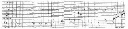
Figure 11.2. Spontaneous negative contraction stress test (no oxytocin needed). The patient is actually found to be in labor.


How to Perform the Contraction Stress Test

External monitors for contraction and FHR measurement are placed on the patient. With the patient in semi-Fowler’s position or left lateral tilt to minimize supine hypotension, an initial monitoring period lasting from 20 to 30 minutes is obtained to assess the FHR baseline, to identify the presence or absence of periodic changes, and to determine if there is evidence of spontaneous uterine activity. If there are three adequate spontaneously occurring contractions within a 10-minute period and the FHR recording is of sufficient quality, the test is concluded (Fig. 11.2). If the contractions are absent or of insufficient frequency, they must be stimulated. Historically, oxytocin infusion intravenously has been used to elicit contractions of the uterus. This is accomplished by beginning oxytocin through an infusion pump at a rate of 1.0 mU/minute. The infusion rate is initially doubled every 15 minutes until the appearance of contractions. Smaller increments for oxytocin increase are then used until three contractions lasting 40 to 60 seconds occur in 10 minutes. Patience and experience are valuable in obtaining an adequate CST and avoiding overstimulation of uterine activity (Fig. 11.3).

A widely used alternative to intravenous oxytocin infusion is that of breast stimulation. Oxytocin is released from the posterior pituitary following breast stimulation, and this technique has been used for both initiation of labor as well as for initiation of the CST (32,33,34,35,36 and 37). When used for fetal testing, breast stimulation has similar efficacy to oxytocin infusion with a shorter testing time with less expense, discomfort, and inconvenience. Breast stimulation has been
associated with uterine tachysystole and fails to achieve adequate uterine activity in approximately 20% of tests (38,39,40 and 41). The test is best performed with the patient initially rolling or tugging on one nipple through her clothing until a contraction occurs. If no contraction results following 2 to 3 minutes, the patient is asked to perform bilateral stimulation following a 5-minute rest period. This cycle of stimulation is then repeated until adequate uterine activity is documented. Figure 11.4 is an example of a reactive negative CST done with nipple stimulation. Following the appearance of adequate uterine activity, oxytocin infusion or breast stimulation is stopped and the patient continued to be monitored until activity significantly diminishes or disappears (42).


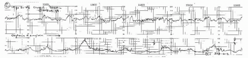
Figure 11.3. Negative contraction stress test result. Three contractions are obtained in 10 minutes, lasting more than 60 seconds each, with adequate quality fetal heart rate recording.


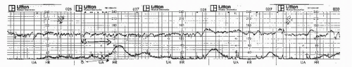
Figure 11.4. Reactive negative contraction stress test using breast stimulation.


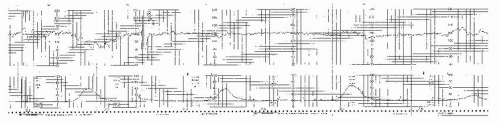
Figure 11.5. Reactive positive contraction stress test using breast stimulation.

If late decelerations are present with every spontaneous contraction, yet the contraction frequency is less than three in 10 minutes, initiation of further uterine activity is not indicated, and the CST result is positive.


Interpretation of the Contraction Stress Test


Negative

No late decelerations appearing anywhere on the strip, adequate contraction frequency (three in 10 minutes), and adequate FHR recording must be obtained (Figs. 11.2,11.3 and 11.4) for the interpretation to be negative.


Positive

In a positive CST result, late decelerations are present with the majority (greater than one-half) of contractions during the period of maximum contraction stress without excessive uterine activity (see “Hyperstimulation”). If persistent late decelerations are present before the contraction frequency is adequate, this is a positive test result and may be concluded. Figure 11.5 shows a reactive positive CST result, Figure 11.6 a minimally reactive positive CST result, and Figure 11.7 a nonreactive positive CST result.


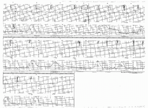
Figure 11.6. Minimally reactive positive contraction stress test using breast stimulation.


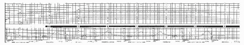
Figure 11.7. This is a nonreactive positive contraction stress test elicited with unilateral breast stimulation. The decelerations are subtle and frequently not appreciated. Note the complete absence of accelerations, which should always lead to a close evaluation of the heart rate.


Equivocal Test Results

Suspicious. A test result is considered suspicious if late decelerations are present with less than half of the contractions. It is sometimes necessary to keep a test going awhile longer to determine whether the late deceleration is persistent or only sporadic (Fig. 11.8).

Hyperstimulation. Decelerations after contractions lasting more than 90 seconds, or with contraction frequency greater than every 2 minutes, constitute hyperstimulation (Figs. 11.9 and 11.10). When such prolonged frequent contractions occur without late decelerations, it is not hyperstimulation (Fig. 11.11). Hyperstimulation may occur with either spontaneous or oxytocin-induced contractions.

Only gold members can continue reading. Log In or Register to continue

Stay updated, free articles. Join our Telegram channel

Jun 22, 2016 | Posted by in CARDIOLOGY | Comments Off on Antepartum Fetal Monitoring

Full access? Get Clinical Tree

Get Clinical Tree app for offline access