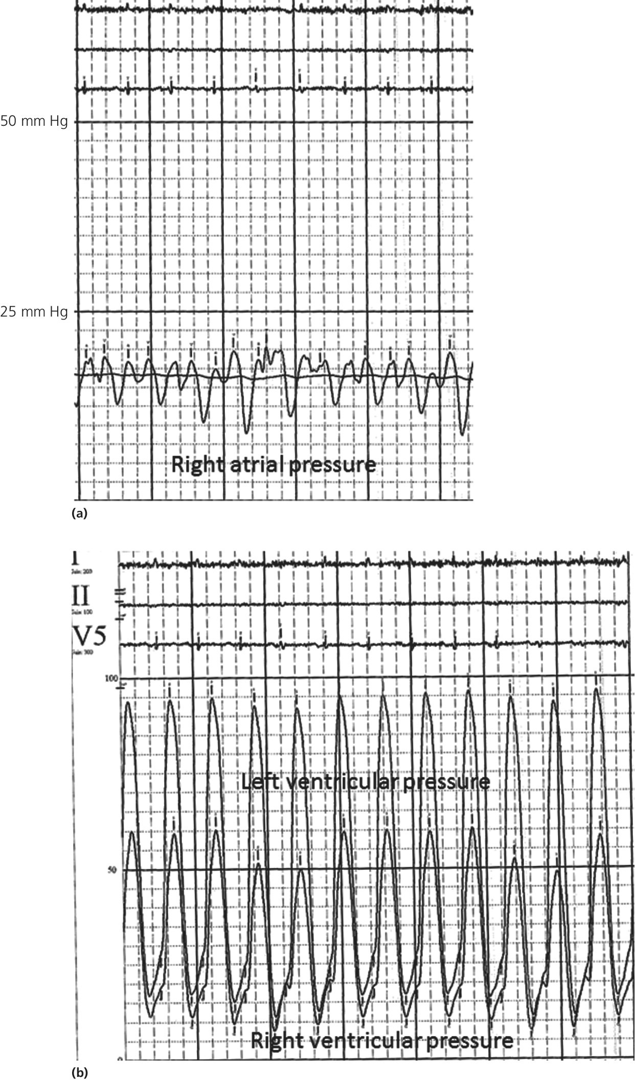
CHAPTER 17 David P. McLaughlin and George A. Stouffer Restrictive cardiomyopathy is a disease of the myocardium characterized by impaired relaxation and diastolic dysfunction of either or both ventricles. The disease is progressive and as the ventricles become less distensible, diastolic filling and cardiac output are impaired and filling pressures increase. A reduction in systolic function may also occur as the disease progresses. Initial symptoms are usually low output–type complaints (e.g., exertional dyspnea or exercise intolerance) because of inability to augment stroke volume. As right atrial (RA) pressure increases, symptoms of systemic venous congestion may predominate. Further increases in RA and pulmonary capillary wedge (PCW) pressures will be accompanied by symptoms of orthopnea and paroxysmal nocturnal dyspnea. The clinical presentation of restrictive cardiomyopathy can be similar to constrictive pericarditis and differentiation of these processes can be difficult. Additionally, some disease processes (e.g., amyloid) can involve both myocardium and pericardium, leading to a mixed hemodynamic profile. Causes of restrictive cardiomyopathy include amyloidosis, hemochromocytosis, metabolic storage diseases, hypereosinophilic syndrome, metastatic malignancies, sarcoid, carcinoid, idiopathic, endomyocardial fibrosis, and mediastinal radiation. The hemodynamic findings of restrictive cardiomyopathy and constrictive pericarditis are similar. Chapter 18 contains a more detailed description of the findings in constrictive pericarditis. Figure 17.1 Pressure tracings from a patient with amyloidosis. Note the elevated RA pressures with an exaggerated Y descent (a). Simultaneous RV and LV pressures shows equalization during diastole (most notable during expiration) and concordance in which RV and LV systolic pressures both decrease with inspiration (b). The hemodynamic effects of restrictive cardiomyopathy are similar to those of constrictive pericarditis, and it is generally difficult to make a correct diagnosis based on hemodynamics alone. Hemodynamic criteria that favor restrictive cardiomyopathy include: A review of 82 cases of constrictive pericarditis and 37 cases of restrictive cardiomyopathy found that the predictive accuracy of a difference between RVEDP and LVEDP >5 mm Hg was 85%, the predictive accuracy of RV systolic pressure >50 mm Hg was 70%, and the predictive accuracy of a ratio of RVEDP to RV systolic pressure of less than 0.33 was 76%. If all three criteria were concordant, the probability of having classified the patient correctly was greater than 90%. However, one‐fourth of patients could not be classified by hemodynamic criteria [1]. A more detailed description of hemodynamic findings that can be used to differentiate restrictive cardiomyopathy from constrictive pericarditis can be found in Chapter 18. The ventricles are usually normal in size. LV hypertrophy is frequently present (of note is that decreased R wave amplitude on ECG coupled with LV hypertrophy on echo is frequently a clue to restrictive cardiomyopathies). Mitral inflow velocity in restrictive cardiomyopathy is typically one of a prominent E wave, decreased A wave, an increased ratio of early diastolic filling to atrial filling (>2), decreased deceleration time (<150 ms), and a decreased isovolumic relaxation time (<70 ms). Cardiac valves may be involved in restrictive cardiomyopathy, leading to regurgitation. Respiratory variation in mitral valve inflow is less in restrictive cardiomyopathy compared to constrictive pericarditis. An echocardiographic study of 30 patients (19 with constrictive pericarditis and 11 with restrictive cardiomyopathy) found that significant respiratory variation in the ventricular inflow peak velocity ≥10% predicted constrictive pericarditis with a sensitivity and specificity of 84% and 91%, respectively [2]. Remember that mitral valve inflow velocity is dependent on preload and that marked elevation in LA pressure may mask respiratory variation [3]. Reducing preload (e.g., by head‐up tilting) may accentuate respiratory variation in inflow velocity.
Restrictive cardiomyopathy
Hemodynamic principles
Differentiating restrictive cardiomyopathy from constrictive pericarditis
Echocardiography
Stay updated, free articles. Join our Telegram channel

Full access? Get Clinical Tree


