Wolff-Parkinson-White Preexcitation Patterns

Chapter 12 Wolff-Parkinson-White Preexcitation Patterns


This chapter is a bridge between the first part of the book dealing primarily with abnormal QRS-T patterns and the second part on arrhythmias. Preexcitation patterns, especially the Wolff-Parkinson-White (WPW) pattern, may be mistaken for bundle branch blocks, hypertrophy, or myocardial infarction (MI) and are also an important substrate for supraventricular tachycardias. Even beginning clinicians need to be familiar with this finding, still a relatively common cause of referral to cardiologists.


Wolff-Parkinson-White Pattern: Preexcitation and Bypass Tracts


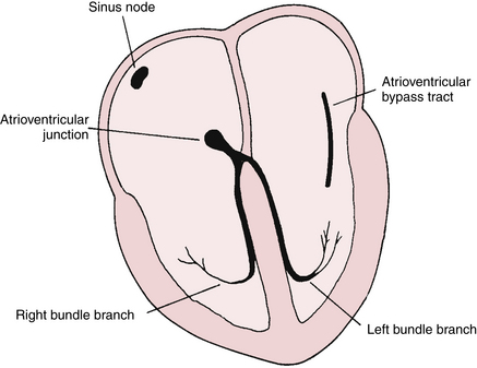
The WPW pattern is a distinctive and important ECG abnormality caused by preexcitation of the ventricles. Normally the electrical stimulus travels to the ventricles from the atria via the atrioventricular (AV) junction. The physiologic lag of conduction through the AV junction results in the normal PR interval of 0.12 to 0.2 sec. Consider the consequences of having an extra pathway between the atria and ventricles that would bypass the AV junction and preexcite the ventricles. This situation is exactly what occurs with the WPW pattern: an atrioventricular bypass tract connects the atria and ventricles, circumventing the AV junction (Fig. 12-1).


Bypass tracts (also called accessory pathways) represent persistent abnormal connections that form and fail to disappear during fetal development of the heart in certain individuals. These abnormal conduction pathways, composed of bands of heart muscle tissue, are located in the area around the mitral or tricuspid valves (AV rings) or interventricular septum. An AV bypass tract is sometimes referred to as a bundle of Kent.


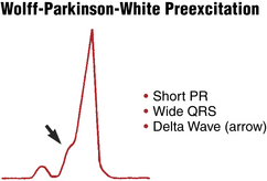
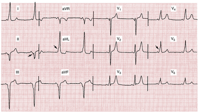
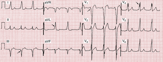
Preexcitation of the ventricles with the classic WPW pattern produces the following characteristic triad of findings on the ECG (Figs. 12-2 to 12-4):


The QRS complex in sinus rhythm with WPW pattern is the result of the competition (fusion) between signals going down the normal conduction system and down the bypass tract. The signal going down the bypass tract usually reaches the ventricles first, while the signal going down the normal conduction system gets delayed in the AV node. The early activation of the ventricles results in a shorter than normal PR interval. Slow conduction through the ventricular muscle from the bypass tract insertion site is responsible for the initial QRS slurring (delta wave). Once the signal going down the normal conduction system passes the AV node, this activation wave “catches up” with the preexcitation wave by spreading quickly through the His-Purkinje system and activating the rest of the ventricles in the usual way. This competitive mechanism produces the relatively narrow second part of the QRS complex. The degree of preexcitation (amount of the ventricles activated through the bypass tract) therefore is dependent on the speed of AV nodal conduction—the longer the delay in the AV node, the larger portion of the ventricles that is activated through the bypass tract and the longer the delta wave is.


Figures 12-2 and 12-3

Only gold members can continue reading. Log In or Register to continue

Stay updated, free articles. Join our Telegram channel

Jun 11, 2016 | Posted by in CARDIOLOGY | Comments Off on Wolff-Parkinson-White Preexcitation Patterns

Full access? Get Clinical Tree

Get Clinical Tree app for offline access