Figure 2.1
Image of the heart showing the right parsternal, parasternal, apical and subcostal windows as they relate to the cardiac structures
Transducer positions and related echocardiographic images for parasternal long axis views are shown in Fig. 2.2.
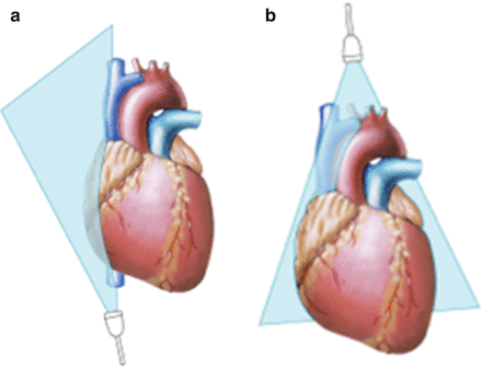
Figure 2.2
Image of the heart showing the suprasternal (b) and subcostal (a) windows as they relate to the cardiac structures
Transducer positions and related echocardiographic images for parasternal short axis views are shown in Fig. 2.3.
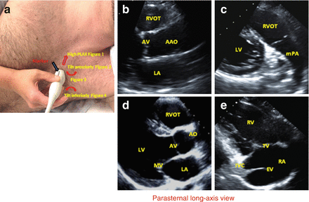
Figure 2.3
Transducer positions and echocardiographic images for parasternal long axis views. (a) Probe position. (b) Ascending aorta view. (c) RV outflow view. (d) Parasternal long-axis view. (e) RV inflow view. Abbreviations: LA left atrium, RCC and NCC right coronary and non-coronary cusps of aortic valve, AAO ascending Aorta, RVOT right ventricular outflow tract, LV left ventricle, mPA main pulmonary artery, AMVL and PMVL anterior and posterior mitral valve leaflets, ATVL and PTVL anterior and posterior mitral valve leaflets, RA right atrium, CS coronary sinus, IVC inferior vena cava, EV Eustachian valve
Transducer positions and related echocardiographic images for apical views are shown in Fig. 2.4.
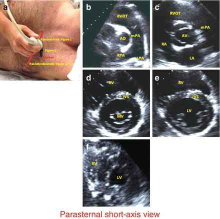
Figure 2.4
Transducer positions and images for parasternal short axis views. (a) Probe position. (b) Pulmonary bifurcation view. (c) AV level. (d) MV level. (e) Papillary muscle level. (f) Apex level. Abbreviations: RVOT right ventricular outflow tract, Ao aorta, mPA main pulmonary artery, RPA and LPA right and left pulmonary arteries, RA right atrium LA left atrium, RCC, LCC and NCC right, left and non-coronary cusps of aortic valve, RV right ventricle, IVS interventricular septum, AMVL and PMVL anterior and posterior mitral valve leaflets, LV left ventricle
Transducer positions and related echocardiographic images for subcostal views are shown in Fig. 2.5
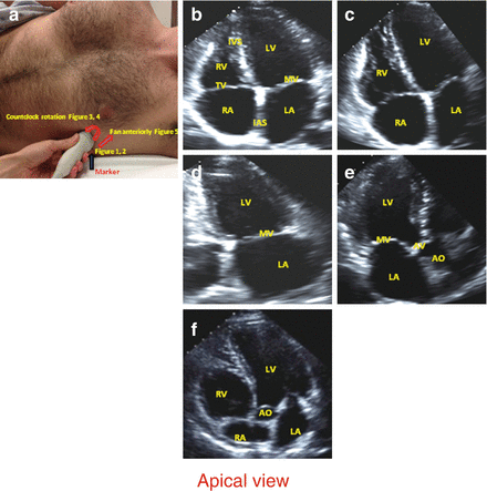


Figure 2.5
Transducer position and echocardiographic images for apical views. (a) Probe position. (b) Apex 4-chamber view. (c) RV focused 4-chamber view. (d) Apex 2-chamber view. (e) Apex 3-chamber view. (f) Apex 5-chamber view. Abbreviations. LA and RA left and right atria, LV and RV left and right ventricles, ias and ivs interatrial and interventricular septi, AMVL and PMVL anterior and posterior mitral valve leaflets, ATVL and PTVL anteropr and posterior tricuspid valve leaflets, MV mitral valve, AV aortic valve, AO aorta
< div class='tao-gold-member'>
Only gold members can continue reading. Log In or Register to continue
Stay updated, free articles. Join our Telegram channel
Full access? Get Clinical Tree
