Figure 8.1
Subcostal view showing heart with significant pericardial thickening (arrow)
Pericardial Effusion (Fig. 8.1)
Determine:
Size (Size criteria differ between publications). Measure in diastole: trivial 1–5 mm, small 5 mm-1 cm, moderate 1–3 cm, large >3 cm
Circumferential vs. loculated (Loculated effusions are more common after surgery, trauma, purulent pericarditis)
Fluid characteristics: look for fibrin strands, echogenic masses
Assess for cardiac tamponade (Figs. 8.2, 8.3, 8.4, and 8.5, Videos 8.1 and 8.2)
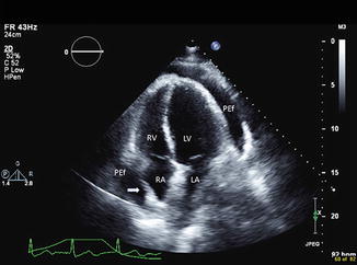
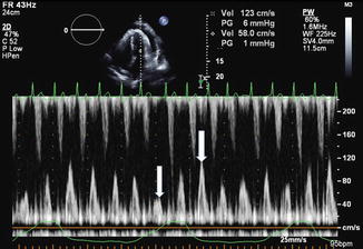
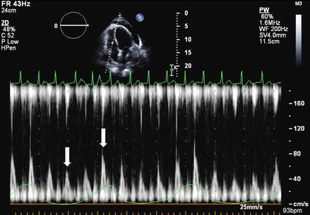
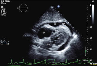


Figure 8.2
Apical 4 chamber view showing circumferential pericardial effusion (PEf). The arrow shows right atrial collapse during late diastole
Figure 8.3
Doppler mitral valve flow profile in patient with pericardial effusion. Note the variability of mitral valve inflow pattern (arrows) during the respiratory cycle (green curve on the bottom of the image represents patients breathing pattern). Significant increase of mitral flow with expiration and decrease with inspiration suggests tamponade physiology
Figure 8.4
Doppler tricuspid valve flow profiles in patient with pericardial effusion (arrows). Note the significant increase in inflow during the inspiration and decrease with expiration suggesting tamponade physiology
Figure 8.5
Subxyphoid view of pericardial effusion (PEf). Note the early diastolic right ventricular collapse (arrow) consistent with tamponade physiology
Exaggerated respiratory variations in mitral and tricuspid inflow profiles. Cut offs 30 % MV, 60 % TV [1]< div class='tao-gold-member'>Only gold members can continue reading. Log In or Register to continue
Stay updated, free articles. Join our Telegram channel
Full access? Get Clinical Tree
