The stress test images consist of (Figs. 17.2, 17.3, 17.4, and 17.5):
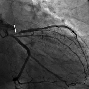
Figure 17.2
Raw images showing the heart (arrows) and breast attenuation in female patients (different angles of rotation are shown in a–c). Please pay attention to the dark shadows over the heart outlines
Figure 17.3
Tomographic images in short, vertical long and horizontal long axis views showing significant anterior ischemia (arrows point to ischemic regions in several view planes). Pay particular attention to the decrease uptake of anterior wall with stress (Str, upper rows) when compared to rest (Rst, lower rows)
Figure 17.4
Polar display showing myocardial perfusion during stress (upper row), rest (middle row) and reversibility of ischemia (right lower image, meshed part of the image)
Figure 17.5
Coronary angiogram of the same patient showing proximal Left Anterior Descending Coronary Artery lesion (arrow)
Raw images (which allow basic visualization of the heart, the surrounding tissue and the motion of the heart due to patient movement which allows quality control check)
Short, vertical and horizontal long axis views (both stress and rest) which allow the comparison of perfusion and calculation of transient ischemic dilatation index
Polar display: short axis views arranged from apex to lateral wall.
Gated SPECT (to evaluate wall motion abnormalities and abnormalities contractility of the heart, and to calculate Left Ventricular Ejection Fraction through end systolic and end diastolic volumes)
There are several basic options for the protocols:
One day vs. two day protocols
Sequence of image acquisition: rest before stress or stress before rest
Single vs. dual isotope imaging
One day vs. two day protocols
Historically two day protocols have been preferred to avoid residual activity from first imaging to affect the second imaging acquisition. However, one day protocols have now become the more common. The two day protocol is preferred for obese patients (BMI >40) or women in whom significant breast attenuation is expected (thus potentially requiring a higher initial dose of radioisotope).
One day protocols are preferred due to practical considerations involving patient convenience, cost reduction and facilitation of decision making. This requires a combination of lower dose first imaging (rest or stress) and higher dose second imaging to differentiate between the first and second Myocardial Perfusion Images (MPI). Enough time for decay after the first dose is required prior to second acquisition and interpretation of MPI images.
Stress-rest vs. rest-stress sequence
Low dose stress followed by high dose rest protocol
Pros: possibility of avoiding extra images in cases where stress images are completely normal.
Cons: increased radiotracer uptake by the myocardium during hyperemia requires either a longer delay between a stress and a rest acquisitions or a higher dose of radioisotope< div class='tao-gold-member'>Only gold members can continue reading. Log In or Register to continue

Stay updated, free articles. Join our Telegram channel

Full access? Get Clinical Tree


