Fig. 25.1
Grade 2 LLSB crescendo/decrescendo murmur after S1
The murmur increases to grade 4 with exertion.
No ejection clicks noted.
No diastolic murmurs are present.
Test Results
An electrocardiogram is normal.
Clinical Basics
Normal Anatomy
Ventricular septum components include the following:
Muscular septum: muscular trabeculae, muscular outlet and muscular inlet.
Membranous septum.
Definitions
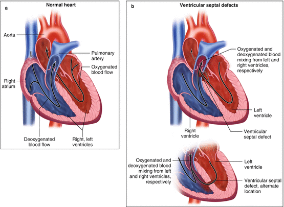
VSD is a ventricular septal opening caused by improper septal formation (Fig. 25.2a, b).


Fig. 25.2
(a) Normal heart anatomy. (b) Heart anatomy with ventricular septal defects
Etiology
Subtypes of VSD are classified by location:
Muscular = apical most common (prevalent in neonates).
Perimembranous = majority of VSD cases (extends into muscular septum).
Outlet = 5–7 % (approx. 30 % in Far East).
Inlet (endocardial cushion) = 5–8 %.
Signs and Symptoms
Signs and symptoms are determined by patient age, size of lesion, and location.
Adults are primarily asymptomatic, especially with small defects.
The most common symptom of a VSD is dyspnea.
Prevalence
VSD is the most common congenital heart defect in adults (20 % of CHD as a solitary lesion) [1].
Key Auscultation Features of VSD
Normal S1 and S2 should be present.
If S2 is split and/or loud, consider that pulmonary hypertension may be present.
Systolic murmur.
Louder with isometric hand grip and transient arterial occlusion (TAO) [2].
TAO: Inflate cuffs on both arms to 20–40 mmHg above peak systolic pressure for 20 s.
Small VSD’s are the loudest and usually includes thrills [3].
The murmur of a VSD begins in early systole and extends through S2.
Mid-lower LSB (Infundibular defects heard best at upper LSB).
Radiates along LSB or to back.
Intensity may vary as the defect changes during contraction with the muscular VSD.
Diastolic murmur.
Diastolic decrescendo murmurs are heard in patients with aortic insufficiency as a complication of the VSD.
Noted best at the LSB when the patient is sitting/leaning forward.
Large VSDs will have a diastolic rumble at the apex from high flow across the mitral valve [3].
No extracardiac sounds should be present with a VSD.
Murmurs may not appear at birth due to equal pressures across the ventricles [3].
Auscultation examples of a VSD:
Click here to listen to a VSD murmur in a 15-year-old boy, as described by Dr. W. Proctor Harvey (Video 25.1).
Click here to listen to auscultation findings in a large VSD: harsh holosystolic murmur and soft mid-diastolic rumble; an image of the phonocardiogram is also present (Video 25.2).
Click here to listen to auscultation findings in a small VSD: high-pitched short SEM; an image of the phonocardiogram is also present (Video 25.3).
Clinical Clues to Detect the Lesion
Clues to Location
Small outlet VSD: second left intercostal space and can radiate into the suprasternal notch.
Small muscular VSD: short in duration, cuts off in mid-systole (systolic contraction of septal musculature closes the defect).
Muscular VSD in apical septum may be heard best towards apex.
Clues to Size
Small VSD
No exam findings of LV volume load or pulmonary hypertension.
Usually normal exam other than murmur.
Moderate VSD
Size 50 % of aortic valve orifice.
Pansystolic murmur, usually without signs of RV volume or pressure overload. Chamber enlargement (left atrium, left ventricle) common.
S2 split, P2 usually normal.
Large VSD
Size approximates aortic valve orifice.< div class='tao-gold-member'>Only gold members can continue reading. Log In or Register to continue
Stay updated, free articles. Join our Telegram channel

Full access? Get Clinical Tree


