Fig. 20.1
EKG demonstrating sinus rhythm and low voltage QRS consistent with pericardial thickening
Chest CT (Fig. 20.2). CT scan of the chest in a patient with constrictive pericarditis demonstrates a thickened pericardium.
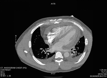
Fig. 20.2
A thickened pericardium was identified on computed tomography of the chest
Hemodynamics (Fig. 20.3). In the right atrial pressure tracing, a deep diastolic Y descent is present. Kussmaul’s sign is present, seen as the rise in RA pressure with inspiration.
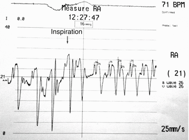
Fig. 20.3
CP presents with elevated JVP with rapid collapsing diastolic Y descent with or without an X wave descent. In the right atrial pressure tracing, a deep diastolic Y descent is present. Kussmaul’s sign is present, seen as the rise in RA pressure with inspiration. The systolic X descent may create an M or W pattern
Clinical Basics
Normal Anatomy
The normal pericardium has a limiting effect on cardiac volume and amplifies the diastolic interaction by transmitting intracavitary filling pressures to adjacent chambers [2].
Definition
CP is the end stage of an inflammatory condition in the pericardium that leads to adhesion of the visceral and parietal peritoneum, calcification and dense fibrosis [2].
This leads to restriction of the myocardium and prevents adequate ventricular filling leading to elevated diastolic pressures in all four chambers [1].
Etiology
Previously, the major cause of CP was tuberculosis [1]. However, recent studies have indicated that idiopathic, prior surgery and irradiation therapy account for the majority of cases in the developed world.
More recently, the cause of CP in 163 patients who underwent pericardiectomy was determined [3].
46 % – viral or idiopathic.
37 % – post surgical.
9 % – secondary to mediastinal irradiation.
8 % – Miscellaneous: tuberculosis, rheumatoid arthritis, systemic lupus erythematosus, prior chest trauma, Wegener’s granulomatosis, or purulent pericarditis.
Signs and Symptoms
A common presentation of CP is right sided heart failure [1].
A preoperative analysis of 135 patients who were diagnosed with CP (Table 20.1) revealed the following clinical characteristics [4].
Table 20.1
Clinical characteristics of patients with CP
1985–1995 cohort (n = 135)
Characteristics
No. or value
%
Age, years
Mean
56 ± 16
Median
61
Range
Nov-78
Male
103
76
Symptom duration, month
Median
11.7
Range
0.1–349
NYHA class
I–II
40
30
III–IV
93
69
Indeterminate
2
1
Elevated JVP
119
93
Peripheral edema
103
76
Hepatomegaly
71
53
Pericardial knock or S3
63
47
Ascites
50
37
Pleural effusion
47
35
Kussmaul’s sign
28
21
Pulsus peradoxus
25
19
Pericardial rub
22
16
Known CAD
26
20
Diuretic use
68
50
Atrial arrhythmia
22
16
Low QRS voltage
37
27
Pericardial calcification
34
25
Common signs and symptoms include:
NYHA grade III–IV heart failure.
Elevated JVP.
Peripheral edema.
Hepatomegaly.
Pericardial knock or S2.< div class='tao-gold-member'>Only gold members can continue reading. Log In or Register to continue

Stay updated, free articles. Join our Telegram channel

Full access? Get Clinical Tree


