Vascular anomalies

20


Vascular anomalies


Introduction


Vascular anomalies are common in infants and children, but their classification is frequently a source of great confusion and, as a result, they are often poorly managed. This is despite the fact that, as long ago as 1982, Mulliken and Glowacki proposed a new way of classifying vascular anomalies1 that is now the most widely recognised system in use. It is based upon differences in the cellular kinetics and natural history of these lesions and describes two separate groups: vascular tumours and vascular malformations, which will be discussed in turn. Vascular tumours include the common infantile haemangiomata as well as rarer congenital haemangiomata and other vascular tumours, many of which are high flow. Vascular malformations comprise low-flow venous and lymphatic malformations and high-flow arteriovenous malformations. An acquired arteriovenous fistula following trauma is a separate entity that may occasionally be misdiagnosed as a vascular malformation.


Vascular tumours


Infantile haemangioma


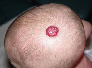
Typically, this lesion is not present on the day of birth but becomes visible within the first 2 months of life. An initial stage of rapid proliferation first brings the lesion to attention, although the clinical appearance of the haemangioma at this time will depend upon its depth of involvement of the skin. Those involving the superficial dermis produce lobulated, bright red lesions that are commonly referred to as ‘strawberry birthmarks’ (Fig. 20.1). A haemangioma involving the deep dermis and subcutis, however, will produce a swelling with either no discoloration or a blueness of the overlying normal skin. The term ‘cavernous haemangioma’ is often used to describe such lesions, but it is important to recognise that their appearance is due solely to their depth of involvement of the skin and not to any variation in their histology.


The proliferative phase of a haemangioma varies in its duration, but rapid growth will usually occur during the first 6 months of life. Growth will often continue at a slower rate after this until about 1 year of age, following which the haemangioma will gradually involute completely in 50% of individuals by 5 years, in 70% by 7 years and 90% by 9 years. Residual skin changes following complete involution are common but in the majority are mild and inconspicuous, and include mild hypopigmentation, focal telangiectases and cutaneous atrophy.


As mentioned above, many haemangiomata are focal nodular lesions but others have a plaque-like configuration involving a larger area of skin and these ‘segmental’ haemangiomata involving the face are important to recognise, as they may be associated with other abnormalities, as outlined below.


PHACE syndrome2


PHACE (an acronym for: Posterior fossa; Haemangioma; Arterial anomalies; Coarctation of the aorta and other cardiac defects; Eye abnormalities) syndrome describes the association between large segmental facial haemangiomata and several structural abnormalities. Affected individuals are nearly always female and the majority will have one or two of the described structural anomalies rather than all of them. The haemangioma most commonly involves the upper face and forehead, but this is not invariable. A child with such a lesion should be carefully examined for signs and symptoms of the syndrome and appropriate investigations should be performed to exclude anomalies of the heart and aorta, including coarctation. Neurological symptoms will prompt magnetic resonance scanning of the brain with or without catheter angiography.


Diagnosis and imaging


The diagnosis of a haemangioma of infancy is usually straightforward based upon the clinical history and findings on clinical examination; imaging is often unnecessary unless there is a concern about associated underlying structural anomalies (see above). It may occasionally be difficult, however, to differentiate between a deep-seated haemangioma and other vascular anomalies or tumours, and in such instances further investigation, and rarely biopsy, may be necessary.


The imaging features during the proliferative phase are usually characteristic: ultrasound demonstrates a well-defined, echogenic lesion that is highly vascular, with large central feeding arteries and draining veins. Magnetic resonance imaging (MRI) will demonstrate a well-defined lobulated tumour that is isointense or hypointense when compared with normal muscle on T1-weighted images and hyperintense on T2-weighted images. If contrast medium is given the tumour will enhance homogeneously and dilated feeding arteries and draining veins will be visualised.


Biopsy may be required in the rare instance that doubt remains as to the diagnosis. Infantile haemangiomata express glucose transporter-1 (GLUT-1), a simple diagnostic marker that may be very helpful in the differentiation between this entity and other vascular tumours. Core needle biopsy is usually straightforward under ultrasound guidance as lesions are typically superficial.


Management of haemangiomata of infancy


The majority of haemangiomata can be managed conservatively but intervention is indicated when a lesion causes significant mass effect or disfigurement, when it involves the airway and when it obstructs the visual axis. Pharmacological management is now the mainstay of treatment in this group, with beta-blockers the preferred first-line therapy.3 Rarely, high output cardiac failure occurs in children with very large infantile haemangiomata, particularly when there is hepatic involvement, and this requires urgent intervention. Management consists of a combination of optimal medical therapy and particle embolisation of the capillary bed, with the aim of reducing the vascularity of the lesion and stabilising the child’s cardiovascular status until the natural history of the lesion causes involution.


The surgical management of haemangiomata can be divided into those procedures performed during the proliferative phase of development and those performed later when partial or complete involution has occurred but a persistent cosmetic deformity remains. Early surgery is generally reserved for lesions obstructing the visual axis when more conservative measures have failed. Later surgery, between the ages of 3 and 5 years, may be considered when a large haemangioma persists that is slowly involuting and which is thought to be having a detrimental effect on the child’s social development because of its appearance. The surgical scar that is likely to result from such an operation should, however, be weighed against the likely outcome if the haemangioma were allowed to involute completely.


Other vascular tumours


Infantile and congenital haemangiomata make up the vast majority of the lesions in the vascular tumour arm of the Mulliken and Glowacki classification of vascular anomalies, but it is important to recognise other much less common lesions. These include the pyogenic granuloma, the tufted angioma and the kaposiform haemangioendothelioma.


The lobular capillary haemangioma, or pyogenic granuloma, is the second most common vascular tumour of childhood after the haemangioma of infancy, although it can occur at any age. It most often involves the head and neck and presents as a rapidly growing, bright red papule varying in size from a few millimetres to about 2 cm, which frequently ulcerates and may bleed profusely. Most can be removed surgically.


The tufted angioma (TA) presents initially as a small red or purple patch involving the skin of the neck or upper trunk, which grows slowly and may reach a large size. Its natural history is unpredictable, however, and some lesions will resolve spontaneously. The kaposiform haemangioendothelioma (KHE) most frequently occurs in the skin of the trunk, shoulder and thigh. Seventy-five per cent of these tumours occur in early infancy and are manifest as a deep reddish-purple discoloration of the skin that may appear tense, shiny and bruised. Recognition of these two vascular tumours (TA and KHE) is important as they are sometimes associated with the Kasabach–Merritt phenomenon (KMP). Both are GLUT-1 negative on biopsy and have characteristic histology, so biopsy is often useful.


KMP describes a pattern of variable but often severe coagulopathy and thrombocytopenia seen in association with a variety of soft-tissue lesions, most commonly KHE, and is associated with a high mortality. It is not a complication of the common haemangioma of infancy5 and should not be confused with the coagulopathy that may be associated with large venous malformations in which there is consumption of clotting factors with elevated D-dimers but a much less profound reduction in platelet and fibrinogen levels. The management of this condition lies outside the scope of this chapter but evolving pharmacological approaches to other vascular tumours may have a role to play in the management of these lesions and embolisation may be helpful in some cases.


Vascular malformations


Low-flow malformations


Stay updated, free articles. Join our Telegram channel

Jul 1, 2016 | Posted by in CARDIOLOGY | Comments Off on Vascular anomalies

Full access? Get Clinical Tree

Get Clinical Tree app for offline access