The Diagnosis and Management of Cardiovascular Disease in Patients with Cancer: Introduction
With the advent of more effective cancer treatments and the increasing likelihood of an earlier cancer diagnosis, patients with many forms of cancer can expect to be either cured of their disease or have their disease stabilized by maintenance therapy. Many forms of cancer should now be thought of as chronic and slowly progressive diseases. Regardless of whether the patient is in the active treatment stage, the chronic maintenance stage, or complete remission cardiovascular specialists are assuming a much greater role in the management of cancer patients. In the active treatment phase, the cardiovascular consultant should manage the acute and chronic cardiovascular complications of cancer therapy such as blood pressure fluctuations, acute coronary syndromes (ACS), congestive heart failure, thromboembolism, and pericardial effusion. Also in this phase, patients who are at high risk for cardiovascular complications during complex cancer surgery can be identified and cardiovascular complications managed in the perioperative period. In the chronic maintenance stage, cardiologists are frequently asked to diagnose potential cardiovascular complications of cancer therapy or manage other developing risk factors for vascular disease. With the development of specific targeted cancer therapy, such as antiangiogenesis therapy and vascular disrupting agents, new cardiovascular complications have emerged.
The incidence of patients diagnosed with both heart disease and cancer is rising, primarily because of the aging population and the length of time that cancer patients survive. Accordingly, cardiologists are increasingly involved in the care of patients with concomitant cardiovascular problems and cancer.
There are several considerations that do not pertain to the care of patients without cancer. A cardiovascular symptom that occurs during cancer treatment may be caused by the administered agent or agents, whether it is a result of an underlying cardiovascular condition or of a progressive malignancy. Frequently, a combination of chemotherapy agents is used, adding to the complexity in determining which drug is responsible for a particular cardiac problem; causal relationships between particular agents and a cardiovascular symptom may not yet be firmly established.
Cardiologists must make every effort to optimize the management of any underlying cardiovascular risk factor such as hypercholesterolemia or hypertension and minimize any thrombogenic condition. Cardiologists must also manage cardiovascular complications that arise acutely as the result of cancer treatment. Also, long-term follow-up of cancer patients with regard to evolving cardiac dysfunction is an important part of such surveillance. This chapter discusses the classic and emerging cardiovascular complications of cancer therapy and describes suggested patterns for managing cancer-related cardiac issues. For this chapter, the cardiotoxicities of chemotherapy and antibody-based therapy are grouped by symptom clusters: heart failure, ischemia, blood pressure changes, and dysrhythmia (Table 90–1). This organization allows for the development of useful guidelines when dealing with cancer patients who have cardiovascular symptoms.1-4
| Heart failure |
| Anthracyclines |
| Mitoxantrone (Novantrone) |
| Alkylating agents |
| Cyclophosphamide (Cytoxan) |
| Ifosfamide (Ifex) |
| Antimicrotubule agent |
| Docetaxel (Taxotere) |
| Monoclonal antibody-based tyrosine kinase inhibitor |
| Bevacizumab (Avastin) |
| Trastuzumab (Herceptin) |
| Proteasome Inhibitor |
| Bortezomib (Velcade) |
| Small molecule tyrosine kinase inhibitors |
| Dasatinib (Sprycel) |
| Lapatinib (Tykerb) |
| Imatinib (Gleevec) |
| Sunitinib (Sutent) |
| Ischemia |
| Bevacizumab (Avastin) |
| Capecitabine (Xeloda) |
| Docetaxel (Taxotere) |
| Erlotinib (Tarceva) |
| Fluorouracil (5-FU; Adrucil) |
| Paclitaxel (Taxol) |
| Sorafenib (Nexavar) |
| Hypotension |
| Alemtuzumab (Campath) |
| All-trans–retinoic acid (ATRA; Tretinoin) |
| Decitabine (Dacogen) |
| Denileukin (Ontak) |
| Etoposide (VePisid) |
| Interferon-α |
| Interleukin-2 |
| Paclitaxel (Taxol) |
| Rituximab (Rituxan) |
| Hypertension |
| Bevacizumab (Avastin) |
| Sorafenib (Nexavar) |
| Sunitinib (Sutent) |
| Bradycardia |
| Paclitaxel (Taxol) |
| Thalidomide (Thalomid) |
| QT prolongation or torsade de pointes |
| Arsenic trioxide (Trisenox) |
| Dasatinib (Sprycel) |
| Lapatinib (Tykerb) |
| Nilotinib (Tasigna) |
| Vorinostat (Zolinza) |
| Thromboembolism |
| Bevacizumab (Avastin) |
| Cisplatin (Platinol-AQ) |
| Erlotinib (Tarceva) |
| Lenalidomide (Revlimid) |
| Sunitinib (Sutent) |
| Thalidomide (Thalomid) |
| Vorinostat (Zolinza) |
Factors that Contribute to the Development of Cardiotoxicity
Many factors contribute to the development of cardiotoxicity in patients being treated for cancer and may include the dose of the drug administered during each chemotherapy cycle, the cumulative dose, the schedule of delivery, the route of administration, the combination of drugs given, and the sequence of administration of these drugs. The age and sex of the patient, the underlying cardiovascular status, and the concomitant or sequential delivery of radiation therapy may also predispose a patient to cardiotoxicity.
Some chemotherapeutic agents induce cardiotoxicity only when administered at high doses. For example, cyclophosphamide, can induce heart failure only at higher doses. Anthracyclines potentially can induce cardiac damage with the first administration but more typically affect cardiac myocytes at higher cumulative doses. Additionally, ifosfamide can induce low-grade arrhythmias at doses of 1.2 to 2 g/m2/d for 5 days but may result in heart failure when administered at a higher dose of 10 to 18 g/m2/d for 5 days.
For some agents, the cardiac side effects depend on the schedule of administration. Interleukin (IL)-2, a T-cell growth factor, causes weight gain when given in a continuous (low-dose) fashion at the rate of 9 × 10⁶ IU/m2/d and causes hypotension when given as a bolus at a dose of 600,000 IU/kg every 8 hours. Anthracyclines and cyclophosphamide also have cardiac side effects, which depend on the schedule of administration. Administering anthracyclines by continuous infusion over 24 to 96 hours rather than by rapid intravenous (IV) infusion probably reduces the cardiotoxicity of these drugs. Similarly, parenteral but not oral busulfan can result in tachyarrhythmias, hypertension, or hypotension, as well as left ventricular (LV) systolic dysfunction.
Changing the sequence in which drugs are administered can also influence the risk or severity of cardiotoxicity. For example, the combination of IL-2 and interferon given simultaneously produces pronounced hypotension, but interferon treatment alone for 2 weeks followed by IL-2 treatment has less effect on blood pressure.
Pathophysiology of Cardiotoxicities
Among the various cardiac complications of cancer therapy, the most prominent one is LV systolic dysfunction as a direct result of myocardial damage. A large number of cancer therapeutics can cause direct myocardial damage (see Table 90–2). More recently, trastuzumab (Herceptin), an antibody directed against the HER2-neu receptor in the treatment of selected patients with breast cancer, has also been associated with an increased incidence of LV systolic dysfunction. Not all forms of cardiac dysfunction related to anticancer treatments are identical, and it is likely that many forms will be identified as newer agents are developed (Table 90–3).5,6
| Drug Class/Name (Generic [Brand]) | Cardial Adverse Events | Relative Frequency of Specific Adverse Effecta | Relative Frequency of Therapeutic Useb | Comments |
|---|---|---|---|---|
| Anthracyclines/Anthraquinolones | ||||
| Doxorubicin (Adriamycin) | CHF or LV dysfunction | +++/++++ | +++ | Risk of CHF is cumulative dose and schedule dependent. LV dysfunction is secondary to free radical production. |
| Daunorubicin (Cerubidine) | ||||
| Epirubicin (Ellence, Pharnorubicin) | ++ | ++ | ||
| Mitoxantrone (Novantrone) | CHF or LV dysfunction | ++ | + | Anthraquinone derivative. Low propensity for free radical production. Myocarditis and arrhythmia occur acutely with infusion. |
| Alkylating agents | ||||
| Busulfan (Myleran) | Endomyocardial fibrosis | + | + | |
| Cardiac tamponade | + | |||
| Cisplatin (Platinol) | Ischemia | + | +++ | |
| Thromboembolism | +++ | |||
| CHF | ++ | CHF risk is increased in elderly individuals and after chest XRT. | ||
| Cyclophosphamide (Cytoxan) | Pericarditis or myocarditis | + | +++ | Rare incidence of hemorrhagic myocarditis; more common in high doses. |
| CHF | +++/++++ | CHF risk is increased with higher doses, in elderly individuals, after chest XRT, after prior anthracyclines. | ||
| Ifosfamide (Ifex) | CHF | ++++ | +++ | CHF risk is increased with cumulative dose and with prior anthracyclines. |
| Arrhythmias | ++ | |||
| Antimetabolites | ||||
| Capecitabine (Xeloda) | Ischemia | +++ | +++ | Mechanism is potentially vasospasm or thrombosis. |
| Cytarabine, Ara-C (Cytosar) | Pericarditis | + | +++ | Rare cases of cardiomyopathy after high-dose therapy. |
| CHF | + | |||
| Fluorouracil (Adrucil) | Ischemia | ++++ | +++ | Risk increased in CAD, prior chest XRT, concomitant cisplatin therapy. Rate and dose dependent. Vasospasm is a possible mechanism. |
| Antimicrotubules | ||||
| Paclitaxel (Taxol) | Sinus bradycardia, | ++/++++ | +++ | |
| Hypotension | +++/++++ | |||
| CHF | + | |||
| Vinca alkaloids | Ischemia | + | ++ | |
| Biologic agents monoclonal antibodies | ||||
| Alemtuzumab (Campath) | Hypotension | +++ | + | In setting of infusion reactions. |
| CHF | + | LV dysfunction rarely seen in patients with mycosis fungoides. | ||
| Bevacizumab (Avastin) | Arterial thromboembolism | +++ | ||
| Hypertension | ++++ | ++ | ||
| CHF | ++ | |||
| Rituximab (Rituxan) | Hypotension | ++++ | ++ | Usually in setting of infusion reactions (hypotension, hypoxia, bronchospasm). |
| Trastuzumab (Herceptin) | CHF or LV dysfunction | +++ | ++ | LV dysfunction is uncommon when given as a single agent, but there is an increased incidence when given with cyclophosphamide, anthracyclines, or paclitaxel. |
| Interleukins | ||||
| Interleukin-2 | Hypotension | ++++ | + | Usually seen at higher doses; associated with capillary leak syndrome |
| Arrhythmias | ++ | |||
| Denileukin diftitox (Ontak) | Hypotension | ++++ | + | In the setting of a capillary leak syndrome. |
| Interferon-α | Hypotension | ++ | +++ | Increased risk with preexisting cardiac dysfunction or prior cardiotoxic therapy. |
| Ischemia | + | |||
| LV dysfunction | ++ | 2% AIDS-related Kaposi’s sarcoma; less than 5% all indications. | ||
| Small molecule tyrosine kinase inhibitors | ||||
| Dasatinib (Sprycel) | CHF | ++ | ++ | |
| QT prolongation | ++ | |||
| Erlotinib (Tarceva) | Ischemia | ++ | +++ | |
| Thromboembolism | ++/+++ | |||
| Imatinib mesylate (Gleevec) | CHF | ++ | + | |
| Lapatinib (Tykerb) | CHF | ++ | + | |
| QT prolongation | ++++ | |||
| Nilotinib (Tasigna) | QT prolongation | ++ | + | |
| Sorafenib (Nexavar) | Hypertension | ++++ | +++ | |
| Ischemia | ++ | |||
| Sunitinib (Sutent) | CHF | +++/++++ | +++ | |
| Miscellaneous | ||||
| All-trans–retinoic acid, (ATRA; Tretinoin) | CHF | ++ | + | May occur in the setting of ATRA syndrome (respiratory distress, fever, pulmonary infiltrates). |
| Hypotension | ++++ | |||
| Arsenic trioxide (Trisenox) | QT prolongation | ++++ | + | Important to maintain normal electrolytes and to discontinue QT-prolonging drugs. Fatal torsade de pointes has been reported. |
| Thalidomide (Thalomid) | Bradycardia | +++ | + | Known severe congenital defects in fetuses. Prescribers should be registered in the STEPS program. Patients with multiple myeloma are routinely given low-dose Coumadin for DVT prophylaxis. |
| Thromboembolism | +++ | |||
| Etoposide (VePesid) | Hypotension | ++ | ++ | Usually seen with rapid infusion. |
| Bortezomib (Velcade) | CHF | ++ | ++ | |
| Vorinostat (Zolinza) | QT prolongation | ++/+++ | + | |
| Thromboembolism | +++ |
| Type I | Type II | |
|---|---|---|
| Characteristic agent | Doxorubicin | Trastuzumab |
| Clinical course, response to therapy | Appears to be irreversible | Likely to be reversible |
| Dose effects | Cumulative, dose related | Not dose related |
| Mechanism | Free radical formation, oxidative stress/damage | Blocked ErbB2 signaling |
| Ultrastructure | Vacuoles; myofibrillar disarray and dropout; necrosis | No apparent ultrastructural abnormalities |
| Effect of rechallenge | High probability of recurrent dysfunction that is progressive | Increasing evidence for the relative safety of rechallenge; additional data needed |
Among the anthracyclines, doxorubicin, daunorubicin, epirubicin, and idarubicin are approved by the US Food and Drug Administration for the treatment of patients with a variety of hematologic malignancies and solid tumors. All anthracyclines are associated with both early and late toxicity. Early toxicity may be manifested as a myopericarditis with nonspecific ST-segment and T-wave abnormalities on the electrocardiogram (ECG); arrhythmias may be part of the clinical presentation. Late anthracycline cardiotoxicity is cumulative dose related, and at sufficiently high dosages, it may result in LV dysfunction, leading to life-threatening heart failure. The mechanism is thought to be direct myocardial injury because of free radical formation. The incidence of cardiomyopathy increases significantly for patients who receive cumulative doses of doxorubicin that exceed 450 mg/m2; it can occur at lower cumulative doses.7 The mortality rate in patients with advanced stages of heart failure secondary to anthracycline cardiotoxicity is as high as 30% to 60%. In addition, the prognosis can be greatly altered if cardiac dysfunction is recognized early, the offending agent is eliminated, and optimal treatment is instituted. Anthracyclines cause a unique pattern of histologic cardiac changes, including the vacuolization of myocardial cells, myofibrillar disarray and loss, and necrosis.8 A direct relationship between biopsy grade of histopathologic change, the cumulative dose of the drug, and the clinical symptoms has been shown. Free radicals cause cardiotoxicity by injuring lipid structures in the myocardial cells, resulting in peroxidation that impairs the function of the sarcoplasmic reticulum and mitochondria, resulting in cellular necrosis. Histopathologic and pathogenic information regarding the cardiotoxicity of other anticancer drugs is sparse.
Considering other classes of agents, the reported incidence of LV dysfunction and heart failure in patients who receive trastuzumab appears to depend on whether the drug is given alone or in combination with other cardiotoxic agents.9 The incidence of cardiotoxicity is also increased in older patients, those with preexisting cardiac disease, and those who previously received chemotherapy and radiation therapy. The incidence of toxicity associated with trastuzumab has been much lower, however, in more recent trials with closer monitoring and avoiding trastuzumab and anthracyclines simultaneously.10
Heart Failure and Left Ventricular Dysfunction
The basic pathophysiology of heart failure is essentially the same in both cancer and noncancer patients. The neurohormonal hypothesis that forms the basis of the diagnosis of heart failure and shapes the strategies for effective treatment is applicable in both groups of patients. Heart failure and cancer progression also share pathophysiologic characteristics. It is imperative to understand the cause of heart failure in these patients because this is one of the most important comorbidities that affects the life span of a cancer patient. Frequently multifactorial scenarios exist, and although the triggering event may be defined, the contribution of preexisting and coexisting additive factors are difficult to quantitate.
Patients who are undergoing therapy for cancer may have many stresses in their lives, any one of which could contribute to the overall excess catecholamine state that further promotes LV systolic dysfunction (Fig. 90–1). Table 90–4 summarizes the typical causes of LV systolic dysfunction and heart failure in cancer patients. All of the major contributors to heart failure need to be investigated and treated appropriately to optimally affect the course of illness.

| Preexisting risk factors or underlying disease |
| Coronary artery disease and ischemia |
| Hypertension |
| Alcohol-related cardiomyopathy |
| Diabetes |
| Nutritional deficiencies |
| Cardiac cachexia |
| Thyrotoxicosis or hypothyroidism |
| Related to cancer diagnosis |
| Amyloidosis |
| Myocarditis |
| Cardiotoxic chemotherapy |
| Radiation |
| Sepsis |
| Capillary leak phenomenon |
| Carcinoid syndrome |
| Other |
| Arterial venous fistula |
| Endocarditis |
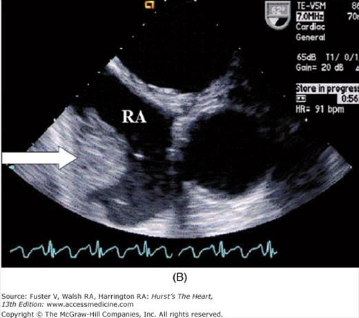
| Pericardial disease, including constrictive pericarditis |
| Pulmonary emboli |
| Pulmonary hypertension |
| Hemochromatosis and iron overload (frequent transfusions) |
It must be kept in mind that diagnosing heart failure can be a challenge, as evidenced by a recent study in a group of patients undergoing chemotherapy in which the examining physician failed to identify shortness of breath 77% of the time and failed to identify fatigue 38% of the time.11 Furthermore, in patients with only suspected heart disease, correct documentation of manifestations that establish the diagnosis were present only 50% of the time.12 Thus, techniques other than history alone must be used to support the diagnosis of heart failure. Recent observations suggest that certain monoclonal antibodies may also produce unexpected heart failure, and this is proposed to be the result of interfering with cellular mechanisms that have protean effects. The importance of the physical examination in confirming the presence of heart failure and LV systolic dysfunction in cancer patients cannot be overemphasized (see Chap. 27). Certain physical findings, such as a third heart sound, pulsus paradoxus, or jugular venous distension, can be highly predictive of heart failure (see Chap. 14).
Among basic laboratory studies, the ECG does not discern heart failure or LV dysfunction, although it can confirm suspected abnormalities or indicate potential underlying cardiac disease, such as ischemia or conduction abnormalities. Additional laboratory testing is commonly necessary to identify those at increased risk for cardiomyopathy or to confirm a diagnosis of heart failure. The most commonly used serologic tests include a complete lipid panel to indicate a risk for vascular disease and blood glucose monitoring to screen for diabetes, both clinical predictors for heart failure. Anemia, especially common in cancer patients, has been directly correlated with outcomes in heart failure patients and should be part of a basic laboratory screen (see Chap. 28). Other specific biomarkers that are crucial in the evaluation of patients with suspected heart failure, especially those with cancer, include troponins (I and T) as well as B-type natriuretic peptide (BNP). Troponins, in conjunction with creatine kinase myocardial band (CKMB), are the standard markers for confirming myocardial infarction (MI). These biomarkers may be of value in screening for LV systolic dysfunction that may occur during chemotherapy.4 In the noncancer population, BNP has also shown promise as a biomarker to detect early cardiotoxicity. Recent data suggest that markedly elevated BNP values do not always correlate with volume overload or LV dysfunction in this population. A careful physical examination is still the most reliable means of determining the presence or absence of volume overload.
Other specific cardiac testing is frequently necessary to evaluate the extent of underlying heart disease that may be present in a patient with cancer. Exercise or pharmacologic stress testing can help confirm that coronary artery disease is present (see Chap. 16), and cardiac catheterization demonstrates angiographically the extent of coronary artery disease, LV systolic dysfunction, valvular abnormalities, and other hemodynamic disturbances. A myocardial biopsy may be appropriate when it is necessary to confirm a diagnosis of amyloidosis or myocarditis. In addition, a myocardial biopsy may reveal the acute changes related to anthracycline administration. The typical histologic changes associated with the anthracyclines dissipate over time. Thus, a myocardial biopsy is not useful in evaluating patients months or years after exposure to an anthracycline. An invasive hemodynamic assessment may be extremely useful in determining the presence or absence of constrictive pericarditis or other causes of heart failure that may be difficult to discern noninvasively.
The Principles of Therapy for Heart Failure
The principles of therapy for heart failure and LV dysfunction in cancer patients are similar to those in patients without cancer. An often overlooked management component is the education of both the patient and family members. This is especially important in cancer patients in whom noncancer issues are often considered noncrucial. One area to be emphasized is compliance with medication prescriptions. Additionally, education is necessary regarding the symptoms of cardiac decompensation.
Guidelines for the management of patients with heart failure are very explicit, and the evidence-based approach is detailed in these guidelines. There is no reason to suspect that standard medical therapy would not be effective in most cancer patients as well. Table 90–5 outlines the therapeutic options for these patients. Angiotensin-converting enzyme (ACE) inhibitors and β-adrenergic blocking agents remain the cornerstone of therapy. Loop diuretics are used for controlling volume overload. In addition, risk factors for heart failure should also be aggressively treated. Other therapies that are recommended in special situations include aldosterone inhibitors, which are indicated in patients with severe heart failure and in patients who recently experienced an MI (see Chap. 28). The usefulness of angiotensin receptor blockers as alternatives to ACE inhibitors for the treatment of heart failure is well established. The combination of nitrates and hydralazine is of special benefit in African American patients, as well as in patients with renal insufficiency.
| Medications | Devices |
|---|---|
| ACE inhibitors | Permanent pacemaker |
| β-Blockers | Implantable cardiac defibrillator |
| Loop diuretics | Biventricular pacemaker |
| ARBs | Ultrafiltration |
| Aldosterone antagonists | |
| Vasopressin antagonists | |
| Nitrates and hydralazine | |
| Digoxin |
There are important concerns for patients who are undergoing active treatment for cancer because treatment for heart failure can heighten the risk for drug–drug interactions. It is imperative that unnecessary medications are avoided in these patients and that the addition of a potentially beneficial heart failure medication is carefully weighed against the risk of possible interactions with anticancer agents. However, these agents may pose a substantial bleeding risk, particularly in patients who have thrombocytopenia. Several pharmacotherapeutic agents are being developed that may be particularly appropriate for use in cancer patients with heart failure. Stem cell therapy for LV dysfunction may become an important strategy in the future; methods of delivery and the enhancement of engraftment remain subjects of intense investigation. There are a variety of other potentially useful agents for treating heart failure in cancer patients. IV immunoglobulin, which is used in patients who have immune deficiencies or even an excessive graft-versus-host response after bone marrow transplantation, may have important future implications. However, one randomized, controlled study did not show any clear benefit. Other therapies that appear promising include sildenafil for right-heart failure because of pulmonary hypertension and a newer class of diuretics, the vasopressin antagonists (eg, conivaptan and tolvaptan). These agents produce water diuresis without significant solute loss, which may be particularly advantageous in patients with hyponatremia who have significant volume overload.
Cardiac devices are an integral part of modern heart failure management. Pacing is appropriate if a patient is unable to tolerate β-blocker therapy because of a low heart rate or disease of the conduction system. The use of a biventricular pacemaker to lessen symptomatic heart failure in patients with a wide QRS complex is an accepted therapy and should not be denied to appropriate cancer patients; the benefit of biventricular pacing devices, however, occurs after 6 months, so their use is not warranted in patients with end-stage malignancy. Similar considerations pertain to the use of implantable cardiac defibrillators (see Chap. 28). Indications include symptomatic ventricular arrhythmias in patients with severe LV dysfunction with or without a prior MI. In patients who are at high risk for life-threatening ventricular arrhythmias, such as those with marked amyloidosis, implantable cardiac defibrillators should be considered more liberally. Ultrafiltration devices are especially useful in patients with evidence of cardiac decompensation who have not responded initially to diuretics.


