Value and Limitations of Electrocardiography
The surface electrocardiogram (ECG), introduced more than 100 years ago by Willem Einthoven, is the most common technique for the study of heart diseases. It is very useful for the evaluation of acute chest pain, palpitations, syncope, and acute dyspnea and the gold standard for the diagnosis of cardiac arrhythmias, conduction disturbances, pre-excitation syndromes, channelopathies, and some aspects of acute ischemic heart disease (IHD). Additionally, it is a fundamental tool to assess the evolution of heart diseases, in particular ischemic diseases, as well as in situations such as electrolytic disorders and drug therapy. It is also useful for epidemiologic studies, screening, checkup of athletes, and other situations.
Despite its invaluable usefulness if used correctly, the interpretation of an ECG recording of normal appearance must be performed with caution. We should bear in mind that a relatively high percentage of patients with coronary heart disease, in the absence of chest pain, show a normal ECG recording. In approximately 5% to 10% of acute coronary syndromes (ACSs), ECG is normal or borderline, especially in the early phases. Furthermore, ECG may appear normal after a myocardial infarction. Therefore, a normal ECG is not completely reassuring, because a patient may die from cardiac causes even on the same day a normal recording was taken. However, in the absence of any clinical symptoms or signs or family history of sudden death, the possibility of this occurring is very remote.
On the other hand, some subtle ECG abnormalities without evidence of heart disease may be observed occasionally. Notwithstanding, one must be cautious in such circumstances, and IHD, channelopathies, and pre-excitation syndromes should be ruled out before considering this as a nonspecific abnormality. Therefore, it is necessary to read the ECG recordings while keeping in mind the clinical setting (eg, family history, chest pain, syncope) and, if necessary, taking sequential recordings.
In addition, normal variants may be observed in the ECG recording that are related to constitutional habits, chest malformations, age, or other factors. Even transient abnormalities may be detected due to a number of causes (eg, hyperventilation, hypothermia, glucose or alcohol intake, ionic abnormalities, effect of certain drugs).
Today, the importance of ECG goes beyond its established role of diagnosing abnormal patterns. It has become a tool to determine prognosis and perform risk stratification in many clinical situations, because it can provide insight into basic electrophysiology by ascertaining abnormalities at the molecular level, such as in channelopathies. Therefore, as has been recently stated,1 it could be said that we face a new renaissance of ECG.
These facts should be borne in mind before starting to learn a technique such as ECG, because the ECG assessment has to be done within the context of a given clinical condition. For example, the occurrence of a small ST-segment depression, in the presence of precordial pain, may be due to ischemia (ACS), or in the absence of pain, it may be explained by the ingestion of drugs or alcohol.
In this chapter, the basis of the normal ECG and of the abnormal morphologies that may occur as a consequence of the enlargement of cavities, atrial and ventricular conduction disorders, pre-excitation, and ischemia will be discussed. After that, the most relevant ECG abnormalities found in ischemic and other heart diseases, as well as in some special situations such as ionic imbalances, hypothermia, and athletes, will be summarized. ECG of arrhythmias and the other noninvasive (exercise ECG, Holter technology, body mapping) and invasive electrical technologies used for study of heart diseases will not be discussed in this chapter because they will be discussed in other chapters of the book, especially in those chapters devoted to cardiac arrhythmias (see Chaps. 41 to 43).
The Normal ECG
The ECG is a lineal recording of the electrical activity of the heart that repeats over time, taken from outside the body by surface electrodes. For each cardiac cycle, different successive morphologies are recorded: the wave of atrial depolarization (P wave); the ventricular depolarization, called the QRS complex; and the wave of ventricular repolarization (T wave). Occasionally, other waves such as U waves may be recorded. The smooth wave of atrial repolarization usually remains hidden in the QRS complex (Fig. 15–1A).
Figure 15–1

A. Temporal relationship between the different ECG waves and nomenclature of the various intervals and segments. Ta wave, T wave of atrial repolarization. B. Schematic representation of the vectors and sense of the phenomenon for each wave and its representation on surface ECG. C. Observe the different spaces of the PR interval. HRA, high right atrium; HBE, His bundle electrogram. PA interval extends from the upper right atrium—onset of the P wave in the surface ECG—to the first rapid lower right atrial deflection; this represents right intra-atrial conduction (Au); its normal value oscillates between 30 and 50 ms. AH interval extends from the first rapid deflection of the lower atrial electrocardiogram (A) until the bundle of His (H) deflection; this represents intranodal conduction (N), and its normal value oscillates between 45 and 100 ms. The value of the HV interval, which is the distance between the bundle of His and the ventricular muscle, ranges from 35 to 55 ms.
Between these waves, there are different intervals. Figure 15–1A shows these different waves and intervals that represent the successive activation (depolarization and repolarization) of atria and ventricles, which repeat one cycle after another. In this figure, the ECG morphology corresponds to the electrical activity measured from the surface of the left ventricle (Fig. 15–1B). The P, QRS, and T waves are positive because the electrode faces the head of the main depolarization vector (→) of the atria and left ventricle (see Fig. 15–1B) that moves toward the electrode and the head of the repolarization vector of the ventricles; yet during repolarization, the sense of the phenomenon (see Figs. 15–1B and 15–5) moves away from the electrode (from epicardium to endocardium). The vector of depolarization and repolarization of the ventricles is the vectorial expression of the surface of a dipole caused by the propagation of depolarization and repolarization fronts (couple of electrical changes, –+, that initiate the process of depolarization and repolarization; see Fig. 15–1B). The small initial and terminal morphologies of QRS are negative because the initial and terminal forces of ventricular depolarization move away from the left ventricle electrode (see Fig. 15–12B).
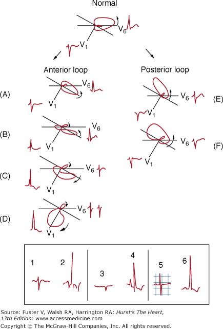
Other forms of recording the electrical activity of the heart may be used, such as vectorcardiography (VCG), body mapping, and intracavitary recordings. VCG records the path that follows the electrical activity of the heart through closed loops that represent atrial depolarization (P loop), ventricular depolarization (QRS loop), and ventricular repolarization (T loop) (see Fig. 15–12). Each loop is formed by the sum of its multiple vectors and represents the pathway followed by the successive dipoles of depolarization of atria and ventricles and of repolarization of the ventricles. The head of these vectors is always located toward the positive part of the depolarization or repolarization dipoles (see Fig. 15–12).
A close correlation exists between VCG loops and the ECG curves. The ECG morphology may be deducted from the morphology of the VCG loops and vice versa. This is due to the loop-hemifield correlation theory (see The Dipole-Vector-Loop-Hemifield Correlation). According to this correlation (see Figs. 15–10 and 15–12), the morphology of different waves (P, QRS, and T) recorded from different sides (leads) varies. Because the heart is a three-dimensional organ, the projection of the loop’s maximum vectors in the frontal and horizontal planes related to the positive and the negative hemifield of each lead (see Leads and Hemifields: ECG-VCG Correlation) is required to ascertain exactly the loop’s location and deduct ECG morphology (Fig. 15–2; see also Fig. 15–10). However, the morphology of ECG not only depends on the maximum vector of a given loop but also on its rotation (see Figs. 15–2 and 15–10). This underscores the importance of considering the loop in its entirety, and not only its maximum vector, as most of the available texts do, to explain the ECG morphology.
Figure 15–2
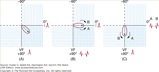
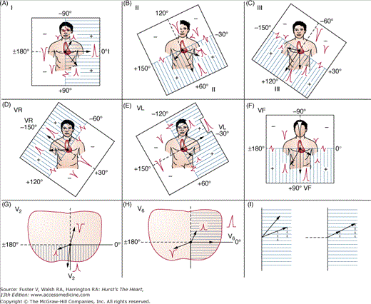
If the maximum vector of a loop falls in the limit of positive and negative hemifield of a certain lead, an isodiphasic deflection is recorded. However, according to the direction of loop rotation, the QRS complex may be positive-negative or negative-positive; see examples for leads VF and I in case of maximum vector directed to 0° (B) and +90° (C). In a loop with maximum vector at +45° (A), the loop always falls in the positive hemifield of I and VF independently of the sense of rotation.
Presently, the recording of the VCG has little practical application,2-4 although it may be of interest for research purposes. In addition, the correlation of the ECG with the VCG loop is extremely useful for educational purposes.
To summarize:
- To have a clear understanding of ECG curves, it is essential to perform a deductive analysis of the electrical activation of the heart under normal and pathologic conditions with the use of VCG loop projection, knowing its clockwise or counterclockwise rotation, on the positive and negative hemifields of different leads (see The Dipole-Vector-Loop-Hemifield Correlation).
- The ECG-VCG correlation is a useful method to understand the ECG curves and is the method that we use for teaching purposes (see Figs. 15–2 and 15–10 and The Dipole-Vector-Loop-Hemifield Correlation).
When we place one microelectrode outside and another inside a myocardial cell, we can record a transmembrane diastolic potential (TDP), which is stable in contractile cells (Fig. 15–3) and ascendant in pacemaker cells (see Fig. 15–11). Then, if we properly stimulate the cell, we record a monophasic curve called the transmembrane action potential (TAP), which corresponds to the depolarization and repolarization of the cell (Fig. 15–4).
Figure 15–3

Two microelectrodes placed on the surface of a myocardial fiber during the resting phase record a horizontal reference line, indicating the absence of differences in potential on the cell surface. When one of the microelectrodes is inserted into the interior of the cell, there is a movement below the baseline corresponding to the difference in potential between the cell exterior (+) (Na, Ca) and interior (−) (predominance of nondiffusible anions [A]). This line, the diastolic transmembrane potential (DTP), is stable in contractile cells and with more or less upsloping in automatic cells.
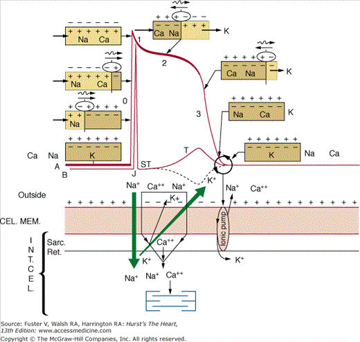
The correlation between the TAP of the contractile cells and the human ECG is as follows (see Fig. 15–4): Phase 0 corresponds with the QRS complex; phase 1 and the initial part of phase 2 correspond with the J point and start of the ST segment; the middle part of phase 2 corresponds with the isoelectric ST line; and the end of phase 2 and phase 3 correspond with the T wave.
The cellular electrogram and the human ECG represent the sum of the cellular or ventricular depolarization plus repolarization. Both cell and ventricle recordings are characterized by having a similar (QRS complex) depolarization ( ), but repolarization (T wave) is different, being negative (
 ) in the cellular electrogram and positive (
 ) in the human ECG. In both cases, the T wave follows a period (ST segment) that in normal conditions is isoelectric.
Since the work by Wilson et al,5 it is accepted that this difference between the cellular electrogram and human ECG may be explained by the opposite direction of the human heart repolarization wave (T wave) with respect to the depolarization (QRS complex), at least in some areas of the ventricles, whereas in the cellular electrogram, the repolarization wave follows the same direction. This means that some areas of the heart that are activated earlier have a longer TAP than other areas that are activated later. There are discrepancies about which areas of the heart present this repolarization gradient.
It is well known6 that the depolarization starts in the endocardium, specifically in three areas of the left ventricle, and that the epicardium depolarizes after the endocardium. Many experimental works performed in animals7,8 and in human beings, through the separate recording of endocardium and epicardium TAPs during catheterization and cardiac surgery,9 have shown that the areas with the shortest TAP, and thus the earliest repolarization, correspond to the epicardium. Therefore, it seems evident that a transmural repolarization gradient exists; although for some authors,10 the middle section of the ventricular wall (M cell zone) takes even longer to repolarize than the endocardium, whereas for other authors,11,12 the repolarization gradient, more than transmural, is transregional (for example, between the apex and the base).
We explain the origin of the human ECG based on the fact that the epicardium repolarizes before the endocardium; this is because repolarization always starts in areas with more abundant perfusion, and physiologically (in normal conditions), the subendocardium is relatively underperfused (Fig. 15–5). At a cellular level, the opposite happens because the zone distal to the electrode (equivalent to the endocardium) does not have this physiologic hypoperfusion, and therefore, the first zone to be depolarized (the zone distal to the electrode) is also the first to be repolarized.
Figure 15–5
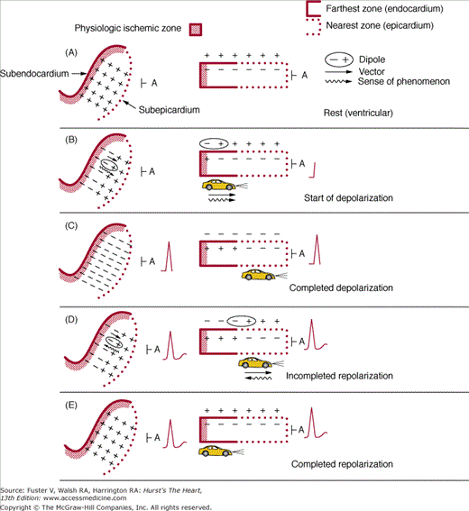
Diagram of depolarization (QRS) and repolarization (T) morphologies in the normal human heart. The figures to the left show a view of the free left ventricular wall from above, and only the distribution of the charges on the external surface of this enormous left ventricular cell is seen. In the right column is a diagram with a lateral view in which the intracellular changes in the electrical charges are observed. With electrode A in the epicardium, the QRS and T are positive because, in both cases (depolarization and repolarization), the electrode A faces the head of a vector. However, during depolarization, the direction of the phenomenon goes toward the electrode (B and C), and during repolarization, the direction moves away from the electrode (D and E). Nevertheless, in both cases, the lights of a car, used here as an example, are the head of the vector of depolarization and repolarization and are directed toward the electrode.
The approach to explain the human ECG arises from two different theories:
- The first theory considers, in light of what has been discussed, that the curves of the human ECG are the sum of the successive dipoles (− +) of depolarization and repolarization recorded by an electrode located on the opposite side (epicardium) relative to the place (endocardium) where the stimulation initiates (depolarization) (see Fig. 15–5).
- The second theory considers that the human ECG is the sum of the TAPs of the subendocardium, the farthest part to the recording electrode (which TAP starts before and ends later), and the subepicardium, the closest part to the electrode (which TAP starts later and ends before; see previous discussion; Fig. 15–6).
Figure 15–6
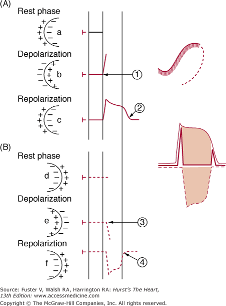
Correlation between transmembrane action potential (TAP) of the farthest (A) and the nearest (B) part of the left ventricle and ECG curve. 1. Beginning of the depolarization in the farthest zone. 2. End of repolarization in the farthest zone. 3. Beginning of depolarization in the nearest zone. 4. End of repolarization in the nearest zone. At the end of depolarization (b), in the farthest zone (A), the electrode is confronted with this part depolarized (ie, negative on the outside and positive on the inside), and as an electrode faces the positive charges of inside an ascendant TAP, phase 0 is recorded. At the end of repolarization (c), the electrode faces internal negativity because repolarization has concluded and the curve returns to the isoelectric line. In the case of subepicardial TAP (B), the opposite occurs. When this TAP has started to be depolarized (e), which occurs later than in the subendocardial zone (compare 1 and 3), this zone presents external negativity. The electrode faces this negativity, and phase 0 is inscribed as negative. When this zone has finished the repolarization (f), because it takes place earlier than in the subendocardial zone, the electrode is confronted with positive external charges because repolarization has concluded, and the subepicardial TAP curve returns to the isoelectric line (point 4). The first and last parts of the sum of both TAPs produce the QRS complex and T wave. The rest of the two TAPs is cancelled and seen as an isoelectric line (ST segment).
This is a simplification of how the ECG is produced because we have considered the left ventricle alone, and thus its depolarization can be represented by a single vector. Actually, this is not the case because there are more vectors in the human ECG. At least one vector is required, as the sum of all the atrial vectors, to explain the pathway that is followed by the stimulus to depolarize the atrium. This is known as the atrial depolarization loop (P wave) (see Figs. 15–1A and 15–12A). Three vectors explain the pathway of the ventricular depolarization loop (QRS complex) (see Fig. 15–1B). The QRS complex is followed by the ventricular repolarization loop (T wave) (see Figs. 15–1C and 15–12C). Three vectors are required to explain the morphology of the ventricular depolarization loop (qRs or rSr′) and depend on the placement of the recording lead, although they actually represent many more vectors (see Figs. 15–1 and 15–12).
To summarize:
There is considerable evidence that in the human heart, the epicardium (the zone nearest to the exploring electrode) is the first part to repolarize; and therefore, the human ECG records a positive T wave (see Fig. 15–5), the opposite of what the cellular electrogram shows.
The depolarization and repolarization dipole is expressed as a vector. The sum of the successive depolarization and repolarization dipoles and the vectors that express them, from an electrode located on the opposite side relative to the site were the stimulus originates, can explain the human ECG curve (see Fig. 15–5).
The human ECG can also be considered as the result of summation of the TAPs of the farthest (the subendocardium) and the nearest (the subepicardium) zones from the exploring electrode (see Fig. 15–6).
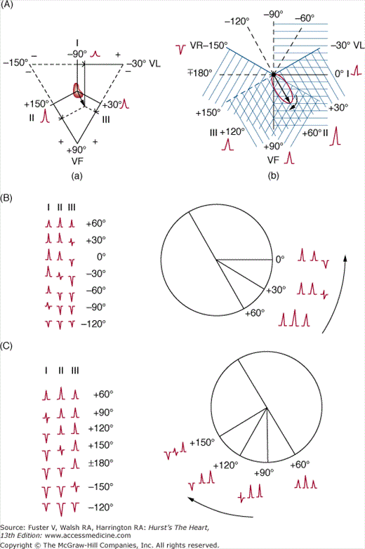
The ECG presents different morphologies as it is recorded from different sites, named leads. We currently use six frontal (I, II, III, VR, VL, and VF) and six horizontal (V1 to V6) leads (Figs. 15–7A,B, and 15–8C).
Figure 15–7
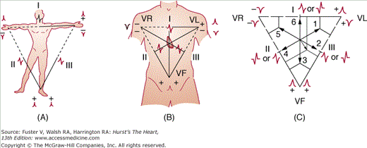
A. The Einthoven triangle. B. The Einthoven triangle superimposed on a human thorax. Observe the positive (continuous line) and negative (dotted line) part of each lead. C. Different vectors (from 1-6) produce different projections according to their location. For example, vector 1 has a positive projection in lead I, diphasic in II, and negative in III, whereas vector 3 is diphasic in I and positive in II and III.
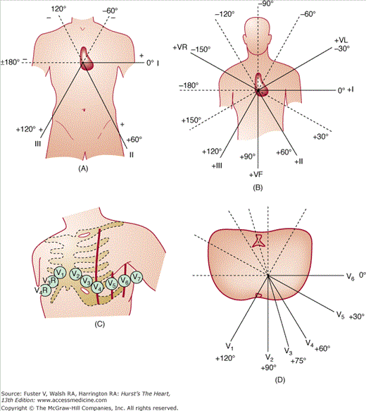
There are three bipolar leads (I, II, and III) in the frontal plane, which, according to the Einthoven law, should accomplish the equation II = I + III. These three leads form what we call the Einthoven triangle (see Fig. 15–7A). Figure 15–7C depicts how the projection of different vectors (or loops) explains the different morphologies found in leads I, II, and III. Bailey, by shifting the three leads toward the center, obtained a reference figure (Bailey triaxial system) (Fig. 15–8A). In the frontal plane, there are also three monopolar leads (VR, VL, and VF) (Fig. 15–7B). By adding these three leads to the Bailey triaxial system, the Bailey hexaxial system is obtained (Fig. 15–8B).
All of the 12 leads located around the circumference in the frontal and horizontal planes have a positive line (Fig. 15–8B,D) that goes from the place where the electrode is located up to the center of the heart. If lines perpendicular to the frontal and horizontal leads are drawn passing through the center of the heart, the positive and negative hemifields of these leads may be obtained (Fig. 15–9). The positive hemifield of lead I extends from +90° to –90° passing through 0°; that of lead II extends from –30° to +150° passing through +60°; that of lead III extends from +30° to –150° passing through +120°; that of VR extends from +120° to –60° passing through –150°; that of VL extends from –120° to +60° passing through –30°; that of VF extends from 0° to ±180° passing through +90°; that of V2 extends from 0° to 180° passing through +90°; and that of V6 extends from –90° to +90° passing through 0°. The rest of the hemifields corresponding to the horizontal plane leads can be obtained in the same manner, drawing lines that are perpendicular to the corresponding lead, passing through the center of the heart (see Fig. 15–9). In all cases, the negative hemifields are opposed to the positive ones.
Figure 15–9
Positive and negative hemifields of the six frontal plane leads and the horizontal plane leads. Depending on the magnitude and direction of the different vectors (that represent the corresponding loops), positive and negative deflections with different voltages are originated (see text).
A loop of P, QRS, or T, or their maximum vectors, located in the positive or the negative hemifield or on the interface between both hemifields in any of the 12 leads, gives rise to, respectively, positive, negative, or isodiphasic deflections of P, QRS, or T waves in that given lead. An isodiphasic deflection has a maximum vector but may have a different morphology; it can be positive/negative or negative/positive, according to the direction of the loop rotation representing the pathway followed by the stimulus (see Fig. 15–2). The degree of positivity or negativity in each morphology depends on two factors: the magnitude and the direction of the loops or vectors. For a given magnitude, the vectorial result will have a greater positivity or negativity according the direction of the maximal vector of the loop; and for a given direction, the vector of the loop with a greater magnitude will cause a greater positivity or negativity (see Fig. 15–9I).
As previously mentioned, a pair of electric charges called a dipole (–+) is formed in both depolarization and repolarization processes of activation of the heart. The existence of these dipoles results from ionic changes and explains the formation of TAP (see Fig. 15–4 and legend). These dipoles have a vectorial expression, with the head of the vector located in the positive part of a dipole. An electrode that faces the head of the vector records positive deflection regardless of whether this dipole approaches the electrode or moves away. It has already been explained how the cellular and ventricular electrograms are formed and that, in human ECG, the repolarization wave (T wave) is positive because physiologically there is less perfusion in the subendocardial zone and the process of repolarization always starts in the more perfused zone. Therefore, in human ECG, this process begins in the subepicardium, the opposite of what occurs at the cellular level (see Fig. 15–5).
It is relevant to highlight that the P, QRS, and T loops are formed from the vectorial sum of depolarization and repolarization dipoles that express the pathway followed by electric stimulus during these processes (see Fig. 15–12). As stated, only the simultaneous projection on two planes, frontal and horizontal, may provide exact information as to the direction of respective electric forces (frontal plane: upward-downward and rightward-leftward; horizontal plane: rightward-leftward and frontward-backward) (see Fig. 15–12). Each of these loops has a maximum vector, which is considered to be the result of all instantaneous vectors (see Fig. 15–12) and expresses the magnitude and the overall direction of a loop. Nevertheless, the morphology of a loop, especially the initial and terminal parts, and the loop rotation (clockwise or counterclockwise) have relevant added value (Fig. 15–10; see also Fig. 15–2).
The tracing of atrial (P) and ventricular (QRS) wave activation explained by the VCG curve may be translated into ECG curves in different leads according to the VCG-ECG correlation. Figure 15–10 shows this correlation with VF and V2 leads.
To summarize:
The following chain of events explains the ECG morphology: dipole → vector → loop → hemifield → ECG.
The P, QRS, and T loops overall have an orientation that may be expressed by a maximum vector. Although these vectors provide important information on ECG morphology in different leads, only the global contour of the loop, its sense of rotation, and the loop-hemifield correlation may explain the total ECG morphology (see Figs. 15–2, 15–10, and 15–12).
The rotation of the loop, clockwise or counterclockwise, explains why, for a given maximal vector, the morphology of the QRS may be either
 or
or  (see Figs. 15–2 and 15–10).
The ECG tracing reflects the activation sequence (depolarization and repolarization) of the heart, starting with the stimulus arising in the sinus node (this is the structure with greater automaticity), up to the ventricular Purkinje network through the specific conduction system. Figures 15–1B and 15–12 show a summary of atrial depolarization and ventricular depolarization and repolarization that explains the ECG according to the dipole → vector → loop → hemifield theory, and Fig. 15–11 shows how the ECG may be also considered as the sum of different TAPs originating along the heart activation sequence from the sinus node to the ventricular muscle. The conduction speeds of different parts of the specialized conduction system are shown at the right in Fig. 15–11.
Figure 15–11
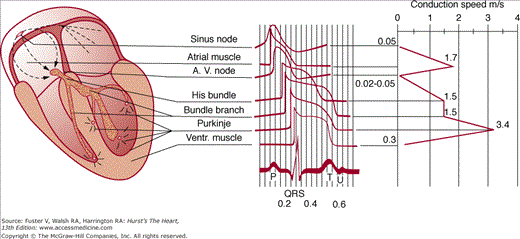
Diagram of the transmembrane action potential (TAP) morphologies of different structures of the specialized conduction system, the atrial and ventricular (Ventr.) muscle and the impulse conduction speed through these structures, and correlation with the curve of the ECG. AV, atrioventricular.
The union of the heads of all atrial depolarization vectors represents the P loop, which is recorded on the ECG as the initial deflection, the P wave (Fig. 15–12A). The loop-hemifield correlation explains the morphology of the P wave in the different leads. Generally, atrial repolarization (Ta wave) is seldom seen, being masked by the significant forces generated by ventricular depolarization that give rise to the QRS complex (see Fig. 15–1A).
Figure 15–12

A. Right and global atrial depolarization vector and P loop. The successive multiple instantaneous vectors are also pictured. The P loop and its projection on frontal and horizontal planes are shown. B. Observe the vectors and ventricular depolarization loop (left) and the projection of the cardiac vectors and loops on the frontal and horizontal planes (right) in a heart with no rotations. C. T loop and its representation in the space.
Once atrial depolarization ends and prior to starting ventricular depolarization (PR segment in ECG), the electric stimulus depolarizes small structures, and therefore, no waves are recorded on the surface ECG (see Fig. 15–1). Nonetheless, depolarization of the bundle of His and its branches can be recorded with intracavitary recording techniques (His recording) (see Fig. 15–1C).
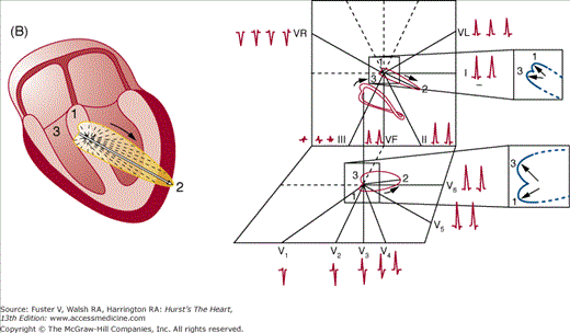
Ventricular depolarization occurs in three successive steps that give rise to the generation of three vectors (the expression of three dipoles) (Fig. 15–12B). Each of the three vectors explains a deflection of the QRS.13 Ventricular depolarization starts at three different sites in the left ventricle6: the areas of the anterior and posterior papillary muscles and a midseptal area (Fig. 15–13A,C). At almost the same time, the right ventricle begins its depolarization. These three initial depolarization sites in the left ventricle (black asterisks in Fig. 15–13A) dominate the small initial forces of the right ventricle and originate a joint depolarization dipole (vector), which is called the first vector (1) (Fig. 15–13B). This first vector is directed front-rightward and, generally, upward (see Fig. 5–12B), although in some patients, especially obese individuals, it may be also directed downward. Once this initial area in the left ventricle is depolarized, most of the right and left ventricular mass is depolarized at the same time, giving rise to a right depolarization vector (2r) and a left depolarization vector (2i). The sum of these vectors is directed toward the left, somewhat backward, and, generally, downward (see Fig. 15–12B) and is known as the second vector. In obese individuals, it is usually located around 0°. Finally, the late areas to depolarize in both ventricles (the areas with fewer Purkinje fibers; ie, the basal septal areas) originate a third vector (3), which is directed upward, somewhat to the right, and backward (see Fig. 15–12B). As stated, the union of the heads of these three vectors, which is merely a simplification of the union of the heads of all the instantaneous vectors originated during ventricular depolarization, represents the pathway that the electrical stimulus follows when it depolarizes the ventricles and is called the QRS loop, which originates the QRS complex in the ECG (see Figs. 15–1B and 15–12). The loop-hemifield correlation explains the morphology of QRS in different leads (see Figs. 15–2 and 15–10).
Figure 15–13
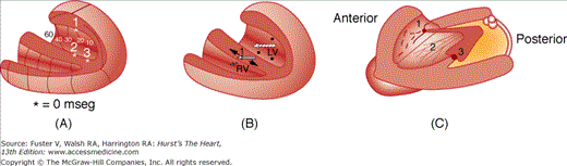
A. The three initial points (1, 2, and 3) of the ventricular depolarization are marked by an asterisk (*). The isochronic lines of the depolarization sequence can also be seen. (Adapted from Durrer et al.6) B. The first vector of the ventricular depolarization indicated by the continuous line arrow (1) is the result of the sum of the initial depolarization vectors of the left and right ventricles (dotted arrows). The first initial vector of the left ventricle corresponds to the sum of depolarization of the three points indicated in panel A, and because it is more potent than the forces of the right ventricular vector, the global direction of vector 1 will be from left to right. C. Left lateral view showing the left papillary muscles and the divisions of the left bundle branch. 1, superoanterior; 2, medioseptal (inconstant); 3, inferoposterior. There is an excellent correlation between the divisions of the left bundle and the three initial points of ventricular depolarization (1 and 3 always and 2 when present).
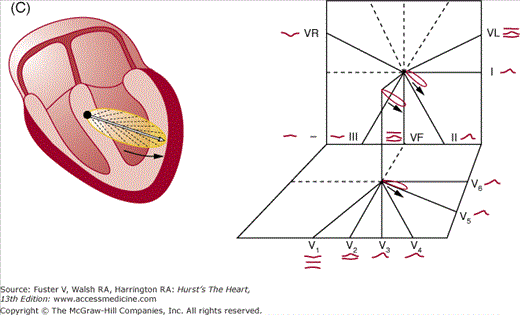
Finally, ventricular repolarization takes place, which is mainly represented by the repolarization of the left ventricular free wall. From a physiologic viewpoint, the subendocardial area is relatively underperfused (physiologic hypoperfusion), and as previously stated (see Fig. 15–1B), this explains the positivity in the last part of repolarization in the leads facing the left ventricle and the negativity in the opposite leads (VR). The initial pathway followed by repolarization does not produce any expression in the ECG and is recorded as an isoelectric ST segment. Later, when a repolarization dipole is formed, the union of the heads of all instantaneous vectors originates the T loop, which is recorded as a T wave in the ECG (see Fig. 15–12C).
After the T wave, which represents the end of ventricular systole, and prior to onset of the next atrial systole, an isoelectric line, corresponding to the resting phase of all cardiac cells, is recorded (see Fig. 15–12C). Sometimes, a small wave, called a U wave, which forms part of the repolarization process, is recorded after the T wave (see Fig. 15–1A).
The equipment for performing ECG is a conventional device that produces a paper recording of tracings. However, we are now immersed in the digital era, and in the field of ECG, this has given rise to the need for functional programs and devices that are portable, versatile, and interactive.
This means working in an integrated fashion with management and quality assurance in the different health institutional settings, independently of the types of technologies used. Currently, these systems allow us to work online and advance rapidly in the implementation of telemedical services, including diagnosis performed by an expert in real time through the Internet, which can be applied to multiple scenarios that, otherwise, would not have the same quality of assistance because of the geographic location or the associated economic costs.
The recording technique includes the following: (1) connection of the device and preparation of the patient; (2) placement of electrodes at their correct locations; (3) adjustment of baseline tracing; (4) checking of calibration; and (5) ensuring that the tracing is correctly obtained according to the Einthoven law (II = I + III). The misplacement of the electrodes may lead to several errors (see Misplacement of Electrodes).
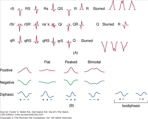
The names given to different waves and intervals are shown in Fig. 15–1, and the different morphologies of P, QRS, and T waves are shown in Fig. 15–14. In some pathologic situations, other waves may be recorded—a delta wave in Wolff-Parkinson-White pattern, an epsilon wave in arrhythmogenic right ventricular cardiomyopathy, and a J wave in hypothermia (Osborne wave) or other situations such as early repolarization.
Different items should be routinely assessed when reading an ECG. The following sections will briefly discuss the most important aspects of the normal limits of these ECG parameters, as well as some relevant aspects of the abnormal values. For more detailed information, consult Bayés de Luna7805045 and McFarlane and Lawrie.14
Sinus rhythm at rest normally ranges from 60 to 90 beats/min. Considering that the recording paper is divided into 5-mm rectangles (when the paper runs at 25 mm/s, it is equivalent to 0.20 second) each containing five smaller (1-mm) rectangles, the following methods may be used to measure the heart rate: (1) Observe the R-R intervals occurring every 6 seconds (every five 5-mm rectangles represent 1 second) and multiply this number by 10 (this is the best method when arrhythmia is present; or (2) use a proper ruler (Fig. 15–15).
Figure 15–15
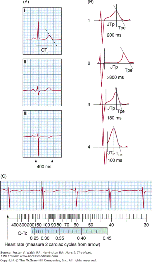
Measurement of PR and QT intervals and the heart rate. A.Leads I, II, and III. PR interval is measured with the three-channel device. The exact PR interval measurement is the distance from the earliest onset of the P wave in any lead (in this case, lead III) to the earliest onset of the QRS complex in any lead (in this case, also in lead III). Measurement of QT interval: The QT interval should be measured from the onset of the Q wave to the latest end of the T wave. The corrected QT (QTc; QT in relation to heart rate) is obtained with a ruler (C), which gives us the result when the end of two R-R cycles coincides with the QTc value obtained on a ruler; in this case, the QTc is 390 ms. The value of QT in this ECG, 400 ms, is equal to a QTc of 390 ms plus 10 ms. It is considered a normal value that does not exceed, as in this case, ± 10% to 15% of the QTc shown on the ruler. Measurement of the heart rate: The same ruler gives the heart rate, in this case approximately 60 beats/min. B. This panel shows (1) the normal QT interval; (2) an example of the long QT interval (LQT3; see Fig. 15–80B); (3) the short QT interval of asymptomatic people; and (4) the QT interval of symptomatic short QT syndrome (the distance between point J and T peak is short—100 ms; see Fig. 15–80A).
Rhythm can be normal sinus rhythm or ectopic rhythm. Sinus rhythm produce P positive waves in I, II, VF, and V2 to V6; positive or positive/negative waves in III and V1; positive or negative/positive waves in VL; and negative waves in VR. Figure 15–16 explains, according to rotation of the loop (counterclockwise in sinus rhythm or clockwise in ectopic rhythm), the different bimodal morphologies in the P wave in both cases. For example, when the axis of the P loop is located around +60°, the morphology of a sinus P wave in VL will be negative/positive (counterclockwise rotation of P wave; see Fig. 15–16) and positive/negative in case of ectopic rhythm (clockwise rotation). The same correlation is useful to explain the morphologies of P, QRS, or T waves seen in other leads.
Figure 15–16
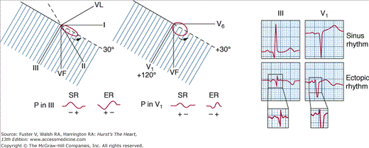
The sinus P wave (counterclockwise rotation in FP and HP and positive/negative morphology in III and V1 and negative/positive morphology in VL) and ectopic P wave (clockwise rotation and negative/positive morphology in III and V1 and positive/negative morphology in VL). ER, ectopic rhythm; SR, sinus rhythm.
The morphology of P wave in reentrant atrioventricular (AV) junctional arrhythmias and the morphology of atrial activation waves in other supraventricular arrhythmias will be discussed in Chap. 41.
PR interval is the distance from the beginning of the P wave to the beginning of the QRS complex (see Fig. 15–1A). The procedure for this measurement is shown in Fig. 15–15. Normal PR interval values in adult individuals range from 0.12 to 0.20 second (up to 0.22 second in the elderly and even <0.12 second in the newborn). Longer PR intervals are seen in cases of AV block, and shorter PR intervals are seen in pre-excitation syndromes and different arrhythmias.
The PR segment is the distance from the end of the P wave to the QRS onset and is usually isoelectric. However, with intracardiac recordings, the depolarization of the bundle of His may be seen. Figure 15–1C shows the different spaces of PR interval taken with this technique (see figure legend).
Sympathetic overdrive may cause a descent in PR segment that forms part of an arch of circumference together with an ascendant ST segment (Fig. 15–17C). In pericarditis and other diseases affecting the atrial myocardium, as in atrial infarction, a depressed or, more frequently, an elevated PR segment may also be seen (see Fig. 15–25).
Figure 15–17
Different morphologies of normal variants of the ST segment and T wave in the absence of heart disease. A and B. Normal variants. C. Sympathetic overdrive: ECG of a 22-year-old male obtained with continuous Holter monitoring during a parachute jump. D. Early repolarization (see p. 317). E. Normal repolarization of a 3-year-old child. F. ECG of a 75-year-old man without heart disease but with rectified ST/T. G. ECG of a 20-year-old man with pectus excavatus. Normal variant of ST elevation (saddle morphology).
QT interval represents the sum of depolarization (QRS complex) and repolarization (ST segment and T wave). Often, particularly in the presence of a flat T wave or a U wave, it is difficult to measure the QT interval properly. It is commonly accepted that this measurement should be performed using a consistent method to ensure that equivalent measurements are taken when the QT interval is studied sequentially.15 The most suitable method entails considering the end of repolarization in a point where the tangent line following the descendent slope of the T wave crosses the isoelectric line (see Fig. 15–15A,B). It is necessary to correct the QT interval by heart rate (QTc). In clinical practice, QTc may be measured with a ruler (see Fig. 15–15C), and it is considered that its duration should not exceed approximately 10% of the value corresponding to heart rate.
A long QT interval may be found in congenital long QT syndrome16 (see Figs. 15–15B and 15–80) and also in diseases such as heart failure and IHD, in some electrolyte imbalances, and after the intake of different drugs. It is considered that a drug should not increase the QTc by >30 ms and that a change of 60 ms may result in torsade de pointes (TdP) and sudden cardiac death. Nevertheless, TdP rarely occurs unless the QTc exceeds 500 ms.17
A short QT interval can be found in cases of early repolarization, digitalis effect, hypermagnesemia, acidosis, and other conditions and, rarely, in some genetic disorders associated with sudden death (short QT syndrome)18 (see Figs. 15–15B and 15–80A). Usually, in the latter case, the QT is <300 ms and the T wave is peaked and tall, especially in V2 to V3. In symptomatic short QT syndrome, the distance from the J point to the T peak is shorter (≅100 ms) compared with asymptomatic short QT (see Fig. 15–15B).
The P wave is the atrial depolarization wave (see Figs. 15–1 and 15–12A). In general, its height should not exceed 2.5 mm, and its width should not exceed 0.10 second. It is rounded and positive, except in VR, but may be positive/negative in V1 and III and rarely in II and VF and negative/positive in VL according to the loop-hemifield correlation (see Figs. 15–1, 15–12A, and 15–16).
The P wave changes seen in atrial abnormalities are discussed later (see Atrial Abnormalities).
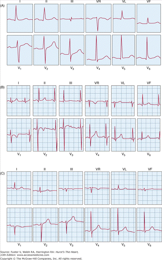
The QRS complex corresponds to ventricular depolarization. Its morphology varies in the different leads according to the loop-hemifield correlation (see Figs. 15–10 and 15–12B). An example of this correlation in a heart without rotations is shown in Fig. 15–12B, and different morphologies of QRS are also presented in this figure. Figure 15–14 shows the different letters used to express these morphologies. The normal values of QRS and T in 12 leads according to age can be found elsewhere.2,14
The width of the Q wave should be less than 0.10 second, and the R wave height should not exceed 25 mm in leads V5 and V6 or 20 mm in leads I and VL, although in VL, heights >15 mm are usually abnormal and suggest left ventricle hypertrophy (see Table 15–3).
Furthermore, the Q wave must be narrow (<0.04 second) and of quick recording and should not exceed 25% of the following R wave, although some exceptions exist, mainly in leads III, VL, and VF. The presence of abnormal Q waves suggests myocardial necrosis but may be also observed in many other circumstances (see Table 15–11).
The T wave, together with the preceding ST segment, is generated during ventricular repolarization (see Figs. 15–1B and 15–12C). According to the loop-hemifield correlation, in the adult, the T wave is positive in all leads, except VR (because the T loop is located in the negative hemifield of that lead) but being the slope up slower that the slope down. The T wave may be negative or flattened or, occasionally, slightly positive in V1, and sometimes, it may also be flattened or slightly negative in V2 and even in V3 in women and black people. In III and VF, the T wave may be flattened or even slightly negative. In children, a negative T wave of characteristic morphology is seen in the right precordial leads (children’s repolarization) (Fig. 15–17E).
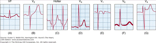
Under normal conditions, the ST segment is isoelectric (see Fig. 15–1) or shows only a small downward slope (<0.5 mm) with ascendant inclination. A small downward slope (1-2 mm) that is convex in relation to the isoelectric line is often seen in V1 and V2 as a normal variant (Fig. 15–17B).
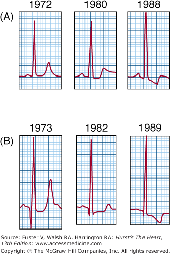
Different examples of normal ST variants are shown in Fig. 15–17, and some of these are discussed here. The saddle-type pattern (Fig. 15–17G) can be observed in V1 in healthy people, especially in those with pectus excavatus or when the V1 and V2 leads are located in a higher position (second intercostal space). This pattern should be differentiated from the type 2 Brugada pattern (see Fig. 15–80C). The pattern of early repolarization (Fig. 15–17D) entailing a 2- to 4-mm ST elevation with downward convexity, often preceded by the J wave, is particularly evident in midprecordial leads. In early repolarization, the ST-segment elevation normalizes with exercise. Recently, it has been described19 that many patients with idiopathic ventricular fibrillation present with this pattern. However, from an epidemiologic point of view, its presence only represents a slight increase in the risk of sudden death (11 in 100,000 vs 4 in 100,000 in a control group).20
The abnormalities of repolarization encompass T wave disturbances and abnormal deviations of ST segment. We will only comment here that these abnormalities may be primary and secondary. Primary T wave disturbances are changes in normal T wave (T wave higher than normal or flat/negative) due to delayed TAP in some regions of the heart or some layers of the left ventricle as a consequence of ischemia or other causes (see Changes in T Wave). Primary ST deviations (abnormal elevations and depressions of ST) are due to shape changes of TAP in some regions of the heart or some layers of the left ventricle as a consequence of ischemia or other causes (see Changes in ST Segment). Finally, secondary abnormalities of repolarization (usually ST depression and asymmetric negative T waves) are due to changes in shape and duration of TAP at the ventricles secondary to conduction delays (bundle-branch block) (see Fig. 15–32) and/or ventricular hypertrophy (strain pattern) (see Fig. 15–30). Sometimes a mixed pattern (primary and secondary) may be seen.
All changes in the normal morphology of the T wave and the abnormal deviations of the ST segment secondary to IHD or present in other heart diseases or situations will be discussed later in this chapter.
Occasionally, a small wave called the U wave is observed after the T wave, particularly in persons with vagal predominance and in the elderly. Normally, it has the same polarity as the T wave (see Fig. 15–1). If not, it is always pathologic. Characteristically, the presence of negative U waves in right precordial leads is suggestive of left anterior descending coronary artery occlusion.
When the QRS axis (ÂQRS) is at +60°, the morphology is positive in I, II, and III because the loop and its main vector fall in the positive hemifield of I, II, and III. However, it is more positive in II according to the rule that II = I + III (the same rule may be followed for the assessment of P and T wave axis) (Fig. 15–18A). When the axis shifts to the left from +60° to +30°, up to –120°, the QRS complexes become negative starting from lead III, changing from positive to isodiphasic and then from isodiphasic to negative for each shift of 30° to the left in the electrical axis (Fig. 15–18B). As the ÂQRS shifts to the right from +60° to +90°, up to –120°, the complexes again become negative, but starting from lead I, they change from positive to isodiphasic and then from isodiphasic to negative with every 30° shift to the right in the electrical axis (Fig. 15–18C). All of these changes may be explained by the correlation between loop location and the projection on different hemifields of I, II, and III in the frontal plane (Fig. 15–18A, right) and the projection of the ÂQRS vector in leads I, II, and III in the Einthoven triangle (Fig. 15–18A, left). Using this procedure, the ÂQRS may be calculated with a precision of 30°. To locate the ÂQRS more precisely, the morphology in the VR, VL, and VF leads has to be checked. For instance, a positive R wave in I, II, and III means that ÂQRS is approximately +60°. The QRS is exactly at +60° if QRS in VL is isodiphasic ( ). According to the loop-hemifield correlation, if the complex in VL is more positive than negative (
 ), the major part of the loop is still located in the positive hemifield of VL, and the ÂQRS is between +30° and +60°; if the QRS complex is more negative than positive (
 ), the greatest part of the loop is in the negative hemifield of VL, and the ÂQRS is between +60° and +90°. The same can be done for VR and VF. With such an approach, at a glance, we can calculate the ÂQRS precisely (with an error <10°).
The normal values of the P, QRS, and T electric axes are as follows. In >90% of normal cases, the P axis (ÂP) is located between +30° and +70°. The ÂQRS generally ranges from 0° to +80°, although it can be shifted somewhat more to the left in obese people and more to the right in very lean people. The T axis (ÂT) generally ranges from 0° to +70°. The ÂT is shifted more to the left when the ÂQRS is also shifted to the left. Nevertheless, on certain occasions, when ÂQRS is shifted to the right, the ÂT is between 0° and –30°.
Figure 15–19A shows a normal ECG with an RS is shifted to the right, theQRS of approximately +30° (the voltage of the QRS complex in lead III is isodiphasic and is positive in I and II).
The most important features of the ECG in healthy children (Fig. 15–19B) compared with normal adults can be summarized as follows:
There is a faster heart rate and a shorter PR interval.
Due to the physiologic right ventricular hypertrophy in infants, the heart is usually vertical, with an ÂQRS shifted to the right and negative or bimodal T waves in V1 to V3-V4 and with a characteristic morphology (children’s repolarization) that may be present until adolescence, particularly in females. The QRS loop goes to the left before going backward, which explains why the V6 but not the V1 morphology (there is higher R in V1 compared with q in V6) looks like the adult’s morphology. Sometimes an rsr′ pattern is observed in V1. In infants, especially if they are postterm, even R or qR patterns can be seen at birth with a somewhat positive T wave. The Rs pattern persists for a time, perhaps for years or even until adulthood (see Table 15–2). However, the T wave usually becomes flattened or negative in the days following birth.
In some adolescents, an R wave with high voltage in precordial leads (SV2 + RV5 >60 mm) without the existence of left ventricular enlargement may be seen.
Sometimes, there is an evident increase in the heart rate with inspiration.
The following phenomena can be considered age-related ECG variants of the elderly (Fig. 15–19C):
A slower heart rate and longer PR interval (normal up to 0.22 second).
Occasionally, an ÂP shifted more to the right is caused by pulmonary emphysema, with the S wave present in lead V6 and an ÂQRS shifted to the left (from 0° to –30°).
A poor r progression from V1 to V3, probably due to septal fibrosis. This can produce differential diagnosis issues with septal necrosis.
Unspecific alterations of repolarization (slightly depressed ST segment and/or flattened T wave). A U wave is frequently present, particularly in the intermediate precordial leads.
In a heart without apparent rotation (intermediate position), the ÂQRS is situated around +30° (between +20° and +40°). The ECG of a heart featuring these characteristics is shown in Fig. 15–19A.
Nevertheless, the heart may present isolated or combined rotations, the most characteristic of which are rotations on the following axes: (1) the anteroposterior axis (vertical or horizontal heart; see VL and VF leads in Fig. 15–20A); and (2) the longitudinal axis (dextro- or levorotation; see precordial leads in Fig. 15–20B).
Figure 15–20
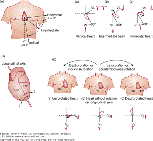
A. Rotation of the heart along the anteroposterior axis. Direction of the QRS axis (ÂQRS) in the vertical and horizontal heart. ÂQRS morphology in the vertical (a), intermediate (b), and horizontal heart (c). B. (a) Rotation of the heart along the longitudinal axis. (b) Scheme of dextrorotation and levorotation. (c) The respective loops and morphologies on the horizontal plane (V2 and V6) in levorotated heart, intermediate heart, and dextrorotated heart.
Certain special types of rotation explain some variants of the normal pattern, such as SI, SII, and SIII (differential diagnosis with right ventricular enlargement [RVE])21 and QR in III (differential diagnosis with previous inferior infarction). In the latter, the Q wave diminishes during the inspiration. For more information consult Bayés de Luna.2
In addition to the knowledge of normal and pathologic patterns, the correct interpretation of ECG recordings requires the use of recording procedures compliant to approved standards.22 The most frequent faults are a consequence of errors in limb and precordial electrodes placement, artifacts, and inadequate filter application.23
Misplacement of electrodes may lead to a diagnosis of ectopic rhythm or dextrocardia (Fig. 15–21A) or to anomalous location of the Q wave myocardial infarction (MI).
As a consequence of some artifacts, a false diagnosis of flutter or ventricular tachycardia may be made (Fig. 15–21B).
Finally, the inappropriate use of some filters may induce abnormal ST elevation in V1 to V2 or may suppress an otherwise visible J wave (Fig. 15–21C).
Figure 15–21

A. (1) Frontal plane leads in a healthy patient. (2) Interchanging left arm and right arm electrodes, the ECG shows the classical pattern of P, QRS, and T as negative waves in lead DI and positive in aVR. B. Patient with Parkinson disease whose ECG simulates atrial flutter in DIII while V4 clearly shows P waves. C. Lead V5 in a healthy 23-year-old man with early repolarization. The J wave is visible on applying the low-pass filter at 150 Hz (arrow), but this disappears on reducing the frequency to 40 Hz.
For more information consult García-Niebla et al.23
For many conditions, such as blocks and arrhythmias, the ECG is the gold standard for the diagnosis. For other conditions, such as enlargement and some types of ischemia, the ECG diagnostic criteria have to be contrasted against some other gold standard techniques (echocardiography, cardiovascular magnetic resonance [CMR], or coronarography). In these cases, it is interesting to know the diagnostic accuracy of the ECG criteria. For that purpose, we have to study the sensibility, specificity, negative predictive value, and positive predictive value of each criterion. For further information, consult Bayés de Luna.2
Abnormal ECG Patterns
The term atrial abnormalities encompasses atrial enlargement, atrial blocks, and abnormalities of atrial repolarization. The characteristics to be emphasised are as follows:
Atria become dilated rather than hypertrophied.
P wave voltage is influenced by extracardiac factors that increase it (eg, hypoxia, sympathetic overdrive) or decrease it (eg, emphysema, atrial fibrosis).
Atrial repolarization wave is usually hidden within the QRS complex (see Fig. 15–1).
Right atrial enlargement (RAE) is mainly present in patients with congenital and valvular heart diseases affecting the right side of the heart and in patients with cor pulmonale (Figs. 15–22B and 15–23B,C). Typically, the P wave is increased in voltage but not in length.
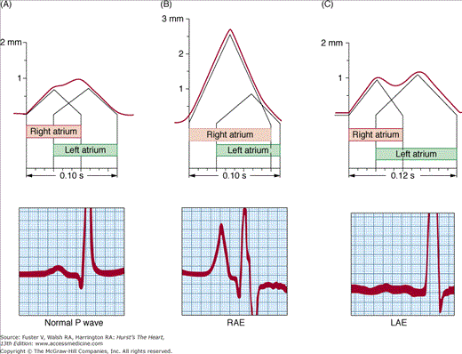
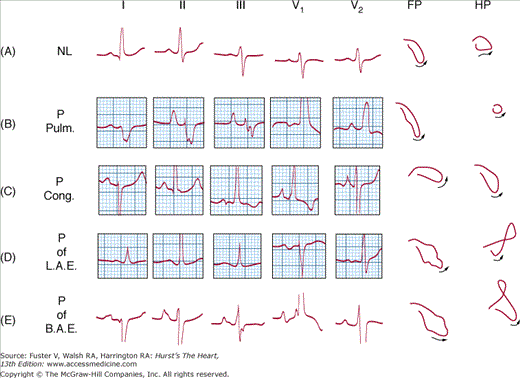
The diagnostic criteria of RAE are based on P wave abnormalities (P ≥2.5 mm in II and/or 1.5 mm in V1). These criteria have low sensitivity and somewhat higher specificity.
Left atrial enlargement (LAE) is seen in patients with mitral and aortic valvular disease, IHD, hypertension and some cardiomyopathies (Figs. 15–22C and 15–23D).
Typically, the P wave is bimodal in some leads and +/− in V1 with evident final negative mode.
The diagnostic criteria of LAE are as follows: P wave with a duration ≥0.12 second especially seen in leads I or II, generally bimodal, but with normal height; diphasic P wave in V1 with evident final negativity of at least 0.04 second of duration because the second part of the loop is directed backward due to LAE (see Fig. 15–23D, horizontal plane). These two criteria have good specificity (close to 90%; few false-positive cases) but discrete sensitivity (<60%; more false-negative cases). The +/− P wave morphology in II, III, and VF with a P ≥0.12 second is very specific and has a high positive predictive value (100% in valvular heart disease and cardiomyopathies). However, it has a low sensitivity and low negative predictive value for LAE.
The diagnostic criteria of biatrial enlargement (Fig. 15–23E) include criteria for RAE and LAE: (1) P wave in II is taller (≥2.5 mm) and wider (≥0.12 second) than normal; on certain occasions, a “peaked“ positive P wave can arise in V1 to V2; and (2) criteria of LAE with an ÂP shifted to the right and/or criteria of RAE based on QRS complex alterations.
The concept of a block means that in a certain part of the heart (sinoatrial junction, atria, AV junction, or ventricles), the electrical stimulus encounters overall significant difficulties for its conduction. A first-degree or partial block occurs when conduction is slow but the stimulus passes through the area with slow conduction; a third-degree or complete block is when the stimulus is completely blocked; and a second-degree block is when the stimulus sometimes passes and sometimes does not.
The conduction delay in interatrial block occurs between the right and left atria. Although usually associated with LAE, it may also exist as an isolated finding in cases of pericarditis, IHD, and other conditions. The block can be partial or advanced.24,25
In partial interatrial block, the stimulus reaches the left atrium via the normal pathway, but with certain delay. The diagnostic criterion of partial interatrial block is a P wave with a duration ≥0.12 second in the frontal plane.2,24 The P wave length and, consequently, the bimodal morphology of the P wave seen in lead II (which is the most typical lead to detect an isolated partial interatrial block) are similar to the P wave occurring in LAE. In fact, the delay in interatrial conduction, rather than the left atrium dilation, generally explains the morphology observed with an LAE. However, the morphology of the P wave in the horizontal plane, especially in V1, is usually different. In case of isolated interatrial block (eg, pericarditis), the second part of the loop is not directed so much backward because there is no LAE, and consequently, the P wave morphology in V1 is positive or only presents a small negative part.
In advanced interatrial block (Fig. 15–24), the stimulus does not reach the left atrium via the normal path, but by retrograde left atrial activation.25 The diagnostic criteria of advanced interatrial block are as follows26: P wave with a duration ≥0.12 second and +/− in II, III, and VF; and often, P wave +/− in V1 to V3 or V4. This type of block is frequently accompanied by supraventricular arrhythmias especially atypical atrial flutter.27,28
Figure 15–24

Top: Example of atrial activation and characteristics of the P loop in the frontal plane and the morphology of P wave in VF in normal conditions (A) and in a case of partial (B) and advanced (C) interatrial block with left atrial retrograde activation. Bottom left: Leads I, II, and III in advanced interatrial block with left atrial retrograde activation, with direction of the activation vectors of the first and second part of the P wave and three consecutive P waves with +/–morphology in VF. Bottom Right: Esophageal and intracavitary recordings demonstrating the sequence of activation in this type of interatrial block (high right atrium [HRA], low right atrium [LRA], and high esophageal [HE] lead with −/+ morphology).
The atrial repolarization wave (ST-Ta) usually has a polarity opposed to the P wave and is not visible because it is hidden within the QRS complex.25,29 In cases of great sympathetic overdrive in normal individuals, an ST-Ta depression may be seen (see Fig. 15–17C). Moreover, in some cases of remarkable atrial enlargement, pericarditis, or atrial infarction, especially in the presence of a long PR interval, shifts of ST-Ta may be observed (Fig. 15–25).

Morphologies of ventricular enlargement are secondary to a hypertrophy rather than to a dilatation, unlike what occurs in the atria. Nevertheless, slight or even moderate degrees of enlargement of either of the ventricles, mainly the right, or of both at the same time, may not produce abnormalities in the ECG.
The superiority of echocardiography over ECG for the diagnosis of ventricular enlargement, mainly of the left ventricle, is evident (the sensitivity is much higher, and the specificity is similar). However, when a ventricular enlargement is diagnosed with an ECG, the accuracy of the ECG is greater than that of the echocardiogram in predicting heart disease evolution and prognosis.
For details on the diagnosis of right and/or left ventricular enlargement combined with ventricular block (QRS complex duration >120 ms), consult Bayés de Luna2 and McFarlane and Lawrie.14
RVE is found in congenital heart diseases, valvular heart diseases, and cor pulmonale. The changes produced by RVE move the loop rightward and either frontward or backward. This is more a result of a delay of activation of the right ventricle, rather than an increase of the right ventricle mass (which generally never overcomes the mass of the left ventricle). Figure 15–26 shows the changes that RVE may produce in ventricular activation expressed as ventricular loops and how these changes may explain the different ECG patterns. Basically, a large part of the QRS loop is going to the right (presence of S in V6) but sometimes with an anterior loop (R or RS in V1) and occasionally with a posterior loop (rS or QS in V1).
Figure 15–26
In right ventricular enlargement (RVE) with electrocardiographic repercussion, the horizontal loop of the QRS is always directed to the right, either forward or backward. When it is directed forward, different morphologies may be recorded (from A to D cases with more advanced degree of RVE). A patient may have a morphology changing from one to another during the course of the disease. However, in general, heart diseases with mild to moderate RVE present with type A or type B morphologies, and those with severe RVE present with type D. If the loop is directed posteriorly, then the morphologies are of types E or F. The QS morphology is seen in the V1 lead in type E, whereas rS or rSr′ is seen in type F, in both cases accompanied by a significant S in V6. The lower part of the figure shows that the morphology of QRS in V1 depends more on the severity and grade of RVE than on the etiology of the disease. Left. Mild mitral stenosis (1) and advanced mitral stenosis with severe pulmonary hypertension (2). Middle. Long-standing chronic cor pulmonale without severe pulmonary hypertension (3) and subacute cor pulmonale with severe pulmonary hypertension (4). Right. Congenital moderate pulmonary stenosis (5) and severe valvular pulmonary stenosis (6).
The lower part of Fig. 15–26 shows three cases of RVE of different etiologies (see legend) in which the ECG pattern in V1 (with more or less R wave) is related more to RVE degree than to RVE etiology.
The ECG criteria most frequently used for the diagnosis of RVE are shown in Table 15–1 (along with their sensitivity, which is low, and specificity, which is high).
- Morphology of V1: (1) Morphologies with dominant or exclusive R wave in V1 are very specific but not very sensitive (<10%) for the diagnosis of RVE because the loop that gives rise to them (anterior and to the right; see Fig. 15–26B-D) is observed in a small number of cases with RVE (especially in some congenital heart diseases such as pulmonary stenosis). In these cases, the prominent R in V1 is also seen in V2 and V3. Moreover, the repolarization presents an ST-segment depression and a negative and asymmetric T wave (strain pattern; Figs. 15–26 and 15–27A), except in newborns because they may present with an exclusive R wave with a positive T wave. On the contrary, in cases of pulmonary stenosis from a tetralogy of Fallot, the morphology in V1 is similar to that in isolated pulmonary stenosis, but in V2, there is an rS morphology. Other causes that may present a dominant R pattern in V1 must be ruled out (Table 15–2). (2) The presence of rsR′ is especially typical of atrial septal defect and sometimes may” also be seen in moderate pulmonary stenosis. (3) The rS or even the QS morphology in V1, with RS in V6, may often be observed in the early phases of RVE, or with rS in V6 in advanced cases of chronic cor pulmonale (see Fig. 15–26).
- Morphology of V6. The presence of evident forces directed to the right, which are expressed as an evident S wave in V5 to V6, is one of the most important ECG diagnostic criteria (see Fig. 15–26 and Table 15–1).
- SI, SII, SIII. This morphology is frequently seen in chronic cor pulmonale with QS pattern in V1 and RS pattern in V6. Note that this pattern may be secondary to positional changes or it may simply be a normal variant.21 Nonetheless, an abnormal P wave favors the diagnosis of RVE (Fig. 15–27B).
- Electrical axis: ÂQRS ≥ +110°. Inferoposterior hemiblock, vertical heart, and lateral infarction must be ruled out. This criterion is quite specific (>95%) but relays low sensitivity.
| Criterion | Sensitivity (%) | Specificity (%) | |
|---|---|---|---|
| V1 | R/S V1 ≥1 | 6 | 98 |
| R V1 ≥7 mm | 2 | 99 | |
| qR in V1 | 5 | 99 | |
| S in V1 <2 mm | 6 | 98 | |
| IDT in V1 ≥0.35 s | 8 | 98 | |
| V5–V6 | R/S V5–V6 ≤1 | 16 | 93 |
| R V5–V6 <5 mm | 13 | 87 | |
| S V5–V6 ≥7 mm | 26 | 90 | |
| V1 + V6 | HV1 + SV5–V6 >10.5 mm | 18 | 94 |
| ÂQRS ≥ 110° | |||
| ÂQRS | SI, SII, SIII | 15 | 96 |
| 24 | 87 |
Figure 15–27
A. An 8-year-old patient with important pulmonary valve stenosis, with a gradient >100 mm Hg. The patient presents a typical morphology of right ventricular enlargement (RVE) with R wave–type systolic overload (strain) from V1 to V3. B. Patient with RVE due to advanced chronic obstructive pulmonary disease with posterior and right QRS loop types SI, SII, SIII.

The combination of more than one of these criteria increases the diagnostic possibilities.
- Differential diagnosis: The differential diagnosis of exclusive or predominant R wave in V1(R, Rs, or rSR′ pattern) is shown in Table 15–2.
The ECG signs indicative of right ventricle acute overload (decompensation of cor pulmonale or pulmonary embolism) are as follows30:
- Change in the ÂQRS (>30° to the right of its usual position).
- Transient negative T waves, sometimes very evident in the right precordial leads.
- SI, QIII with negative TIII pattern (McGinn-White pattern) in the frontal plane and an RS or rS pattern in V6 (Fig. 15–28).
- Appearance of a complete right bundle-branch block morphology often with ST-segment elevation.
Figure 15–28
A. A 59-year-old patient presenting a typical McGinn-White pattern (SI, QIII with negative T wave in lead III and rSr′ in V1) in the course of pulmonary embolism. B. The ECG findings after the recovery of the patient still show negative T waves in the precordial leads, but the McGinn-White pattern and the r′ in V1 have disappeared.
The latter two criteria are highly specific but have low sensitivity for important pulmonary embolism (see Cor Pulmonale). Nevertheless, the clinical setting and comparison with previous ECGs are important to make a differential diagnosis of both processes.
Left ventricular enlargement (LVE) is found in hypertension, IHD, valvular heart disease, cardiomyopathies, and some congenital heart diseases. In general, in patients with LVE, the maximum QRS vector of the loop increases its voltage and is directed more posteriorly than normal (Fig. 15–29). This explains why the QRS complex negativity predominates in the right precordial leads (see Fig. 15–29A-C). Occasionally the maximum vector is not directed posteriorly (it is located close to 0°). This implies a tall R wave that is seen even in V2, especially in cases of apical hypertrophic cardiomyopathy (see Fig. 15–29E).
Figure 15–29

The most characteristic loops of left ventricular enlargement (LVE): (A) with the initial forces to the right and a positive T wave; (B) observed in cases of LVE that are not long-standing and with mild septal fibrosis; (C) QRS loops initially to the left and with counterclockwise rotation or figure-of-eight rotation on horizontal plane; corresponds to significant LVE seen in advanced heart diseases with significant septal fibrosis; (D) QRS loop with q wave of pseudonecrosis that occurs in cases of hypertrophic cardiomyopathy due to the presence of important septal vector; (E) QRS loop pointed approximately 0° on the horizontal plane with a very peaked T loop pointed upward, backward, and rightward characteristic for the apical type of hypertrophic cardiomyopathy. Bottom: Two examples of aortic valve disease, one (left) with mild septal fibrosis and normal ECG and VCG (presence of q wave in V6 as expression of first vector) and the other (right) with important septal fibrosis and abnormal ECG (ST-T with strain pattern) and VCG (absence of q wave in V6). See in the HP (H) with amplification of the loop (SE = 16) now in the left the qR in V6 coincides with initial vector forces of the loop in the negative hemifield of V6, and in the right with R in V6 the initial forces go directly to the left.
The ECG pattern changes during disease evolution. The pattern of “strain“ appears more in relation with the duration of the disease than with the presence of different types of hemodynamic overload. However, in aortic valve disease, a q wave in V5 to V6 is found more frequently in long-standing aortic regurgitation than in aortic stenosis (Fig. 15–30). The disappearance of the q wave in V6 is probably more related to interstitial septal fibrosis, a substrate of partial left bundle-branch block, than to hemodynamic overload31 (see Figs. 15–29 and 15–30).
The presence of noticeable signs that are suggestive of LVE (high voltage of the QRS together with inverted ST-T wave—strain pattern) in an asymptomatic patient without heart murmur or hypertension suggests hypertrophic cardiomyopathy. The ECG does not correlate with the gene mutation,32 but there are two ECG changes suggestive of hypertrophic cardiomyopathy: (1) the striking negative T wave (see Fig. 15–29E) and (2) the presence of a deep and narrow q wave (see Fig. 15–29D).
Sometimes the LVE pattern, at least partially, and especially the changes in ST-T may be resolved in a few months with pharmacologic treatment, as occurs in hypertension or after surgery (valvular heart disease).
Various diagnostic criteria exist for LVE,33-36 with different sensitivity and specificity, as shown in Table 15–3. It is usually possible to diagnose an LVE through an ECG in patients with severe hypertension, whereas this diagnosis is difficult in asymptomatic normotensive adults. However, the value of the ECG diagnostic criteria shown in Table 15–3 is lower in hypertensive patients. For these patients, the following criterion is useful: sum of the QRS voltage of 12 ECG leads >120 mm.36
| Voltage Criteria | Sensitivity (%) | Specificity (%) |
|---|---|---|
| 1. RI + SIII>25 mm | 10.6 | 100 |
| 2. RVL > 11 mm | 11 | 100 |
| 3. RVL > 7.5 mm | 22 | 96 |
| 4. SV1 + RV5-V6 ≥35 mm (Sokolow-Lyon) | 22 | 100 |
| 5. RV6-V6>26 mm | 25 | 98 |
| 6. RVL + SV3>28 mm (men) or >20 mm (women) (Cornell voltage criterion) | 42 | 96 |
| 7. Cornell voltage duration measurement QRS duration × Cornell voltage >2436 mm/seg | 51 | 95 |
| 8. In V1-V6, the deepest S + the tallest R >45 mm | 45 | 93 |
| 9. Romhilt-Estes score >4 points | 55 | 85 |
| 10. Romhilt-Estes score >5 points | 35 | 95 |
The ECG diagnosis of biventricular enlargement is even more difficult than that of the enlargement of just one ventricle. This is because the increased opposing forces of both ventricles often counterbalance each other or the notable predominance of the enlargement of one ventricle completely masks the enlargement of the other. Therefore, even the better criteria that have a good specificity only have a moderate sensitivity.37
The following ECG patterns are used for the diagnosis of biventricular enlargement:
- Tall R wave with s in V5 and V6, rSR′ pattern in V1, and P wave of biatrial enlargement.
- Tall R wave in V5 and V6, with an ÂQRS shifted to the right (≥90°). The presence of an inferoposterior hemiblock associated with LVE and an asthenic body build must be ruled out.
- Small S wave in V1, deep S wave in V2, predominant R wave in V5 and V6, and an ÂQRS shifted to the right in the frontal plane or an SI–, SII–, SIII–type morphology.
- The presence, especially in the elderly, of QRS complexes within normal limits but with significant repolarization abnormalities (negative T wave and depression of the ST segment), mainly when the patient presents with atrial fibrillation. This type of ECG can be found in the elderly with advanced heart diseases and biventricular enlargement.
Blocks at the ventricular level can occur on the right side (Table 15–4) or on the left side (Table 15–5). They can affect the entire ventricle (global block) or only part of it (zonal or divisional block). The blockage of electrical impulses, in the ventricles or in the whole heart is called a first-degree (partial) block when the impulses pass through the area but with a delay; second-degree block when the stimulus sometimes passes and sometimes does not; and third-degree (advanced) block when the passage of stimulus is completely blocked.2 Second-degree block is known as aberrancy of conduction and is explained in Part 6).
| Global (right bundle-branch block) |
| 1. Third degree (advanced): Morphologies corresponding to type III of the Mexican school13: slurred rSR′ in V1 and qRS with slurred S in V6 with QRS ≥0.12 s. |


