Chapter 3 Role of Intact Biological Models for Evaluation of Radiotracers
INTRODUCTION
The development of radiolabeled tracers for clinical application in humans has been critically dependent on evaluation and testing in biological models. Although in vitro models have been important in the evaluation of radiotracers, this chapter will focus only on the use of intact models. Initial evaluation of a radiotracer generally focuses on assessment of in vivo organ selectivity as determined by biodistribution and pharmacokinetics.1 Often, radiolabeled compounds are evaluated in various species to ensure that behavior of a compound does not demonstrate important species differences. More important, radiotracers must be evaluated for specific organ pharmacokinetics in different disease processes.2
This chapter will define the role of intact biological models in the evaluation of radiotracers for use in the diagnosis and management of cardiovascular disease. Important species differences will be defined as they relate to cardiovascular disease. The selection of short-term versus long-term animal models will be discussed, with particular attention to the need for conscious or sedated models. Approaches for determination of myocardial radiotracer kinetics will be reviewed, including dynamic imaging (planar imaging, single-photon emission computed tomography [SPECT],3 or positron emission tomography [PET]), miniature detectors, serial biopsies, and postmortem tissue well counting or autoradiography. Special consideration will be given to imaging in small animals and the utilization of transgenic models. Approaches for evaluation of radiotracer biodistribution and myocardial radiotracer uptake and clearance will be reviewed briefly, with emphasis on the effect of cellular viability, flow, metabolic inhibitors, and pharmacologic stress on radiotracer kinetics.
SELECTION OF ANIMAL MODEL
Use of Small Mammals for Definition of Biodistribution
The first step in the evaluation of any radiopharmaceutical involves determination of the normal biodistribution of a radiotracer over time. These initial screening studies are usually performed in mice, rats, or rabbits to minimize cost. A measured dose is injected intravenously, and the percentage of uptake per gram of tissue is determined for critical organs.4–7 Uptake in the heart, the target organ, is compared with adjacent background structures at multiple time points after injection. Blood clearance can be easily derived by serial sampling of blood for gamma well counting. Tissue clearance curves can be derived either by sacrificing animals at different time points or by performing serial imaging. Segregation of tissue into cell fractions can provide information on tracer localization in specific cellular fractions.8 This information may offer insight into the mechanism of uptake and retention. The smaller hearts in these animals lend themselves to microautoradiography.
Use of Small Mammals for Evaluation of Specific Disease Processes (See Chapter 11)
A limited number of natural biological models are currently available in smaller mammals for evaluation of specific cardiovascular disease processes. The Syrian hamster model is an example of a small model for evaluation of radiotracer kinetics under cardiomyopathic conditions. Other researchers have developed rabbit models of congestive heart failure.9,10 Investigators have also created a chronic volume-overloaded condition by mechanically disrupting the aortic valve, instigating aortic insufficiency.11 It is more difficult to create a small-animal model of regional ischemia or infarction. However, this has been accomplished by placing a ligature or small clamp around a proximal coronary artery. In the mouse and rat, this occlusion can be done blindly with a small needle and suture through a thoracotomy.12,13 Unfortunately, this approach generally necessitates disruption of both venous drainage and arterial inflow. Regarding rat models of infarction, the use of Lewis rats may be preferable over other strains because of the improved survival and greater uniformity of myocardial injury.14 Several investigators have used a rabbit model of regional myocardial ischemia.15,16 In the rabbit, it is possible to selectively isolate and occlude a proximal coronary artery. Fujita et al. recently established a chronic rabbit model of myocardial infarction without the need for endotracheal intubation, which significantly improves survival.16 Production of graded ischemia in smaller animals is nearly impossible. The use of radiolabeled microspheres for independent assessment of regional myocardial blood flow is more difficult in these preparations. In addition, these preparations do not permit regional arterial-venous balance measurement for the independent assessment of regional metabolism. However, with the advent of dedicated small-animal imaging systems, quantitative evaluation of radiotracers in these physiologic small-animal models will become more feasible.17,18
Use of Genetically Engineered Small Mammals for Evaluation of Specific Biological Processes and Radiotracers (See Chapter 45)
The ability to selectively alter gene expression permits the evaluation of the significance of certain gene products for structure-function studies of cardiac proteins and their role in cardiovascular disease. These transgenic models facilitate the evaluation of targeted radiotracers in relevant biological models. Hundreds of mutant mouse strains and also a few mutant rat and rabbit strains have been generated. The number of genetically engineered mouse lines for cardiovascular research is continuously growing. Recent reviews have provided a summary of important new genetically modified animals with cardiovascular disease.19–27 Transgenic mice are most frequently used for this purpose, since mice breed rapidly, the maintenance costs are lower, and our knowledge of mouse genetics is far more advanced. Although the small size of these animals leads to significant challenges in the evaluation of radiotracers, there have been significant advances in small-animal SPECT and PET instrumentation that make imaging of mice feasible.17,18
Approaches for creation of transgenic animals are nicely reviewed by Williams and Wagner in their application for evaluation of integrative biology.28 However, the use of transgenic animals will be equally useful for the development and testing of biologically targeted radiotracers. Once targeted radiotracers, are established, they could also be used to evaluate new transgenic strains in conjunction with radiotracers which provide information about physiologic processes (i.e., perfusion, metabolism). Thus, the use of radiotracer imaging would also permit phenotyping of transgenic animals.
There are two basic approaches to mouse genomic manipulation: random chromosomal integration, which can be used for addition of an exogenous transgene, and homologous recombination of foreign DNA, which leads to targeted mutation of an endogenous gene.28
Random chromosomal integration is based on addition of DNA into fertilized oocytes in order to generate “gain-of-function” mutations, in which the transgene is overexpressed.28 Another common application of random chromosomal integration of transgenes is to identify transcriptional control elements that respond to physiologic stimuli. In this application, the coding region of a so-called reporter gene is linked to the regulatory elements of interest. By definition, reporter genes encode biologically innocuous proteins that are easily detected. The goal of this approach is to avoid perturbation of the cell in which the reporter is expressed while assessing the function of transcriptional regulatory sequences that are attached to the reporter gene. For application with radiotracer imaging, these reporter genes are linked with a radiolabeled “reporter probe” that can be detected in vivo in mice with microPET or microSPECT imaging systems.
Gambhir et al. reported methods for monitoring reporter gene expression by using herpes simplex virus type I mutant thymidine kinase (HSV1-sr39tk) as a PET reporter gene and 9-(4-[18F]-fluoro-3 hydroxymethylbutyl) guanine ([18F]-FHBG) as a PET reporter probe in animal models.29 Wu et al. applied this technology using microPET imaging to noninvasively monitor cardiac reporter gene expression in rats.30 More recently, Wu et al. applied this approach for the noninvasive assessment of myocardial response to cell therapy using embryonic cardiomyoblasts expressing HSV1-sr39tk and/or firefly luciferase.31 The location, magnitude, and survival duration of the transplanted cells were monitored noninvasively using PET and bioluminescence optical imaging.
Gene targeting via homologous recombination in embryonic stem cells is frequently used to create “loss-of-function” mutations, known as knockouts. Targeted inactivation can be accomplished by introducing a positive selection marker, which will disrupt gene structure. A conditional gene targeting approach is preferred, since this approach provides cell-type-specific and/or inducible gene targeting. One conditional gene targeting approach involves Cre/loxP-mediated targeted mutagenesis.32 This approach is based on the ability of Cre recombinase to recognize a unique nucleotide sequence (loxP site), allow the introduction of mutations in the gene of interest, and by the controlled expression of Cre also control the expression during different time points and avoid germline mutations that may be lethal. Alternatively, temporal or cell-type-specific transcriptional control can be accomplished using a tetracycline-responsive promoter.33
Use of Large Mammals for Evaluation of Specific Disease Processes
Dogs, pigs, and sheep are the large mammals most commonly used for the evaluation of cardiovascular physiology. These models have been used for many years, and the methodologies of their use are well established. The higher cost of large-animal models almost precludes their use for simple biodistribution studies; rather, they are mostly used to evaluate radiotracer kinetics in specific disease processes. In the evaluation of radiotracers, dogs and pigs have been most commonly used. Important interspecies differences in radiotracer uptake have been observed over the years. The most notable example is the uptake of technetium-99m (99mTc) 1,2-bis(dimethylphosphino)ethane. This 99mTc-labeled perfusion tracer showed favorable uptake characteristics in rats, dogs, rabbits, and monkeys; however, in phase I trials, no appreciable uptake was noted in the hearts of pigs or humans. Although there was transient uptake of this radiotracer by human heart, trapping was not observed because of a reductive mechanism that is prominent in human myocardium.34
The dog has a dominant left coronary system. In almost all cases, the left circumflex coronary leads to the posterior descending coronary artery; very rarely does the right coronary artery provide any of the blood supply to the left ventricle. The coronary vascular tree in the dog is also highly collateralized. Therefore, some investigators claim that the dog is the ideal model of chronic coronary artery disease in humans, because patients with longstanding critical coronary disease frequently are highly collateralized. In addition, the dog is very tolerant of ischemic injury. In contrast, pigs and sheep have limited native collaterals and tolerate ischemic insults less reliably. When conducting radioisotope experiments in the dog, it may sometimes be necessary to tie off superficial epicardial collaterals to minimize the nearly instantaneous opening of epicardial collateral circuits. Dogs can also be trained to walk on a treadmill, providing an effective model for evaluation of pharmacologic stress agents.35
The pig has a coronary circulation that is more similar than that of the dog to human coronary circulation in that both the right and left coronary artery supply the left ventricle with blood. Pig coronary circulation systems have less native coronary collaterals, a characteristic akin to normal coronaries in humans. Therefore, the pig may be a better model of acute coronary occlusion in humans, because under conditions of acute occlusion, coronary collaterals may not have yet developed. In recent years, porcine models of ischemia, infarction, stunning, and hibernation have been established. However, sheep may provide the best model for studying chronic infarction and left ventricular remodeling.36
In all of these large-animal models, acute coronary occlusion may be created surgically37 or with a balloon angioplasty catheter under fluoroscopic guidance.38,39 In acute and chronic models, partial occlusions can be created with the aid of a hydraulic occluder or partially occluding ligature. A better way to achieve chronic partial or total occlusion may be placement of an ameroid occluder.40,41 An ameroid occluder is a small ring that is surgically placed around a vessel. This device slowly swells over 3 to 4 weeks. A complete or partial coronary occlusion is thus created very gradually, allowing for the development of coronary collaterals.
Large-animal models offer important advantages over studies in smaller animals. First, studies in larger animals allow for more complete instrumentation and regional interventions.42–47 Second, the distribution of a radiotracer can be easily evaluated with standard radionuclide imaging equipment.48,49 Finally, independent regional measures of flow, function, and metabolism are much easier to obtain in larger animals.50
Many physiologic issues can be addressed by using acute animal models. However, to evaluate changes over an extended period of time, chronic models must be used. Chronic models also offer the ability to evaluate radiotracer behavior under conscious conditions. Animals are usually surgically instrumented and allowed to recover for at least 1 week after surgery. After full surgical recovery, an ischemic insult is implemented or stress is applied while the animal is conscious.35,51
SELECTION OF ANESTHESIA
The cardiovascular effects of anesthesia in experimental animals have been previously reviewed.52 These effects vary with different species and with dosages. Opioids tend to decrease preload, contractility, afterload, and heart rate.52 Benzodiazepines induce taming effects in animals, facilitating imaging of chronically instrumented animals. These drugs may affect the cardiovascular system as a result of their central nervous system effects. Narcotic analgesics tend to have less cardiovascular effects than do general anesthetics. However, these agents require additional use of neuromuscular blocking agents. Inhalant anesthetics tend to decrease contractility, aortic pressure, and cardiac output and cause a compensatory increase in heart rate. Halothane is the most commonly used inhaled anesthetic for research purposes. Halothane at low end-tidal concentrations (1%) has little effect on the coronary circulation.
METHODS OF MEASUREMENT
Dynamic Imaging
Planar Imaging
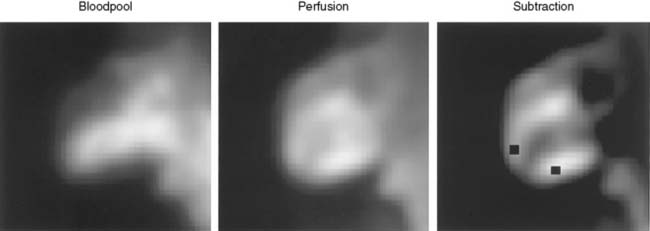
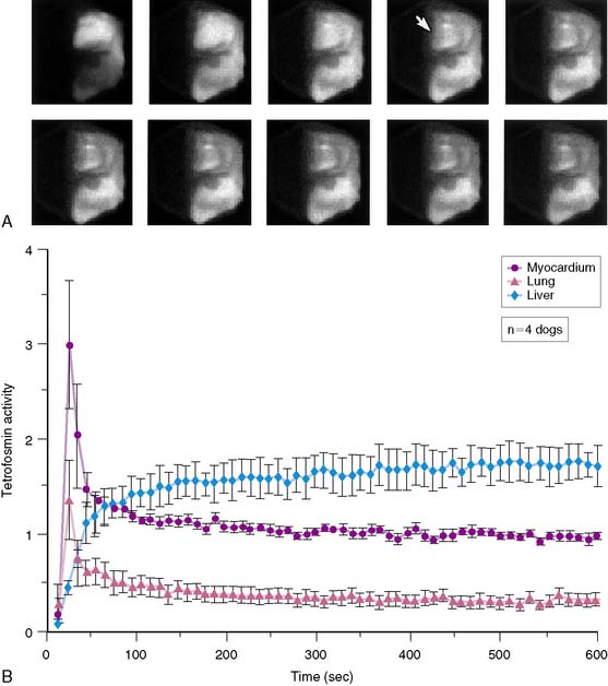
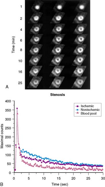
Planar imaging offers some advantages for evaluation of radiotracer pharmacodynamics, including high temporal and spatial resolution. Planar imaging of the heart is complicated by superimposition of activity in adjacent extracardiac structures. This can be obviated in open-chest models by isolation of the heart from other structures by using a flexible lead sheet. It is critical that the shielding material is placed directly under the heart, thereby completely isolating the beating heart from extracardiac structures. Time-activity curves can be derived for determination of blood and regional myocardial pharmacodynamics. In this type of kinetic analysis, placement of the regions of interest is critical. We have found that moving the myocardial region of interest one pixel toward or away from the blood pool can greatly alter the myocardial clearance curves. To facilitate optimal placement of the myocardial region of interest, we generate a reference image by subtracting an early blood-pool image from a later image (Fig. 3-1). An initial left ventricular blood-pool image can be derived by reconstruction of the left ventricular phase of the first-pass list mode acquisition. A myocardial perfusion image can be derived by summing the last 1 minute of the dynamic image sequence. The initial blood-pool image is then subtracted from the final perfusion image, providing an index myocardial image that is minimally contaminated by blood pool. The myocardial clearance curves generated by using this planar imaging approach can provide useful information on myocardial uptake and clearance of radiotracer in carefully controlled experimental models of regional ischemia or infarction. This model allows for selective arterial and venous sampling for simultaneous determination of regional metabolism or radiotracer extraction. Figure 3-2 shows a dynamic image series and corresponding clearance curves that can be derived from it.
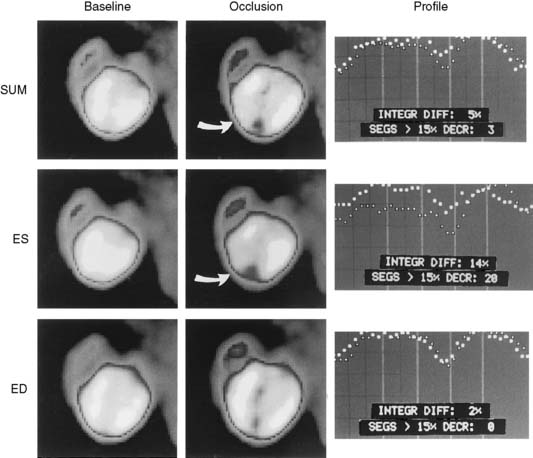
In 1979, it was recognized that distortion of global and regional left ventricular geometry could cause artifact defects in perfusion images obtained with thallium-201 (201Tl) or intracoronary administration of 99mTc-labeled albumin microspheres.53 Sinusas et al. subsequently demonstrated that changes in regional function may confound analysis of planar 99mTc-sestamibi images.44 Thus, changes in regional myocardial thickening may lead to misinterpretation of myocardial tracer uptake or clearance because of partial volume effects.44 Partial volume effects refers to the underestimation of count density from a structure that is thinner than twice the resolution of the imaging system (usually 12 to 20 mm for a gamma camera).54 In models of regional ischemia, systolic thinning may occur in ischemic or postischemic stunned regions. Differences in wall thickness would be most prominent at end systole. The potential impact of these partial volume errors are illustrated in Figure 3-3. We found that analysis of end-diastolic images may compensate somewhat for the partial volume effect associated with regional dyskinesis.
Dynamic planar imaging has been used by several investigators to establish the clearance kinetics of 201Tl,55 99mTc-sestamibi,56 99mTc-tetrofosmin,45 and 99mTc-teboroxime.57 Serial planar imaging permits nontraumatic serial assessment of changes in regional activity. However, dynamic planar imaging is limited by camera resolution, partial volume effects, and potential motion artifacts and may be influenced by background scatter and tissue cross-talk.
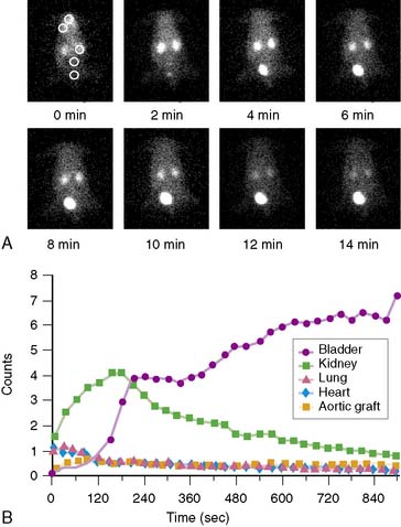
Dynamic planar images can also be obtained in animals as small as a mouse using high-resolution pinhole collimators.58 Figure 3-4 illustrates a series of planar pinhole images in a mouse injected with a 99mTc-labeled compound targeted at the αvβ3 integrin, and corresponding tissue clearance curves. Using an external radioactive reference source (~10 μCi), the regional counts within selected organs can be converted to μCi and expressed as a percentage of injected dose.
Single-Photon Emission Computed Tomography
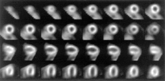
High-quality tomographic images can be acquired to evaluate the biodistribution of a radiotracer. Imaging by SPECT provides better separation of organs than planar imaging. Dynamic SPECT imaging has only recently become feasible with the development of multidetector cameras. Several investigators have used dynamic SPECT imaging for kinetic modeling.59–61 Stewart et al. performed dynamic tomographic imaging with 99mTc-teboroxime in closed-chest dogs by using a single-photon ring tomographic system.61 These investigators demonstrated monoexponential clearance of 99mTc-teboroxime with this approach. By using a triple-detector SPECT system, Smith et al. demonstrated the feasibility of acquiring dynamic 10-second tomographic data with a continuous acquisition protocol.60 Analysis of wash-in of 99mTc-teboroxime in myocardial tissue measured with this dynamic SPECT approach, in conjunction with compartmental modeling, provided an index of regional myocardial blood flow. Smith et al. also demonstrated that the use of attenuation correction in conjunction with dynamic SPECT was critical for compartmental analysis of 99mTc-teboroxime. Figure 3-5 provides an example of a series of high-quality images and corresponding clearance curves derived from dynamic SPECT in a dog. The potential advantage of kinetic modeling with SPECT over PET is the greater availability of SPECT equipment and reduced cost. However, the accuracy of the dynamic SPECT approach is not established.
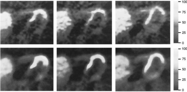
Pinhole SPECT systems have been developed to evaluate regional properties of radiopharmaceuticals in small animals.62,63 Unfortunately, the ten-fold gain in resolution of these systems is at the expense of a 100-fold loss in sensitivity.64 This limitation has been overcome by using multidetector SPECT systems.62 Optimal reconstruction of the pinhole images will probably require more computer-intensive, three-dimensional, maximum-likelihood expectation-maximization (ML EM) algorithms.64 Although these systems cannot compete with the high resolution of microautoradiography, they may provide an alternative to macroautoradiography or well counting. Hirai et al. demonstrated the feasibility of serial SPECT with pinhole collimators for estimation of flow and metabolism in vivo in rat hearts.65 Wu et al. performed myocardial perfusion imaging of mice using 99mTc-sestamibi and demonstrated the feasibility for measurement of perfusion defect size from pinhole SPECT images (Fig. 3-6).31 Other investigators recently demonstrated the feasibility of electrocardiographic (ECG)-gated pinhole SPECT imaging in mice66 and rats67 for the evaluation of regional left ventricular function by applying standard clinical image analysis software to reconstructed ECG-gated microSPECT images.
High-speed and high-sensitivity SPECT cameras have recently become available that employ cadmium zinc telluride (CZT) detectors and novel schemes for reconstruction and collimation.68,69 The novel design of these new systems significantly increases sensitivity and reduces imaging time while providing higher spatial resolution than the conventional Anger camera approach.70,71 These systems will permit dynamic SPECT imaging, facilitating evaluation of the biodistribution and uptake and the clearance kinetics of new radiotracers under evaluation. They also offer the potential for absolute quantification of radiotracers. The increased sensitivity is at the cost of the size of the field of view, but for experimental imaging, these systems may be ideally suited for dynamic imaging of rabbits or focused imaging of larger animals. These systems should expand the horizons of SPECT imaging in experimental animal models.
Positron Emission Tomography (See Chapter 11)
Dynamic PET imaging in large-animal models plays an important role in evaluation of the kinetics of positron-emitting radiopharmaceuticals. However, this chapter will not review this topic. Technetium-94m (94mTc), a positron emitter with a 53-minute half-life, can be used to evaluate the biodistribution and pharmacokinetics of 99mTc radiopharmaceuticals for SPECT.72,73 Performing PET in animal models with Tc-labeled compounds allows in vivo evaluation of regional myocardial uptake and clearance kinetics. Figure 3-7 provides an example of 94mTc-sestamibi PET images. Imaging with PET offers great advantages in the evaluation of SPECT tracers: It provides high temporal and spatial resolution and reliable attenuation correction. Labeling of SPECT tracers with 94mTc allows the direct comparison of SPECT radiotracers with PET compounds.
Recently, several microPET imaging systems have become commercially available and are capable of performing dynamic PET imaging in mice and rats.74 However, accurate kinetic modeling with microPET requires a reliable arterial input function, which is difficult to obtain in mice. Some investigators have attempted to derive an input function using high-sensitivity detectors placed over an extracorporeal circuit, while others are designing precision devices for serial blood sampling.
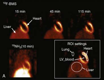
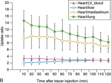
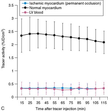
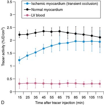
Serial microPET imaging was recently applied in rats following myocardial infarction and transient ischemia, as well as in control rats, to (1) evaluate the biodistribution and uptake and the clearance kinetics of a new 18F-labeled pyridazinone analog (18F-BMS-747158-02) under a range of physiologic conditions, and (2) define the changes in the uptake ratio of heart to adjacent organs (Fig. 3-8A-B). Serial microPET imaging was also used to define the regional uptake and clearance of normal myocardium and myocardium following permanent (see Fig. 3-8C) and transient (see Fig. 3-8D) coronary occlusion.
Miniature Detectors
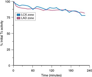
Myocardial radiotracer kinetics can also be derived in large open-chest animal preparations with the use of miniature radiation detectors. In many studies by Okada et al., miniature cadmium telluride radiation detectors were used to evaluate myocardial uptake and clearance of several radiopharmaceuticals, including 201Tl- and 99mTc-sestamibi.56,75,76 These probes allow continuous monitoring and display of regional myocardial activity.77 High-quality myocardial clearance curves derived by using a cadmium telluride detector are shown in Figure 3-9. The use of these probes can be complicated by changes in myocardial thickness and underlying blood pool beneath the probe and in the stability of the instrumentation. In some studies, probes have been placed on the myocardium in combination with a device to measure wall thickening. This system allows online correction of changes in counts associated with cyclic wall thickening. More recently, Stewart et al. measured myocardial clearance of 99mTc-teboroxime with a 1-inch collimated sodium-iodine (Tl) probe.57 The first component of 99mTc-teboroxime washout derived by using this probe was related to microsphere blood flow. This miniature probe approach allows continuous monitoring of regional myocardial activity under different ischemic conditions or during pharmacologic stress. Despite the potential limitations of these miniature probes, much insight into the in vivo behavior of 201Tl-labeled and newer 99mTc-labeled compounds has been derived from probe studies.
Serial Myocardial Biopsy
Investigators have obtained serial myocardial biopsy specimens after injection of a radiotracer to evaluate changes in absolute myocardial activity over time.78–80 This approach is the most accurate method to serially determine changes in absolute myocardial counts per gram. However, serial myocardial biopsy results in myocardial injury, necessitates the analysis of very small pieces of tissue, and has a potential sampling error.
Postmortem Imaging
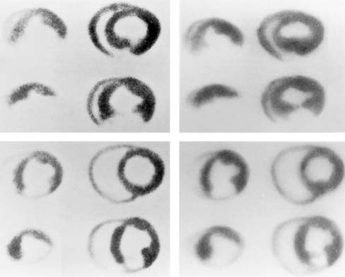
Imaging of excised organs or tissues can be very useful to establish the final distribution of a radiotracer that has been injected in vivo under a specific physiologic condition. Some investigators have acquired high-resolution, tomography-like images by placing slices of the heart on the surface of a gamma camera (Fig. 3-10).43 Images acquired by using this method are similar to those obtained using macroautoradiography. The gamma camera approach enables the simultaneous acquisition of high-resolution images by using multiple energy windows. Investigators have used this method to compare the distribution of perfusion and metabolic tracers labeled with different radioisotopes. Effective application of this method necessitates sectioning the heart in slices of uniform thickness, which requires an automated slicing device. Alternatively, a complete three-dimensional set of short-axis images of the intact heart can be acquired ex vivo with a SPECT camera (Fig. 3-11). This eliminates the need for precision slicing of the heart and avoids the problems of attenuation and motion. To simulate the in vivo geometry of the heart, hearts are generally stuffed with gauze, frozen in a distended state, or filled with inert dental molding material. All methods of postmortem imaging eliminate partial volume errors associated with cardiac deformation. Images derived using this method can be easily quantified and readily compared with postmortem histochemical stains.
