Reentry within the AV junction can result in a single junctional premature beat or in sustained RJT (see Fig. 18.1C). These tachyarrhythmias may be more difficult to understand and identify than those originating in the atria and ventricles because the AV junction is not represented by any waveform in the ECG.
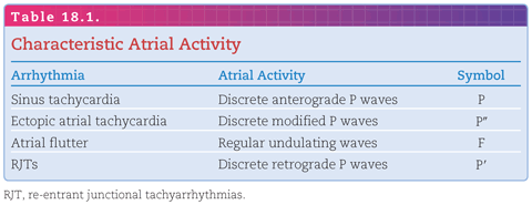
In the normal heart, the only electrically conducting structures of the junction between the atria and ventricles are the AV node and the common (His) bundle. However, congenitally anomalous accessory AV (Kent) bundles, either located centrally in the region of the AV node and His bundle or peripherally, may serve as part of the circuit of RJTs. Usually, the accessory pathway is identified by ECG evidence of ventricular preexcitation during sinus rhythm (see Chapter 7). The combination of ventricular pre-excitation and RJT is called the Wolff-Wolff–Parkinson–White syndrome (see Chapter 7; Fig. 18.1).1 The younger the individual with RJT, the more likely that an accessory AV conduction pathway is present. The definitions of the P, P′, and P″ symbols appearing in Figure 18.1 are presented in Table 18.1.

Variations in Accessory Pathway Conduction
In most individuals, the accessory pathway is capable of conduction in both the anterograde and retrograde directions. However, the accessory pathway may be capable of conduction only in one direction, causing either ventricular preexcitation or RJT:
1. If only anterograde conduction is possible along an accessory AV pathway, there is preexcitation during sinus rhythm.
2. If only retrograde conduction is possible along an AV accessory pathway, there is no preexcitation during sinus rhythm but there is the potential for RJT. In this instance, a concealed fast AV-bypass pathway is present.
Natural History of the Reentrant Junctional Tachyarrhythmias
There have been several follow-up studies of children and young adults with RJT, both with and without evidence of ventricular preexcitation.2,3 A high percentage of neonates with RJT have evidence of ventricular preexcitation, but many of these spontaneously lose their accessory pathway during the first year of life. Some lose only the capability for anterograde conduction through their accessory AV pathway but retain the capability for retrograde conduction, as evident from recurrent episodes of RJT. There is a decreasing incidence of evidence of preexcitation among progressively older individuals with RJT.
One study reported that 85% of adults with RJT did not have evidence of an accessory pathway and that those without accessory pathways were older than those with pathways (a mean of 55 versus 40).4 There was also a much higher incidence of underlying heart disease in adults without accessory pathways (50% versus 10%).
Terms That Characterize the Reentrant Junctional Tachyarrhythmias
Many different terms used to characterize an RJT fall into three categories:
1. Those that apply to the clinical behavior of the RJT: paroxysmal, persistent, permanent, incessant, sustained, nonsustained, chronic, relapsing, and repetitive;
2. Those that describe the site of origin of the RJT: supraventricular, atrial, AV nodal, AV bypass, and junctional; and
3. Those that describe the mechanism of the RJT: reentrant, reciprocating, circus movement, slow–fast, fast–slow, orthodromic, and antidromic.
VARIETIES OF REENTRANT JUNCTIONAL TACHYARRHYTHMIAS
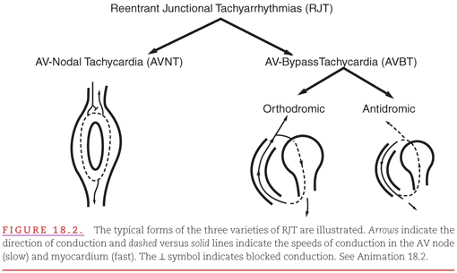
The mechanism that produces RJT may be either micro–re-entry occurring totally within the AV node (AV nodal tachycardia; Fig. 18.2A) or macro–re-entry involving one atrium, an accessory pathway, one ventricle, and the AV node (AV-bypass tachycardia; see Fig. 18.2B). The AV node is the slowest conducting structure with the longest refractory period in the heart. If an impulse entering from the atria encounters a part of the node that has not yet completed its refractory phase, the condition for AV nodal reentry occurs as shown in Figure 18.2A and may lead to either single premature atrial contractions or to AV nodal tachycardia. The presence of an accessory AV conduction pathway creates the potential for the development of re-entry circuits in which the impulses travel in either the normal or reverse direction through the AV node and ventricular Purkinje system. Although its re-entry circuit also includes atrial and ventricular myocardium, AV-bypass tachycardia is included as an RJT because its existence depends on the presence of a congenital anomaly in the junction between the atria and ventricles. The term orthodromic (AV-bypass) tachycardia is used when the impulse proceeds in the normal direction, and the term antidromic (AV-bypass) tachycardia is used when the impulse proceeds in the reverse direction. (This reverse impulse direction results from a premature beat that occurs in close proximity to the AV node and therefore finds the node still in its refractory phase.) The AV node also must be far away from the accessory pathway as seen with the left-side pathway. A concealed AV-bypass pathway is capable only of participating in an orthodromic tachycardia.


CONDUCTION THROUGH THE ATRIA AND VENTRICLES
Because the AV junction is distal to the atria, RJT produces retrograde atrial activation (P′) resulting in inversion of the P waves in the ECG (see Fig. 18.1). The P′ waves are therefore negative in the limb leads (e.g., lead II).
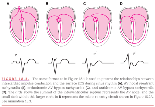
Two of the varieties of RJT—AV nodal tachycardia (Fig. 18.3B) and orthodromic AV-bypass tachycardia (see Fig. 18.3C)—produce anterograde ventricular activation that can result in normal-appearing QRS complexes, and these two varieties of RJT are therefore considered supraventricular tachyarrhythmias. Like all supraventricular tachyarrhythmias, however, an RJT can result in abnormal QRS complexes if it encounters “aberrant conduction” within the bundle branches or fascicles. Conversely, antidromic AV-bypass tachycardia can produce only abnormal QRS complexes because the impulses have “aberrant conduction” to the ventricles via the accessory pathway (see Fig. 18.3D). Note the normal P-wave appearance and timing in Figure 18.3A, the absence of a P′ wave because atrial activation occurs during the QRS complex in Figure 18.3B, the inverted P′ wave immediately following the QRS complex in Figure 18.3C, and the inverted P′ wave following the wide QRS complex in Figure 18.3D.


DIFFERENTIATION FROM OTHER TACHYARRHYTHMIAS
When the QRS complex is normal, RJT superficially resembles sinus tachycardia, ectopic atrial tachycardia, and flutter (with a 2:1 AV conduction ratio). If atrial activity is visible in the ECG, its appearance should be diagnostic because it differs markedly in RJT, sinus tachycardia, ectopic atrial tachycardia, and flutter (Table 18.1).
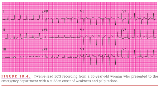
However, there may be no visible atrial activity in any of the ECG leads, as in the example shown in Figure 18.4. The ventricular rate may also be diagnostically helpful because sinus tachycardia rarely exceeds 150 beats per minute in a nonexercising adult, whereas RJT almost always exceeds this rate (about 155 beats per minute in Fig. 18.4).

Stay updated, free articles. Join our Telegram channel

Full access? Get Clinical Tree


