26
Recent Advances
 Speckle Tracking Echocardiography
The ability to measure myocardial contractile function has always been the “Holy Grail” of echocardiography. A number of parameters and indices (e.g., fractional shortening, ejection fraction, etc.) have been developed to achieve this goal, but each one of them is significantly influenced by the loading conditions and is not really a true measure of myocardial contractility. Tissue velocity imaging, developed in 1989, allowed measurement of myocardial motion directly and was rapidly embraced as a tool for assessment of myocardial systolic and diastolic function. 1 However, it too was an imperfect measure of myocardial contractility because it could only measure displacement of myocardium relative to the transducer, not the actual contraction. With further advancements in Doppler techniques, an innovative approach was developed to quantify actual myocardial deformation. This technique, known as myocardial strain imaging, involved simultaneous recording of velocities from two adjacent myocardial regions, and the difference between the two was the rate at which myocardial shortening or lengthening occurred (strain rate). Integration of these instantaneous velocity gradients over time provided the net extent of myocardial shortening or lengthening taking place during a specific phase of the cardiac cycle (strain).2,3
Although Doppler-based strain imaging represented a major technical advancement in echocardiographic assessment of myocardial function, it still had considerable challenges. 3 Being a Doppler-based technique, it was highly dependent on the insonation angle and therefore allowed measurement of myocardial contraction only in the direction parallel to the ultrasound beam. Consequently, only longitudinal strain and strain rate could be measured, with virtually no information about the radial, circumferential, rotational, and torsional components of the complex multidirectional process of myocardial contraction.
Speckle tracking echocardiography is a newer alternative technique for characterizing myocardial motion and contraction.4,5 Unlike the Doppler-based technique, speckle tracking echocardiography relies on grayscale imaging and thereby permits angle-independent measurement of myocardial contraction. Speckle tracking software identifies natural acoustic reflectors, or speckles, within the myocardium and then uses an algorithm (sum of absolute differences) to track these speckles frame by frame throughout the cardiac cycle. The data are then analyzed to derive information about myocardial motion in three-dimensional (3D) space, from which the displacement, velocity, and deformation in any direction can be computed ( Fig. 26-1). Longitudinal and transverse strain can be calculated from the apical views, whereas short-axis views are used for deriving circumferential strain, radial strain, rotation, and torsion. The technique has been shown to have a high degree of reproducibility and a much better signal-to-noise ratio than Doppler-based strain imaging techniques. 6
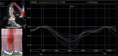
Figure 26-1 Measurement of longitudinal strain from apical two-chamber view using speckle tracking echocardiography. Lower-left segment of image shows parametric display of segmental strain values; right side of display shows graphic depiction of same.
Since its advent, speckle tracking echocardiography has rapidly evolved into a useful modality for quantifying myocardial function in a wide variety of clinical conditions.5,7,8 The greatest advantage of this technique is that it provides a quantitative and objective measure of myocardial contraction and therefore can be used for identifying subtle changes in myocardial function that cannot be detected otherwise with conventional measures such as ejection fraction. The technique can accurately estimate global left ventricular ejection fraction, quantify the extent of myocardial damage in patients with coronary artery disease, and differentiate a subendocardial infarct from a transmural infarct.9–13 When used as an adjunct to dobutamine echocardiography, it has been shown to have incremental value over wall motion analysis for detection of myocardial ischemia and viability.14,15 It has also been successfully used for detecting subclinical left ventricular systolic dysfunction in patients with valvular heart disease, cardiomyopathies, pericardial diseases, congenital heart diseases, and also in patients receiving chemotherapeutic drugs.16–25 It can help differentiate physiologic from pathologic hypertrophy and restrictive cardiomyopathy from constrictive pericarditis.26,27 Radial dyssynchrony identified on speckle tracking echocardiography appears to be a more robust measure of intraventricular dyssynchrony than the conventionally used Doppler-based parameters.28,29 The technique has also been employed for evaluating left atrial and right ventricular function, with initial studies showing great promise.30–32
Despite the rapidly unfolding array of potential clinical applications for speckle tracking echocardiography, there are several limitations associated with this technique.4,5 The accuracy of speckle tracking is highly dependent on grayscale image quality and frame rates. The best results can be obtained at frame rates between 50 and 70 frames/s; lower frame rates compromise tracking the speckles across frames, and higher frame rates adversely affect image resolution. Out-of-plane motion produced by heart movement during the cardiac cycle is another major limitation of two-dimensional (2D) speckle tracking echocardiography and is particularly relevant for short-axis views. Fortunately, 3D speckle tracking echocardiography, which is currently being developed, seems to overcome this limitation to a large extent.33–35
Finally, even though speckle tracking echocardiography has been shown to have good accuracy and reproducibility for overall left ventricular function, there is still a large degree of variability at the segmental level. 36
 Cardiovascular Flow Visualization
For decades it was believed that blood flow through the cardiovascular system was predominantly laminar and unidirectional. However, recent insights gained from in vitro and in vivo experiments, fuelled largely by rapid advancements in imaging techniques, have revealed that blood flow through the cardiac chambers is actually multidirectional, nonlaminar, and vortical. 37 As the bloodstream moves through the cardiac chambers, multiple fluid layers are created that slide against each other and against the myocardial wall and tend to curl or spin to produce spiral streamlines known as vortices
Stay updated, free articles. Join our Telegram channel

Full access? Get Clinical Tree


