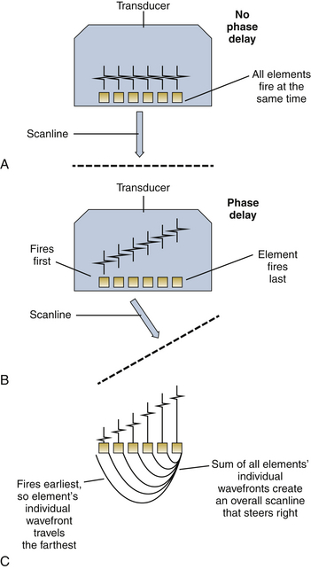
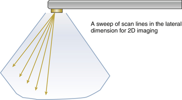
5 As discussed in Chapter 2, piezoelectric crystals convert ultrasound (pressure waves) into electrical signals and vice versa. Specifically, when presented with high-frequency electrical signals, these crystals produce ultrasound energy and, conversely, produce electrical alternating current signal from ultrasound energy. During most ultrasound applications, a 1- to 2-microsecond ultrasound pulse is emitted from the piezoelectric crystal, which is directed toward the areas to be imaged, after which the crystal “listens” for the returning echoes for a given period of time. The time delay between the pulse emission and its detection is proportional to the distance of the objects of interest, which allows image display ( Fig. 5-1). Figure 5-1 Beamforming controls delay of transmit events and creates scanlines. If all elements fire at the same time, the effective wavefront travels straight ahead of the elements. The sum of each element’s wavefront creates a scanline. If elements to one side are phase delayed, beam steers in that direction. A, Scanline steers straight ahead. B, Scanline steers to right, since element on figure’s right are delayed the longest. C, Effective summation of elements’ individual wavefronts create an effective overall wave called a scanline. Current two-dimensional (2D) systems transmit and receive acoustic beams in a 2D scanning plane. Typically, a conventional transducer consists of 64 to 128 elements spaced according to the ultimate frequency (and hence wavelength) of the acoustic vibrations; these propagate radially along the direction of the scanline. This array of elements steers the outward ultrasound beam by utilizing interference patterns generated by varying the spatiotemporal phase of each element’s transmit event ( Fig. 5-2). More simply stated, if all of the crystals are stimulated simultaneously, the summated beam is steered directly forward; if the crystals are stimulated sequentially from right to left, the summated beam is steered leftward. As opposed to M-mode (one spatial and one temporal dimension), 2D scanning systems sweep a scanline within this 2D imaging plane; the angular position of the beam is said to vary in the “azimuthal” dimension. Even though traditional 2D scanning consists of two spatial dimensions plus one temporal dimension, we do not call this “three-dimensional (3D) imaging.” These principles are common to any phased array system. 1 Figure 5-2 A two-dimensional image is created by sweeping ultrasound scanlines at an angular line spacing; the less the spacing, the higher the resolution. However, firing more scanlines per frame (for constant depth) lowers frame rate, so frame rate must be balanced against resolution. Resolution is the ability to distinguish two point targets as distinct. A scanline is not a perfectly “thin” line but actually has width or “fuzziness.” The thickness of the line varies with depth, focus, and other physical parameters. 2 Physicists call this the point spread function. Focus
Principles and Physics
Transducer Characteristics
 Transducers
![]()
Stay updated, free articles. Join our Telegram channel

Full access? Get Clinical Tree


Thoracic Key
Fastest Thoracic Insight Engine
