Fig. 15.1
Auscultation of pulmonic stenosis. Pulmonic stenosis is characterized by an ejection click, crescendo decrescendo murmur loudest at the left sternal border, and sharp, narrowly split S2
Test Results
Echocardiogram (Fig. 15.3) shows valvular doming and thickening with supravalvular dilation of the left pulmonary artery.
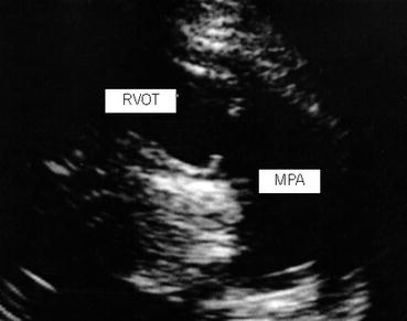
Fig. 15.3
Pulmonic stenosis echocardiogram: echocardiogram showing valvular doming, and thickened pulmonic valve
Clinical Basics
Anatomy
In a normal heart, blood flows from the right ventricle through the pulmonary valve, into the pulmonary artery and into the lungs.
Pulmonic valvular stenosis (Fig. 15.4a, b) occurs when the valve is narrowed and the valve does not open as wide as a normally functioning valve. The valvular cusps can be fused, thickened, or there may be a failure of all three leaflets to form in utero. The result is less blood flow to the lungs, increased pressure in the right side of the heart, and a mid-systolic murmur on auscultation. It is most commonly a congenital condition and can range from trivial to severe.
Fig. 15.4
(a, b) Pulmonic valvular stenosis: (a) Normal pulmonic valve leaflets vs. thickened leaflets with reduced mobility resulting in PS. (b) 4-chamber view of the heart showing relationship of the pulmonic valve, affected by PS, to other cardiac structures
Valvular stenosis is the most common form. Stenosis in the pulmonary infundibulum, below the valve, is rare.
Severity of the lesion is categorized by the peak pressure gradient across the valve:
<25 mmHg: trivial.
25–49 mmHg: mild.
50–79 mmHg: moderate.
≥80 mmHg: severe.
Pulmonic artery stenosis is a narrowing of the pulmonary artery that carries blood from the right ventricle to the lungs. It results in a decreased volume of blood reaching the lungs and increased pressure in the right side of the heart.
Pulmonic stenosis accounts for 10 % of congenital heart disease and is found with an increased prevalence (25 %) in patients with other forms of congenital heart disease.
Etiology
Congenital heart disease.
Isolated pulmonic stenosis.
Multiple Lentigines Syndrome (formerly called Leopard Syndrome) is an autosomal dominant condition most often caused by a deletion of the PTPN11 gene. Multiple Lentigines Syndrome is characterized by multiple dark skin markings called lentigines, wide-set eyes, abnormal genitalia, growth retardation, deafness, ECG abnormalities, and pulmonic stenosis. It is also associated with cardiomyopathy and coronary heart disease.
Noonan Syndrome is an autosomal dominant condition that is most commonly caused by a mutation in the PTPN11 gene or, less commonly in the SOS1 gene. It is characterized by short stature, developmental delays, cryptorchidism, inferior pectus excavatum, and congenital heart abnormalities including pulmonic valve stenosis and pulmonary artery stenosis.
Situs Solitus with cardiac dextroversion is a condition in which the visceral organs are all in the appropriate positions within the body however the heart is located on the right side as opposed to the left and is associated with pulmonic stenosis.
Tetralogy of Fallot is characterized by ventricular septal defect, a right ventricular outflow obstruction, overriding aorta, and right ventricular hypertrophy. In this condition, the most common pulmonic valve defect is a bicuspid stenotic valve.
Rheumatic inflammation of the pulmonic valve is a rare occurrence that is usually associated with other valvular abnormalities as well.< div class='tao-gold-member'>Only gold members can continue reading. Log In or Register to continue
Stay updated, free articles. Join our Telegram channel

Full access? Get Clinical Tree


