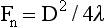
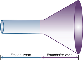
Principles and Physics: Principles of Ultrasound

2


Principles and Physics


Principles of Ultrasound


An ultrasound beam is a continuous or intermittent train of mechanical or pressure waves emitted by a transducer or wave generator. These waves can exist in any solid medium (i.e., not in a vacuum.) As the waves travel past any fixed point along an ultrasound beam, the pressure cycles regularly and continuously between a high and a low value. The number of cycles per second (Hertz [Hz]) is called the frequency of the wave. Perceptible sound waves have frequencies from 20 to 20,000 Hz. Ultrasound is sound with frequencies above 20 kilohertz (kHz), while medical ultrasound often uses frequencies between 2 and 12 megahertz (MHz). In addition to frequency, ultrasound waves are characterized by their wavelength and velocity. 1 Wavelength is the distance between the two nearest points of equal pressure or density along an ultrasound beam, and velocity is the speed at which the waves propagate through a medium. The relationship among the frequency (f), wavelength (λ), and velocity (v) of an ultrasound wave is defined by the formula:


image


The velocity of ultrasound waves varies with the properties of the medium it travels through. In low-density gases, molecules must traverse long distances before encountering the adjacent molecules, so ultrasound velocity is relatively slow. In air, the velocity of ultrasound is 330 m/sec. In contrast, molecules are constrained in solids, and ultrasound velocity is relatively high. For soft tissues, this velocity is approximates 1540 m/sec, but varies from 1475 to 1620 m/sec and approaches 3360 m/sec in bone. Because the frequency of an ultrasound beam is determined by the properties of the emitting transducer, and the velocity through soft tissue is approximately constant, wavelengths are inversely proportional to the ultrasound frequency.


Ultrasound waves transport energy through a given medium; the rate of energy transported per time is expressed as “power” (P), which is expressed in joules per second or watts. 1 Since medical ultrasound is normally concentrated in a small area, the strength of the beam is usually expressed as power per unit area or “intensity” (W/m2). In most circumstances, intensity is expressed with respect to a standard intensity. For example, the intensity of the original ultrasound signal may be compared with the reflected signal. Since ultrasound amplitudes may vary by a factor of 105 or greater, amplitudes are expressed using a logarithmic scale, the decibel, which is defined as:


image


where P1 is the power of the wave to be compared, and Pref is the power of the reference waves.


Since this is a logarithmic scale, positive values imply a wave of greater intensity than the reference wave, and negative values indicate a lower intensity. Increasing the wave’s intensity by a factor of 100 yields 20 dB. Increasing by a factor of 10 yields 10 dB. Doubling the intensity yields 3 dB.


image Ultrasound Beam


In physics, transduction is the conversion of one form of energy to another. A heating element is a transducer that converts electrical energy into heat energy. Piezoelectric crystals convert between ultrasound (pressure) and electrical signals. When presented with a high-frequency electrical signal (pulse), the crystals oscillate to produce ultrasound energy; when they are presented with an ultrasonic vibration, they produce an electrical alternating current signal. Most piezoelectric crystals used in clinical applications are manufactured ceramic ferroelectrics, the most common of which are barium titanate, lead metaniobate, and lead zirconate titanate. Some modern piezoelectric materials are more homogeneous in the solid state and hence are more efficient at generating broader bandwidth pulses (i.e., with more high- and low-frequency content). Pulses with a broader bandwidth have a broad spectrum in the frequency domain and are shorter pulses in the time domain. During most ultrasound applications, a brief ultrasound signal is emitted from the piezoelectric crystal, which is directed toward the areas to be imaged. This pulse duration is typically 1 to 2 microseconds. After this ultrasound burst emission, the crystal “listens” for the returning echoes for a given period of time and then pauses prior to repeating this cycle. This cycle length is known as the pulse repetition frequency (PRF) and must be of sufficient duration to allow a signal to travel to and return from a given object of interest. Typically, PRF varies from 1 to 10 kHz, which results in 0.1 to 1 milliseconds between pulses. When reflected ultrasound waves return to these piezoelectric crystals, they are converted into electrical signals that may be appropriately processed and displayed. Electronic circuits measure the time delay between the ultrasound emissions and their echoes. Since the speed of ultrasound through tissue is a constant, this time delay may be converted into the precise distance between the transducer and tissue. The amplitude or strength of the returning ultrasound signal provides information about the characteristics of the insonated tissues.


The three-dimensional shape of the ultrasound beam is dependent upon both the physical aspects of the ultrasound signal and the design of the transducer, especially its aperture. Further details of ultrasound transducers will be discussed in Chapter 5. An unfocused ultrasound beam may be thought of as an inverted funnel where the initial straight columnar area is known as the “near field” (also known as the Fresnel zone), followed by a conical divergent area known as the “far field” (also known as the Fraunhofer zone) ( Fig. 2-1). The length of the near field is directly proportional to the square of the transducer diameter and inversely proportional to the wavelength; specifically:


image


where Fn is the near-field length, D is the diameter of the transducer, and λ is the ultrasound wavelength.


Increasing the frequency of the ultrasound increases the length of the near field. In this near field, most energy is confined to a beam width no greater than the transducer diameter. Long Fresnel zones are preferred with medical ultrasonography, which may be achieved with large-diameter transducers and high-frequency ultrasound. The angle of the far-field convergence (θ) is directly proportional to the wavelength and inversely proportional to the diameter of the transducer, and is expressed by the equation:


image


where D is the diameter of the transducer.


Further shaping of the beam geometry may be adjusted using acoustic lenses or the shaping of the piezoelectric crystal. Ideally, imaging should be performed within the near-field or focused aspect of the ultrasound beam, since the ultrasound beam is most parallel and of greatest intensity, and the tissue interfaces are most perpendicular to these ultrasound beams.

Stay updated, free articles. Join our Telegram channel

Jun 12, 2016 | Posted by in CARDIOLOGY | Comments Off on Principles and Physics: Principles of Ultrasound

Full access? Get Clinical Tree

Get Clinical Tree app for offline access