Pericardial Diseases
Massimo Imazio, MD
 General Considerations
A. Normal Pericardial Anatomy & Physiology
The pericardium consists of two layers: a serous visceral layer, which is intimately adherent to the heart and epicardial fat, and a fibrous parietal layer. The pericardium encloses the greater part of the surface of the heart, the juxtacardial portions of the pulmonary and systemic veins, and the proximal segments of the great vessels. A significant portion of the left atrium, however, is not enclosed within the pericardium. The pericardium is not essential for sustaining life or health, as evidenced by preservation of cardiac function even if the pericardium is congenitally absent or surgically removed. The pericardium plays a role in normal cardiovascular function, however, and can be involved in a number of important disease states. The normal functions of the pericardium include maintaining an optimal cardiac shape, promoting cardiac chamber interaction, preventing the overfilling of the heart, reducing friction between the beating heart and adjacent structures, providing a physical barrier to infection, and limiting displacement during the cardiac cycle.
B. Pericardial Pressure & Normal Function
The bulk of current evidence indicates that with normal cardiac volumes, the effective pericardial pressure ranges from 0–1 mm Hg to (at most) 3–4 mm Hg. The pericardial space between the parietal and visceral layers normally contains 15–50 mL of fluid, and the reserve volume of the pericardium is relatively small. The pericardium has a limited distensibility essentially determined by the histologic composition of the parietal pericardium with a limited amount of elastic fibers and more collagen fibers. However, if pericardial fluid accumulates slowly, a remodeling of pericardial connective tissue may allow pericardial distension with accumulation of 1000–1500 mL of fluid, and occasionally up to 2000 mL. Normally, acute tamponade occurs with accumulation of < 250 mL. The pressure–volume relation of normal pericardium is a J-shaped curve. After an initial short shallow portion, which allows the pericardium to prevent cardiac chamber dilatation in response to physiologic events such as posture changes, there is a minimal increase in pericardial pressure. Thereafter, the pressure increase is extremely steep for sudden, acute changes of volume. Thus, an acute increase of 100–200 mL may greatly elevate pericar-dial pressure to 20–30 mm Hg and be responsible for cardiac tamponade. On the contrary, a slowly increasing pericardial volume is accompanied by only modest increase of pericar-dial pressure until 1000–2000 mL before the development of cardiac tamponade.
 Etiology & Pathogenesis
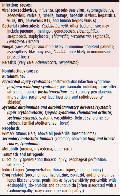
Pericardial diseases are relatively common in clinical practice and may have different presentations either as isolated disease or manifestation of a systemic disorder. Although the etiology is varied and complex (Table 28–1), the pericardium has a relatively nonspecific response to these different causes. On this basis, there are few main clinical presentations including pericarditis, pericardial effusion, cardiac tamponade, and constrictive pericarditis. Causes are essentially divided as infectious or noninfectious.
Table 28–1. Etiology of Pericardial Diseases

A. Infectious Pathogens
1. Viruses—Viruses are considered the most common causes of pericardial diseases in developed countries. An unidentified virus almost certainly underlies most cases of acute idiopathic pericarditis. The possibility of a viral cause is suggested when pericarditis occurs in the absence of other factors. Frequently, a prodromal syndrome consistent with a viral infection is present (ie, upper respiratory tract syndrome, pneumonia, or gastroenteritis). The viral agents most commonly associated with pericarditis include coxsackie viruses, Epstein-Barr virus, parvovirus, and human immunodeficiency virus (HIV). Although a wide range of viral agents have been implicated, no specific antiviral therapy has been shown to be effective in immunocompetent patients.
2. Bacterial pericarditis—Bacterial infection of the pericardium can occur following thoracic surgery; as a result of a contiguous pleural, mediastinal, or pulmonary infection; as a complication of bacterial endocarditis; or as a result of systemic bacteremia. Direct extension from pneumonia or empyema with staphylococci, pneumococci, and streptococci accounts for most cases. However, the most important bacterial agent is Mycobacterium tuberculosis, especially in developing countries. In developed countries, bacterial pericarditis is rare (< 5%). Preexisting pericardial effusions and immunosuppressed states are important predisposing factors. HIV infection is often associated with tuberculous pericarditis and effusions in underdeveloped countries (eg, countries in Africa).
3. Tuberculous pericarditis—Although several decades of effective antituberculous therapy and public health measures have brought about a declining rate of tuberculous pericarditis, this condition remains a major problem in immunocompromised persons and in developing countries, especially in association with HIV infection. Thus, HIV-associated tuberculosis is a common cause of symptomatic pericardial effusion.
4. HIV and acquired immunodeficiency syndrome (AIDS)—The most common pericardial abnormality encountered in AIDS is an asymptomatic pericardial effusion. Before highly active antiretroviral therapies, symptomatic pericardial effusion with or without chest pain, friction rub, and electrocardiogram (ECG) changes was caused by a variety of opportunistic infections and neoplasms in patients with HIV infection and AIDS. The most common infectious pathogens identified in symptomatic pericardial effusion are M tuberculosis and Mycobacterium avium-intracellulare. The HIV virus itself can cause an effusion. Lymphomas and Kaposi sarcoma are the most common neoplasms associated with effusion. Pericarditis or symptomatic pericardial effusion in a patient with AIDS should therefore prompt an immediate search for infection or neoplasm. Pericardial effusion in HIV disease usually occurs in the context of fullblown AIDS and is strongly associated with a shortened survival time independent of the CD4 count. The mortality rate at 6 months for patients with effusion was nine times greater than for patients without effusion. After the introduction of highly active antiretroviral therapy, the etiologic spectrum of the HIV-infected patient has become similar to that of noninfected patients.
B. Iatrogenic Causes
Iatrogenic causes of pericardial diseases are emerging in developed countries due to the aging of the population and widespread use of percutaneous interventions. Such forms are usually reported as postcardiac injury syndromes, reflecting the fact that the offending agent may involve not only the pericardium but also the myocardium and whole heart.
1. Surgery-related syndromes—Several distinct pericar-dial syndromes may occur after heart surgery.
Cardiac tamponade may occur during in-hospital recuperation, most commonly in the first 24 hours due to hemopericardium. It is identified by the hemodynamic perturbations typical of tamponade. The sudden cessation of previously brisk bleeding from drains should alert the physician to the possibility of clogging. Therapy consists of prompt surgical exploration and evacuation.
Cardiac tamponade is less common after the first 24 hours, with fewer typical clinical manifestations, and symptoms may consist largely of nonspecific generalized complaints. Two-dimensional echocardiography establishes the presence of a significant effusion and may delineate its anatomic distribution. Pericardial effusions in this setting are often loculated and may compress only one cardiac chamber. The approach to drainage is largely dictated by the location of the effusion.
Early pericarditis, consisting of fever, chest pain, pericardial friction rubs, and typical ECG features, is common. In most cases, the syndrome resolves spontaneously, and nonsteroidal anti-inflammatory drugs (NSAIDs) with or without colchicine are effective treatment. Corticosteroids are also very efficacious in this setting.
Postpericardiotomy syndrome is reported in 10–40% of patients, depending on the type of surgery and the adopted diagnostic criteria. This syndrome, which usually occurs during the first several postoperative weeks, consists of fever, pleuritis, and pericarditis. Diagnosis proceeds by exclusion, and treatment consists of administering NSAIDs and colchicine; sometimes corticosteroids are used, especially to avoid interference of aspirin or a NSAID with oral anticoagulant therapy.
Constrictive pericarditis occurs rarely as a complication of cardiac surgery. Its incidence is estimated to only be 0.2–0.3% of cardiac operations. However, cardiac surgery is emerging as an important cause of constrictive pericarditis because so many cardiac surgeries are performed in the United States annually. The risk of constriction has been estimated at 2–5% in patients with acute pericarditis due to a postcardiac injury syndrome. Constrictive pericarditis has been reported to occur at times ranging from 2 weeks to 21 years after the surgery. Because of the relative rarity of this complication, it has been difficult to identify specific predisposing procedural factors; however, bleeding in the pericardium is presumed to be a major trigger. Occasionally, constrictive pericarditis appears within days or weeks after surgery. These cases appear to respond well to a course of corticosteroids. With this exception, the mainstay of therapy for postsurgical constrictive pericarditis is pericardiectomy.
2. Trauma—Traumatic hemorrhagic pericardial effusions, which can result from blunt or penetrating injuries of the chest, can also be caused by a variety of iatrogenic causes such as cardiac catheterization and coronary interventional procedures, pacemaker insertion, arrhythmia ablation procedures, endoscopy, and closed chest cardiac massage. The rapidity with which pericardial fluid can accumulate can quickly cause hemodynamic compromise. Hypotension in this setting should prompt both an immediate echocardio-graphic search for pericardial fluid and swift evacuation of any significant effusions. Delayed manifestations may include recurrent pericardial effusions and, in rare cases, constrictive pericarditis.
3. Radiation therapy—The incidence of pericardial injury from therapeutic radiation is related to dose, duration, and technical features. Pericardial damage from radiation may appear during the course of therapy or following it. The syndrome that appears during radiation therapy is acute pericarditis. The onset of clinical manifestations in the delayed form is usually within 12 months but may take many years. The clinical features of the late form range from asymptomatic pericardial effusions to acute pericarditis or constrictive pericarditis. Radiation therapy is now one of the leading causes of constrictive pericarditis.
C. Connective Tissue Disorders
Connective tissue vascular diseases are a common cause of pericardial diseases. A number of rheumatic diseases can involve the pericardium. This is most likely to occur in patients with known systemic lupus erythematosus, Sjögren syndrome, or rheumatoid arthritis, but can also occur in progressive systemic sclerosis, mixed connective tissue disease, polyarteritis, giant cell arteritis, polymyalgia rheumatica, other systemic vasculitides, acute rheumatic fever, and familial Mediterranean fever. Rarely, occasional patients with gout may suffer attacks of pericarditis during an episode of arthritis.
Symptomatic pericarditis can occur with all of these disorders, during the active phase of the systemic disease. In other cases, pericardial involvement may be subclinical with a clinically silent pericardial effusion. In unselected series of patients, acute pericarditis occurred in 2–7% of cases. However, in many cases, the diagnosis of a connective tissue disease was already known at the time of the diagnosis of pericarditis.
D. Other Causes
1. Myocardial infarction—Clinical evidence of pericarditis can be found in 7–20% of patients within the first week after myocardial infarction, although autopsy series suggest a significantly higher incidence of clinically silent, localized fibrinous pericarditis. The incidence is greatest with large, ST segment elevation myocardial infarctions. Anticoagulant therapy and antiplatelet therapy administered in conjunction with percutaneous revascularization procedures do not appear to increase the incidence of pericardial effusions after myocardial infarction. While it is theoretically possible that these drugs may increase the chance of hemorrhage into the pericardial space if an effusion does occur, they are not generally considered to be contraindicated in the setting of early postmyocardial infarction pericarditis. Thrombolytic therapy has been associated with a decreased incidence of pericarditis in placebo-controlled studies. In contemporary series of patients with ST segment elevation acute myocar-dial infarctions (AMIs) treated with primary percutaneous coronary intervention, pericarditis has become less common. Early post-AMI pericarditis has been reported in 4–5% with an increasing prevalence according to presentation delay: < 2% for < 3 hours, 5–6% for 3–6 hours, and 10–15% for > 6 hours. Identified risk factors for early post-AMI pericarditis include presentation times > 6 hours and primary percutaneous coronary intervention failure. Although pericarditis is associated with a larger infarct size, in-hospital and 1-year mortality and major adverse cardiac events are similar in patients with and without pericarditis.
Dressler syndrome (postmyocardial infarction syndrome) occurs from 1 to 6 weeks after myocardial infarction and consists of fever, pleuropericardial pain, malaise, and evidence of pleural and pericardial effusions. The syndrome has been considered a contraindication to anticoagulant therapy since a real increase of the likelihood of pericardial hemorrhage and tamponade has not been demonstrated. However, there is little data to support this precaution. The incidence of this syndrome has been decreasing in recent years. Dressler syndrome has become rare in the primary percutaneous coronary intervention era and is reported in < 0.5% of cases. It is believed to have an autoimmune cause due to sensitization to myocardial cells at the time of necrosis. Antimyocardial antibodies have been demonstrated in patients with Dressler syndrome.
2. Malignancy—A variety of hematologic and solid malignancies (especially lung and breast cancer, lymphomas) can cause pericardial metastases that are more frequently revealed during autopsy rather than during life. Malignant tumors can spread to the pericardium through the lymphatics (mainly lung and breast carcinomas, and other carcinomas with lung metastases), through the coronary arteries (leukemia, lymphomas, and melanomas), or by direct local invasion (lung or mediastinal neoplasms). Neoplastic involvement of the pericardium can cause pericarditis, pericardial effusion with or without cardiac tamponade, and constrictive pericarditis. Primary tumors of the pericardium are rare; most are mesotheliomas and may be relatively frequent in some areas where asbestos is extracted or used in industry. Neoplastic pericarditis is responsible for 4–7% (approximately 5%) of unselected cases of pericarditis.
Typically, the diagnosis of malignancy has already been established in the patient with a malignant pericardial effusion, and other sites of metastatic spread are evident. On rare occasions, tamponade from the malignant effusion is the first manifestation of tumor. It is important to distinguish malignant effusions from other causes of effusion, such as radiation, infection, and uremia, because the management and prognosis of malignant and nonmalignant effusions in cancer patients differ substantially.
3. Renal failure—Pericardial involvement in patients with renal failure can take several forms. Pericardial effusions can be found on an echocardiogram in many patients with chronic kidney disease who are asymptomatic. These effusions, which are typically small, are related more closely to the patient’s volume status than other variables and usually warrant no intervention beyond clinical vigilance.
The incidence of uremic pericarditis has been decreasing for years, a trend attributable to earlier and more intensive dialysis. Uremic pericarditis typically occurs before the initiation of long-term dialysis; its development is related, in part, to the elevation of absolute levels of blood urea nitrogen (BUN) and serum creatinine, and it almost always responds to dialysis. Although uremic pericarditis can lead to tamponade, it is more commonly associated with the slow accumulation of large, low-pressure pericardial effusions.
4. Drug-related causes—A number of pharmacologic agents have been implicated in pericardial disease. Pericarditis can occur as a feature of drug-induced systemic lupus erythematosus syndrome caused by procainamide, hydralazine, diphenylhydantoin, reserpine, methyldopa, and isoniazid. In addition to their propensity for causing myocardial inflammation, anthracycline antineoplastic agents can cause acute pericarditis. Methysergide can cause pericardial constriction as part of the syndrome of mediastinal fibrosis, and pericarditis can be part of a hypersensitivity reaction to penicillin. Minoxidil has been reported to cause pericarditis and tamponade; the mechanism is unknown.
5. Hypothyroidism—Pericardial effusion can be found in one-third of patients with myxedema. The frequency of pericardial involvement is related to both the severity and duration of hypothyroidism. The accumulation of pericardial fluid in this condition appears to be a result of a combination of increased capillary permeability and retarded lymphatic drainage. Because the pericardial fluid accumulates slowly, tamponade is rare. If pericardiocentesis is required before the diagnosis of hypothyroidism is made, the diagnosis can be suspected if the fluid is yellow and contains a high level of cholesterol. Pericardial disease in hypothyroidism reliably responds to thyroid hormone replacement therapy.
Imazio M. The post-pericardiotomy syndrome. Curr Opin Pulm Med. 2012;18(4):366–74. [PMID: 22487945]
Imazio M. Pericardial involvement in systemic inflammatory diseases. Heart. 2011;97(22):1882–92. [PMID: 22016400]
Imazio M, et al. Aetiological diagnosis in acute and recurrent pericarditis: when and how. J Cardiovasc Med (Hagerstown). 2009;10(3):217–30. [PMID: 19262208]
Imazio M, et al. Medical therapy of pericardial diseases: part II: noninfectious pericarditis, pericardial effusion and constrictive pericarditis. J Cardiovasc Med (Hagerstown). 2010;11(11): 785–94. [PMID:20925146]
Imazio M, et al. Post-cardiac injury syndromes. An emerging cause of pericardial diseases. Int J Cardiol. 2012;S0167-5273(12):01158–8. [PMID: 23040075]
Imazio M, et al. Risk of constrictive pericarditis after acute pericarditis. Circulation. 2011;124(11):1270–5. [PMID: 21844077]
ACUTE PERICARDITIS
 ESSENTIALS OF DIAGNOSIS
![]() Central chest pain aggravated by coughing, inspiration, or recumbency.
Central chest pain aggravated by coughing, inspiration, or recumbency.
![]() Pericardial friction rub on auscultation.
Pericardial friction rub on auscultation.
![]() Characteristic ECG changes (widespread ST segment elevation or PR depression).
Characteristic ECG changes (widespread ST segment elevation or PR depression).
![]() Pericardial effusion (new or worsening).
Pericardial effusion (new or worsening).
 General Considerations
Acute pericarditis is an inflammatory condition of the pericardium that can be caused by virtually any of the conditions just discussed. As discussed earlier, a viral infection is the most common cause in developed countries (North America and Western Europe), while tuberculosis is the most common cause of pericarditis and pericardial diseases in developing countries and all over the world. This epidemiologic background should be kept in mind when managing a single patient, since immigration may lead to an increase of less common infectious causes, especially tuberculous pericarditis.
 Clinical Findings
A. Symptoms & Signs
The primary symptom of acute pericarditis is chest pain whose location, intensity, and nature are variable. The pain may be described as sharp or dull. Most often it is precordial or retrosternal in location and may be referred to the trapezius ridge, which is almost pathognomonic for pericarditis. It is characteristically aggravated by inspiration, coughing, or recumbency and lessened by sitting upright and leaning forward. Although it typically takes an hour or two to develop fully, the pain can sometimes appear remarkably abruptly. Many patients relate prodromal symptoms suggestive of a viral infection (respiratory infections or gastroenteritis). Bacterial pericarditis may present with fever, chills, night sweats, and dyspnea.
B. Physical Examination
Patients with pericarditis may be febrile (but high fever > 38°C is usually associated with a bacterial or immune-mediated form) and show tachycardia. The pericardial friction rub—the characteristic auscultatory finding—is typically scratchy and may have three components corresponding to atrial contraction, ventricular systole, and early diastole. It is not unusual for only one or two components to be audible; the systolic component is most consistently present. It has been reported in about one-third of patients. Exercise may facilitate the identification of all three components. Because the friction rub may be evanescent, varying widely in intensity even in the course of a single day, repeated auscultation is important. Furthermore, because posture can affect the pericardial rub, auscultation with the patient in several positions (eg, supine, sitting) is often helpful. When the intensity of the rub is modulated significantly by respiration, it is termed a “pleuropericardial friction rub.”
C. Diagnostic Studies
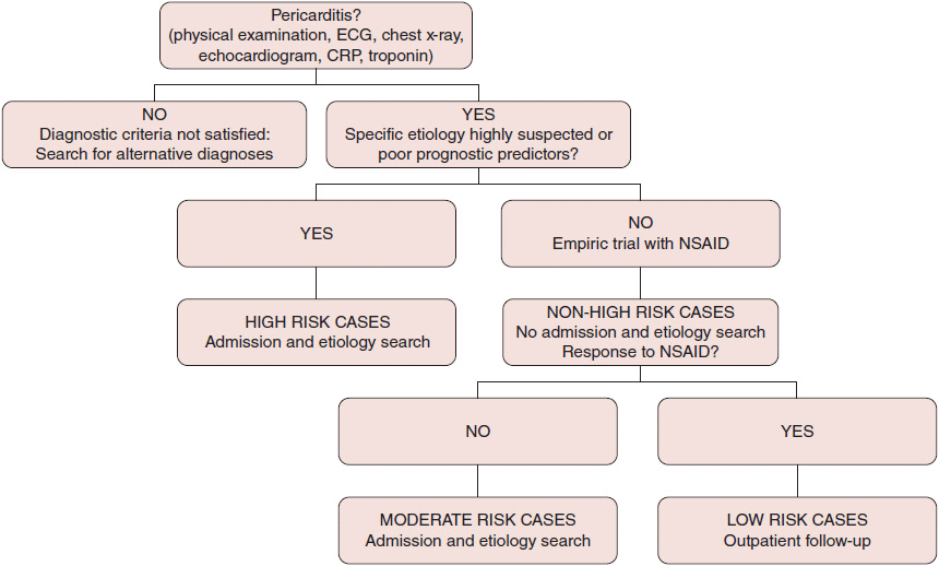
Evaluation of a patient with suspected pericarditis should routinely include an ECG, complete blood count, markers of inflammation (ie, erythrocyte sedimentation rate and C-reactive protein), markers of myocardial lesion (ie, troponins), echocardiography, and a chest radiograph. Additional diagnostic laboratory tests should be tailored to the clinical presentation. High-risk features at presentation include high fever (> 38°C), subacute course, large pericardial effusion, cardiac tamponade, and lack or incomplete response to empiric anti-inflammatory therapy after 7–10 days (Figure 28–1). Echocardiography is a sensitive test for detecting pericardial effusion; however, pericardial effusion can occur in the absence of pericardial inflammation, and pericarditis may occur without a pericardial effusion. About 60% of patients with acute pericarditis show pericardial effusion at presentation, generally mild (< 1 cm). The clinical diagnosis of acute pericarditis is made with the presence of at least two of four clinical diagnostic criteria: (1) pericarditic chest pain, (2) pericardial rubs, (3) typical ECG findings (widespread ST segment elevation or PR depression) (Figure 28–2), and (4) pericardial effusion.

![]() Figure 28–1. Triage of acute pericarditis. Red flags include high fever (> 38°C), subacute course, large pericardial effusion, cardiac tamponade, and lack or incomplete response to empiric anti-inflammatory therapy after 7–10 days. If at least one is present, patient should be admitted to hospital and complete etiology search performed. Otherwise, low-risk pericarditis may be managed as outpatient. CRP, C-reactive protein; ECG, electrocardiogram; NSAID, nonsteroidal anti-inflammatory drug. (Reprinted from Imazio M, et al. Circulation. 2010;121(7):916–28.)
Figure 28–1. Triage of acute pericarditis. Red flags include high fever (> 38°C), subacute course, large pericardial effusion, cardiac tamponade, and lack or incomplete response to empiric anti-inflammatory therapy after 7–10 days. If at least one is present, patient should be admitted to hospital and complete etiology search performed. Otherwise, low-risk pericarditis may be managed as outpatient. CRP, C-reactive protein; ECG, electrocardiogram; NSAID, nonsteroidal anti-inflammatory drug. (Reprinted from Imazio M, et al. Circulation. 2010;121(7):916–28.)
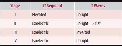
1. Electrocardiography—Serial ECGs are valuable in diagnosing pericarditis. Four stages of ECG changes have been described (Table 28–2). In stage I, the changes accompany the onset of chest pain and consist of widespread ST segment elevation (Figure 28–2). The ST segment is concave upward (in distinction to the elevation in myocardial infarction). ST segment elevation is typically present in all leads except aVR and V1, where ST segment depression is often present. The T waves are upright in the leads with ST elevation. The stage I pattern of pericarditis may be difficult to distinguish from the normal variant of early repolarization. A differentiating point that may be useful is the ST:T ratio in V6. A T-wave apex four times (or greater) higher than the height of the ST segment is more likely to indicate early repolarization; if this ratio is less than 4, pericarditis is more likely. In addition, pericarditis causes changes in the ECG that distinguish it from early repolarization. In stage II, typically occurring several days later, the ST segments return to baseline, and the initially upright T waves flatten. In stage III, the T waves invert, and the ST segments may become depressed—changes that may persist indefinitely. Finally, in stage IV, which may occur weeks or months later, the T waves revert to normal. All four stages can be serially identified in about 50% of patients.
Table 28–2. Serial Electrocardiographic Changes in Pericarditis

Stay updated, free articles. Join our Telegram channel

Full access? Get Clinical Tree


