Figure 6.1
Shows types of prosthetic valves. (a) Bovine pericardial stented valves; (b) Mechanical Bileaflet (St Jude Medical Regent, Carbomedics Standard, Medtronic Advantage); (c) Single tilting disk (Bjork Shirley, Medtronic-Hall); (d) Ball and Cage (Starr-Edwards)
The first valve implantation was performed by Charles Hufnagel in 1952.
Hufnagel CA, Harvey WP, Rabil PJ, McDermott TM. Surgical correction of aortic insufficiency. Surgery 1954,35:673–83
Prosthetic Valves
Aortic Prosthesis Stenosis
Parameter | Normal | Possible stenosis | Significant stenosis |
|---|---|---|---|
Peak V(m/s) | <3 | 3–4 | >4 |
Mean gradient (mmHg) | <20 | 20–35 | >35 |
DVI (VLVOT/VPrV vs VTIs) | ≥0.3 | 0.25–0.29 | <0.25 |
ERO (cm2) | >1.2 | 0.8–1.2 | <0.8 |
Contour of jet velocity | Triangular, early peaking | Triangular to semi rounded | Rounded, symmetrical |
Acceleration time (ms) [2] | <80 | 80–100 | >100 |
Videos 6.1 and 6.2 show images of a 12 year old aortic bioprosthesis. The prosthetic aortic leaflets are significantly thickened and calcified and color Doppler flow shows turbulent flow across prosthesis consistent elevated gradients (Fig. 6.2). Figure 6.3 shows spectral Doppler profile of elevated peak gradient of 80 mmHg with an acceleration time of 160 ms across a bioprosthetic aortic valve consistent with significant prosthetic valve stenosis.
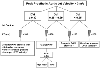
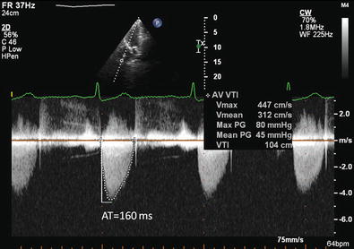
Figure 6.2
Algorithm of assessment of peak prosthetic aortic jet velocity >3 m/s (Reproduced with permission from Zoghbi et al. [1]. *, ** – PW sample located too close and too far from the valve, respectively. ɸ compare derived EOA to the reference values for the valve for further analysis. http://dx.doi.org/10.1016/j.echo.2009.07.013. © 2009 American Society of Echocardiography. Published by Elsevier Inc)
Figure 6.3
shows elevated peak gradient of 80 mmHg with an acceleration time of 160 ms across a bioprosthetic aortic valve consistent with significant prosthetic valve stenosis
Aortic Prosthesis Regurgitation
Parameter | Mild | Moderate | Severe |
|---|---|---|---|
Valve structure | Nl | Abnormal | Abnormal |
LV size | Nl | Enlarged | |
Jet width/LVOT diameter | ≤25 % | 26–64 % | ≥65 |
Jet density | Faint | Dense | |
Jett deceleration PHT (ms) | >500 | 200–500 | <200 |
LVOT flow vs. pulmonic flow (PW) | Slightly increased | Greatly increased | |
Diastolic reversal in descending aorta | Absent or early diastolic | Prominent holodiastolic | |
Regurgitant volume (ml/bt) | <30 | 30–59 | >60 |
Regurgitant fraction | <30 | 30–50 | >50 |
Example of a paravalvular aortic prosthetic valve regurgitation as seen on 3D transesophageal imaging is demonstrated in Video 6.3.
Patient prosthesis mismatch: EOAindex = Stroke Volume/(VTIPrV × BSA)
< div class='tao-gold-member'>
Only gold members can continue reading. Log In or Register to continue
Stay updated, free articles. Join our Telegram channel

Full access? Get Clinical Tree


