Normal
Mildly enlarged
Moderately enlarged
Severely enlarged
LVEDD (cm)
4.2–5.8
5.9–6.3
6.4–6.8
>6.8
LVEDD/BSA(cm/m2)
2.2–3.0
3.1–3.3
3.4–3.6
>3.6
LVESD (cm)
2.5–4.0
4.1–4.3
4.4–4.5
>4.5
LVESD/BSA (cm/m2)
1.3–2.1
2.2–2.3
2.4–2.5
>2.6
Septal/posterior wall thickness(cm)
0.6–1.0
1.1–1.3
1.4–1.6
≥1.6
Women
Normal | Mildly enlarged | Moderately enlarged | Severely enlarged | |
|---|---|---|---|---|
LVEDD (cm) | 3.8–5.2 | 5.3–5.6 | 5.7–6.1 | >6.1 |
LVEDD/BSA(cm/m2) | 2.3–3.1 | 3.2–3.4 | 3.5–3.7 | >3.7 |
LVESD (cm) | 2.2–3.5 | 3.6–3.8 | 3.9–4.1 | >4.1 |
LVESD/BSA (cm/m2) | 1.3–2.1 | 2.2–2.3 | 2.4–2.6 | >2.6 |
Septal/posterior wall thickness(cm) | 0.6–0.9 | 1.0–1.2 | 1.3–1.5 | ≥1.5 |
LV Function
linear dimension method (Fig. 3.1)
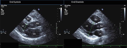
Figure 3.1
left panel: parasternal long axis view end-systole; right panel: parasternal long axis view end-diastole. LV internal dimensions (green line) are measured at mitral leaflet tips. The posterior wall (PW) and septal wall (SW) thickness are measured at end diastole. LA left atrium, LV left ventricle, RV right ventricle, Ao aorta
biplane method of disks (Fig. 3.2)
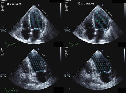
Figure 3.2
Biplane method of disks method of measuring LVEF. The left column shows end systolic apical four and two chamber views, respectively. The right column shows end diastolic apical and four chamber views
Left ventricular systolic function parameters [3]
Normal | Mildly decreased | Moderately enlarged | Severely decreased | |
|---|---|---|---|---|
Ejection fraction (%) | ||||
Men | 52–74 | 41–51 | 30–40 | <30 |
Women | 54–74 | 41–53 | 30–40 | <30 |
Left Atrium
measure in end systole
exclude pulmonary veins and atrial appendage
measure anterior-posterior dimension from parasternal long axis view (Fig. 3.3)
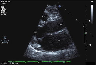
Figure 3.3
Parasternal long axis view – measurement of anterior-posterior dimension of left atrium performed at end-systole. LA left atrium, LV left ventricle, RV right ventricle, Ao aorta
LA volume can be measured by area-length or biplane method of discs is recommended method
Area-Length Method (Fig. 3.4):
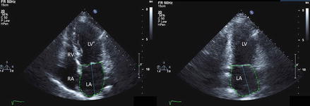
 where A1, A2 – areas of LA from 4 and 2 chamber views, L – length (choose a shorter vertical diameter (mid mitral annulus to superior axis of LA) between 4 and 2 chamber views).
< div class='tao-gold-member'>
Figure 3.4
Apical four (left) and two chamber (right) views are used for left atrial volume calculation. LA left atrium, LV left ventricle, RV right ventricle, RA right atrium
Only gold members can continue reading. Log In or Register to continue
Stay updated, free articles. Join our Telegram channel

Full access? Get Clinical Tree


