Fig. 48.2 This monitoring strip shows four sinus beats initially, and then, after a pause prior to the succeeding sinus beat, a run of a very broad complex tachycardia, showing substantial axis shift from sinus rhythm, and clear-cut independent P wave activity (arrowed). Indisputably, this is ventricular tachycardia.
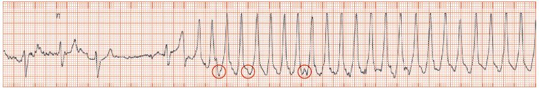
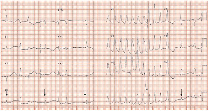
Fig. 48.3 Long QT interval dependent non-sustained ventricular tachycardia (NSVT). The arrowed beats (rhythm strip) are sinus. They have a very long QT interval, difficult to fully measure, as, before they fully finish, a ventricular ectopic arises or a run of 12 beats of NSVT. The sinus rhythm ECG looks diffusely abnormal (non-specific QRS broadening, T wave flattening) but it is difficult to be categorical, as not enough of the ECG is visualized. The patient had syncope, and was on psychotropic drugs; the diagnosis was long QT dependent ventricular tachycardia, induced by drugs.
Non-sustained ventricular tachycardia (NSVT) is ventricular tachycardia (VT) (i.e. ≥ 3 ventricular beats strung together with no intervening supraventricular beat) that terminates spontaneously within 30 s (Table 48.1). It is a common and important rhythm disturbance; it may give rise to no symptoms, near or actual fainting and/or palpitations. It is associated with sustained VT and sudden cardiac death. The ECG appearance of NSVT is the same as sustained VT, except that it does not last as long!
Stay updated, free articles. Join our Telegram channel

Full access? Get Clinical Tree


