Natural History and Open Treatment of Isolated Iliac Artery Aneurysms


Natural History and Open Treatment of Isolated Iliac Artery Aneurysms


Edward Kang, Anatolie Usatii and Steven Santilli


The Ad Hoc Committee on Reporting Standards of the Society for Vascular Surgery and the International Committee on Reporting Standards of Cardiovascular Surgery, North American Chapter, define iliac artery aneurysms (IAAs) as any permanent localized dilation of the iliac artery larger than 1.5 cm in diameter. Normal ranges are reported to be from 0.95 to 1.10 cm in women and 1.04 to 1.30 in men, gradually increasing with age. Because of the relative paucity of isolated IAAs, our current understanding and consequent recommendations are derived from several retrospective studies and case series that have surveyed the natural history and clinical outcomes of IAAs.


Epidemiology


IAAs, first described by Valentine Mott in 1827, are rare and are commonly associated with abdominal aortic aneurysms (AAAs). IAAs constitute 0.4% to 1.9% of all aneurysmal disease, and isolated IAAs account for less than 1% of all aortoiliac aneurysms. In a large population-based study spanning 15 years in which 84% of all deceased inhabitants underwent autopsy, isolated IAAs were identified in only 9 (0.03%) of 35,265 cases.


Of isolated IAAs, 70% to 89% involve the common iliac artery, 10% to 25% involve the internal iliac artery, and 1% involve the external iliac artery. Approximately two thirds of IAAs occur in multiple segments of the iliac system. IAAs are more commonly found in the right side: 44% involve the right iliac artery, 24% involve the left iliac artery, and 32% involve bilateral iliac arteries. Older studies suggested a more significant laterality, with 62% involving the right iliac artery.


IAAs have a 7:1 male-to-female preponderance. Their incidence increases with age and is greatest in the 7th to 8th decades of life, although some case series have reported IAAs in patients in their 3rd and 4th decades of life.


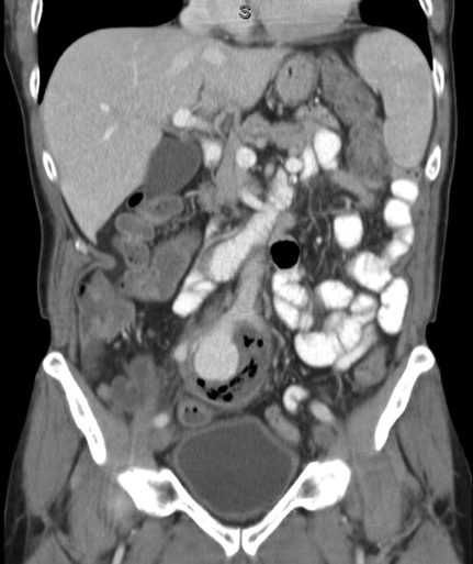
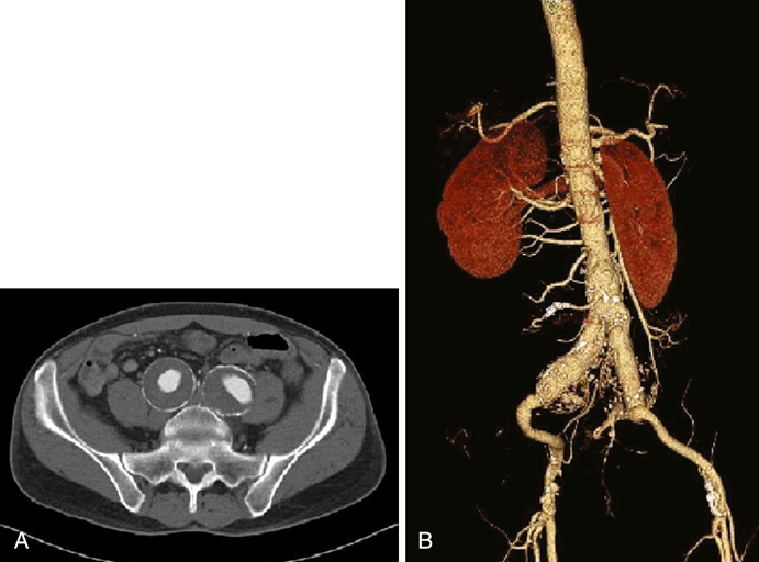
The most common etiology for IAAs is atherosclerotic disease. Other etiologies that have been reported include mycotic (Figure 1) or false aneurysms secondary to syphilis, tuberculosis, and osteomyelitis. Other conditions associated with IAAs include trauma, fibromuscular dysplasia, Behçet’s disease, collagen defects (such as Marfan’s and Ehlers–Danlos syndromes), cystic medial necrosis, and Kawasaki’s syndrome. Reports of IAAs have also been reported after pregnancy, particularly following traumatic childbirth or forceps delivery.


The majority of IAAs are discovered incidentally during abdominal or pelvic imaging for unrelated reasons, especially with the increasing use of cross-sectional imaging.


Clinical Findings


Earlier studies reported symptoms present in up to 50% of IAAs. However, more recent retrospective studies report that only 3% to 16% of patients are symptomatic at the time of diagnosis, of whom more than half experience rupture. Patients with IAA rupture often complain of abrupt onset of abdominal, groin, or flank pain. Microscopic hematuria is sometimes present.


When symptoms are present in nonruptured IAAs, they often consist of vague local pressure symptoms or discomfort attributable to compression of nearby structures. Patients might complain of lower abdominal, groin, or flank discomfort. Symptoms can also follow kidney failure from ureteral obstruction or an ensuing pyelonephritis. Others might complain of painful defecation attributable to rectal compression by the IAA. Some have radicular pain or mild leg paresis from nerve compression. Rarely, patients come to the hospital with thrombosis, emboli, or fistulas.


Physical examination may be unreliable, as the aneurysmal expansion extends into the pelvis. Depending on the size of the aneurysm, a pulsatile mass may be detectable on abdominal or rectal examination. In a retrospective review, all aneurysms greater than 4 cm were detectable by physical examination, whereas only 45% of patients with IAAs between 3 and 3.9 cm had palpable masses. Only 4.8% of IAAs less than 3 cm were palpable. In one case series, a pulsatile abdominal mass was detected in 78% of cases of IAA rupture.


Diagnosis


For those in whom IAAs are suspected, several imaging modalities can aid in diagnosis, long-term monitoring, and preoperative planning. Although no prospective trials have been conducted comparing various imaging modalities in evaluation of IAAs, a large natural history study comparing computed tomography (CT) and B-mode ultrasound scanning found that ultrasound is as accurate as CT scanning in measuring the size of IAAs and provides an accurate means of monitoring expansion. Although ultrasound has several limitations, its low cost, lack of radiation exposure, and widespread availability make it a reasonable means of initial diagnosis, baseline measurement, and monitoring.


CT and magnetic resonance imaging (MRI) provide a more accurate and detailed anatomy of IAAs, have less interobserver variability, and provide anatomy of surrounding structures. Multislice CT scanning (Figure 2) has rapid and accurate image acquisition, although it may be affected by foreign objects (such as metal hip prostheses) and it requires intravenous contrast agents. MRI is limited by its longer acquisition time, a patient’s ability to tolerate an MRI (e.g., claustrophobia, pacemakers, implanted metallic objects), and potential adverse events such as systemic nephrogenic sclerosis from the use of contrast agents. Nevertheless, CT and MRI are particularly useful in preoperative planning to determine if a patient may be a candidate for an endovascular intervention. Formal catheter-based angiography has little role in diagnosing IAAs, especially those with mural thrombus, although it may be useful in preoperative planning.


Stay updated, free articles. Join our Telegram channel

Aug 25, 2016 | Posted by in CARDIOLOGY | Comments Off on Natural History and Open Treatment of Isolated Iliac Artery Aneurysms

Full access? Get Clinical Tree

Get Clinical Tree app for offline access