Percutaneous Arterial Dilation for Fibrodysplastic Renovascular Hypertension


Percutaneous Arterial Dilation for Fibrodysplastic Renovascular Hypertension


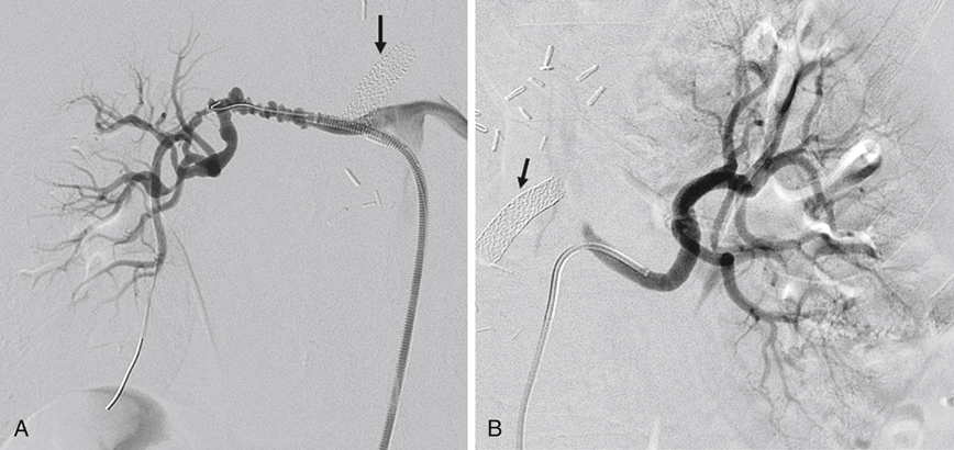
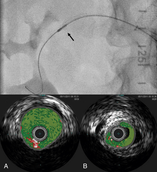
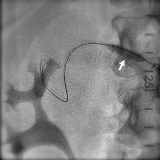
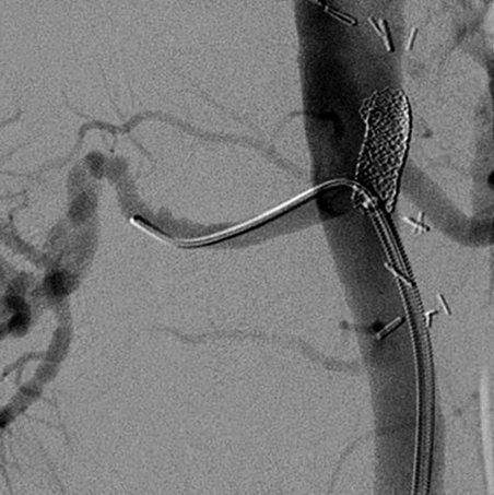
Javier E. Anaya-Ayala, Daynene Vykoukal, Alan B. Lumsden and Mark G. Davies


Fibromuscular dysplasia (FMD) is a nonatherosclerotic, noninflammatory, segmental vascular disease of unknown etiology that can involve all three layers of the arterial wall. Renal FMD generally affects the distal main renal artery and its branches. This entity accounts for less than 10% of renal artery stenosis and rarely leads to vessel occlusion or ischemic nephropathy; however, it can cause significant arterial hypertension.


The natural history of renal FMD is progression in up to 37% of cases (defined by the occurrence of a new focal lesion, worsening arterial stenosis, or the enlargement of a mural aneurysm), but this progression only rarely results in complete occlusion of the renal artery. It is often difficult to assess disease progression according to angiographic criteria, especially in patients with medial fibroplasias, in whom it is difficult to gauge the degree of stenosis. Monitoring for changes in the renal parenchyma can provide another means of assessing disease progression. Patients with FMD demonstrate a significant decrease in mean cortical thickness and reduced kidney length compared with patients with essential hypertension, suggesting that it is associated with parenchymal effects. Whereas up to 63% of patients with FMD experience a loss of kidney mass, interestingly, the incidence of kidney failure remains remarkably low.


At times, it may be difficult to distinguish FMD from vasculitis. FMD is by definition a noninflammatory process and is therefore not associated with anemia, thrombocytopenia, or abnormalities of acute-phase reactants, except when it is associated with an acute kidney infarction. Because large-vessel vasculitis can occur in the absence of changes in acute-phase reactants, in up to 40% of cases it may be impossible to distinguish these entities owing to the similarity of their angiographic appearance.


Treatment


Percutaneous transluminal renal angioplasty (PTRA) has become the treatment of choice for patients with renal artery FMD that is limited to the main renal artery (Figures 1 to 4). The majority of these patients have medial fibroplasia. Before the advent of percutaneous transluminal angioplasty, open surgical revascularization was the primary therapeutic modality for these patients. Overall, the technical success rates of the open surgical approach ranged from 89% to 97%; arterial hypertension was cured in approximately 33% to 63% of patients and improved in 24% to 57%. Several factors such as prolonged duration of hypertension, concomitant atherosclerotic disease, and complex branch-vessel repair can all adversely affect the outcomes of surgical revascularization. Although stents have been used extensively to treat atherosclerotic renal artery stenosis, the use of these devices for FMD has been reserved as a bailout procedure in cases in which there are suboptimal results with balloon angioplasty or in which renal artery dissection occurs.


Complications are rare after PTRA but include renal artery perforation, dissection, or segmental kidney infarction. Special attention must be taken to not overdilate the lesion, which is a risk factor for rupture of the artery. Cutting balloon angioplasty (CBA) for managing renal artery FMD lesions that are resistant to conventional angioplasty should not be considered as safe as previously thought. Small series and single case studies have reported vessel rupture while attempting to treat FMD. Because salvage of the renal artery rupture with the use of stent graft depends not only on their immediate availability but also on the renal artery anatomy, some authors have suggested that patients with FMD lesions resistant to conventional angioplasty should be given the option of open surgery, and cutting balloon angioplasty should be considered a second-line treatment for this group of patients.


Restenosis rates after PTRA for FMD range from 7% to 27%. Clinical factors associated with restenosis following initial PTRA include increased body mass index and duration and degree of hypertension. Oertle and colleagues have shown that about one third of patients who were treated with PTRA for FMD and who return during follow-up with recurrent arterial hypertension have no angiographically demonstrable restenosis, whereas 15% of patients without recurrent arterial hypertension have angiographically demonstrable restenosis.

Stay updated, free articles. Join our Telegram channel

Aug 25, 2016 | Posted by in CARDIOLOGY | Comments Off on Percutaneous Arterial Dilation for Fibrodysplastic Renovascular Hypertension

Full access? Get Clinical Tree

Get Clinical Tree app for offline access