Fig. 11.1
Unadjusted Kaplan-Meier cardiac mortality comparing patients undergoing Stress-Only SPECT imaging and patients undergoing conventional Rest-Stress imaging who had normal studies. No difference in mortality was observed suggesting that the rest scan can be omitted if the stress scan is normal (From Duvall et al. [9]. Reprinted with permission Springer)
Recently, a greater number of vasodilator stress SPECT imaging procedures are being performed using regadenoson, a selective adenosine A2a receptor agonist [10]. This agent can be given as a bolus injection over 10 s without the requirement of an infusion pump. Its vasodilator effect persists for about 30 min after IV administration. The incidence of high degree of atrio-ventricular heart block is markedly reduced compared to adenosine with a lower composite side effect profile [11]. Regadenoson can be combined with low-level exercise to yield a more favorable hemodynamic response and no increase in adverse events [12].
It should be pointed out that the sensitivity of stress SPECT MPI is greater than exercise ECG testing alone for CAD detection when exercise-induced 1.0 mm or more of horizontal or downsloping ST segment depression is used as the marker for a positive test. Sensitivity for the ECG stress test averaged 68 % in 147 consecutive published reports in patients who had exercise testing and coronary angiography [13]. The specificity for the ECG treadmill test was 77 % in this pooled analysis, which is comparable to SPECT MPI.
There have been major advances in gamma camera technology with emergence of a new generation of high-speed cameras for SPECT which employ cadmium zinc telluride (CZT) semiconductor detectors [14]. This technology has enhanced spatial resolution than the conventional cameras and permits the use of lower doses of radiopharmaceutical for imaging. This decreases the scan acquisition time and reduces the radiation dose to the patient. Maintaining quality of imaging has become a high priority for nuclear cardiology laboratories, particularly when incorporating new hardware and software for image processing, display and analysis [15].
Prognostic Applications of Stress SPECT Imaging
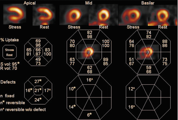
The major objective of stress MPI in symptomatic patients is assessment of prognosis, and to separate those at high risk for future cardiac events from those who have a good outcome because of normal or low risk scintigraphic findings [16, 17]. The latter group of patients can be spared unnecessary premature referral for cardiac catheterization. MPI variables that indicate high risk CAD include: (1) multiple perfusion or wall motion abnormalities in multiple myocardial segments; (2) defects in two or 3 coronary supply regions reflective of multivessel or left main CAD; (3) ≥10 % LV ischemia; (4) transient ischemic cavity dilation (TID) where the LV cavity is larger on stress than rest images although total cardiac size in unchanged; (5) a LV ejection fraction of ≤40 %; (6) greater wall motion abnormalities and/or a lower LVEF on stress compared to rest images suggestive of stress-induced myocardial stunning. TID is indicative of high risk CAD only when associated with a high probability of CAD indicated by concomitant presence of reversible defects, or presence of ischemic ST depression on exercise ECG testing. It has a low association with severe CAD in patients with otherwise normal SPECT studies [18]. Figure 11.2 shows an example of a high risk SPECT stress and rest MPI scan in a patient with an anteroseptal defect and TID from this study [18].


Fig. 11.2
Stress and Rest SPECT images showing apical, mid ventricular and basilar views in a patient with reversible defects in the apex and anteroseptal segments. Transient ischemic cavity dilation (TID) is observed. Below the images are quantitative count data with the stress values (% highest activity) depicted as the numbers on top, and the resting values on the bottom of each pair. Note that the regional counts are reduced to 63 % of the highest activity in the upper posterior wall and improves to 77 % of peak activity on the resting scan. This difference is more than 2 standard deviations (asterisk ) for the normal variation between stress and rest studies. (Abbreviations: Svol stress volume, Rvol rest volume) (From Mandour Ali et al. [18]. Reprinted with permission from Springer)
Prognosis of Patients with Normal and Abnormal MPI SPECT Studies
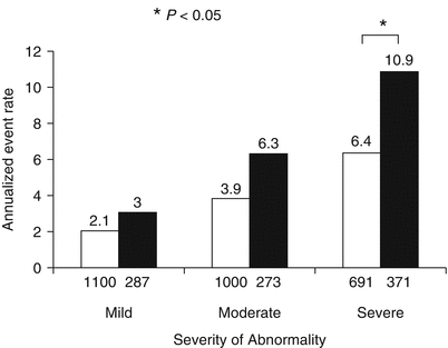
One of the most valuable clinical aspects of stress MPI studies in symptomatic patients is its excellent negative predictive value for predicting a low combined cardiac death or nonfatal myocardial infarction (MI) rate in patients with a normal gated SPECT result. In a pooled analysis of 29,173 patients with low risk SPECT MPI scans, the annual death or nonfatal MI rate was 0.6 % per year with an average follow-up of 2.3 years [17]. In the pooled analysis of 69,655 patients from 39 published studies in patients with either low risk or abnormal scans, those with a high-risk SPECT study had an annual cardiac death or MI rate of 5.9 % per year compared to 0.85 % event rate per year in those with a low risk SPECT study [17]. The average follow-up for this larger pooled analysis was 2.3 years. Patients with normal or abnormal pharmacologic SPECT studies have a higher annual cardiac event rate than patients with normal or abnormal exercise SPECT scans [19]. The annualized hard event rate was 6.4 % in patients with severe exercise SPECT abnormalities compared to 10.9 % for those with similar severity of SPECT MPI studies observed on pharmacologic SPECT studies (Fig. 11.3). This is most likely due to the higher clinical risk for cardiac events in patients deemed unable to exercise. Such patients are older, have more chronic pulmonary disease, a greater prevalence of prior stroke and more peripheral vascular disease. For both exercise and pharmacologic SPECT, the greater the extent and severity of perfusion abnormalities, the higher the subsequent cardiac event rate [20].


Fig. 11.3
Annualized cardiac death or infarction rates in patients with exercise (open bars) and pharmacologic (solid bars) stress according to the severity of perfusion abnormalities. The numbers of patients in each subgroup are shown beneath the bars (From Navare et al. [19]. Reprinted with permission from Mosby, Inc)
Functional variables added to perfusion imaging variables on gated SPECT MPI enhance the identification of high risk patients [21]. Those with a low LVEF have a greater event rate with mild to moderate ischemia than patients with normal LV function and similar perfusion defects [22]. Semiquantitation of SPECT perfusion abnormalities using indices such as the summed stress score (SSS) and summed difference score (SDS) provides a more objective assessment of risk than merely visual interpretation of scans. A 17-segment model has been accepted as the norm for this type of semi-quantitative analysis. Similarly, the extent of LV ischemia can be derived semiquantitatively from stress and rest studies. Patients manifesting ≥10 % LV ischemia appear to have a better outcome with revascularization than medical therapy [23]. Multivariate Cox proportional hazards model were employed to estimate CAD death or MI in 4,575 patients prospectively enrolled in the Myoview (Tc-99m-tetrofosmin) Prognosis Registry [24]. In this multivariable model, 34 % of the model chi-square was contributed by MPI ischemia. Adjustments were made for age, sex, presenting symptoms, stress type, CAD history and risk factors. In risk-adjusted models stress MPI ischemia, rest and post-stress LVEF were significant estimators of CAD death or MI. The net reclassification improvement (NRI) was 0.358 with MPI ischemia was added to a model with the Duke Treadmill Score and pre-test CAD likelihood. In contrast, the NRI was only 0.112 when the Duke Treadmill Score was added to the clinical pretest risk estimate. Thus, this analysis clearly shows that stress-induced ischemia provides independent prediction of CAD outcomes.
Hachamovitch and colleagues have reported from a large patient database, that patients who exhibit 10–15 % LV ischemia benefitted more from early revascularization than medical therapy, whereas patients with lesser amounts of inducible myocardial ischemia seemed to have similar outcomes with revascularization or medical therapy [25].
It is preferable to perform exercise stress as opposed to pharmacologic stress for MPI whenever possible. This is because important prognostic information can be gleaned from the exercise ECG results, particularly the workload achieved. Patients who can achieve 10 metabolic equivalents (METS) or greater have a markedly low prevalence of ≥10 % LV ischemia and have an excellent outcome regardless of scinitgraphic results [26–28]. The value of radionuclide imaging in such patients with excellent exercise tolerance is questionable. The results of the WOMEN study supports the notion that good functional capacity is associated with a good prognosis and that exercise MPI seems to offer little additional prognostic information over the standard exercise ECG stress test [29]. In this study, 824 women with good pretest functional capacity were randomized to exercise ECG testing alone versus exercise MPI. At follow-up, the major adverse cardiac event (MACE) rates were comparable in women with normal test results (0.4 % for exercise ECG versus 1.2 % for exercise MPI). In a more heterogenous group of 2,225 women, ages 55–75 years, patients with abnormal scans had significantly higher death rates compared with patients with normal scans (13.1 % versus 4.0 %, respectively) [30]. In this study, women with severe reversible defects reflective of extensive ischemia had a significantly higher adjusted all-cause mortality rate compared to women with normal scans and women with lesser degrees of SPECT ischemia.
Underestimation of Coronary Artery Disease with SPECT Myocardial Perfusion Imaging
It has been recognized for decades that stress SPECT utilizing the Tc-99m-labeled perfusion agents underestimates the extent of CAD compared to coronary angiographic findings. In the study by Lima et al. [21], only 25 % of patients with 3-vessel CAD had perfusion or wall motion abnormalities in the supply regions of the 3 major coronary arteries. Only slightly more than one-third of the patients with angiographic 3-vessel CAD had perfusion or functional abnormalities in 2 coronary territories. One striking finding was that 12 % of the patients with 3-vessel CAD had normal perfusion scans. This is most likely attributed to balanced ischemia where flow reserve is reduced to a comparable degree in all 3 coronary artery supply regions. This is attributed to the fact that only relative uptake of injected tracer is evaluated. To see a focal defect, at least one myocardial region has to have increased perfusion compared to other regions. If there is a homogeneous diffuse decrease in coronary flow reserve due to 3-vessel disease and balanced ischemia, uniform tracer uptake will be observed on SPECT images. Berman et al. reported [31] that among patients with ≥50 % stenosis of the left main coronary artery, 40 % had a low risk SPECT scan with <10 % of the myocardium rendered ischemic post-stress. The underestimation of extent of CAD using MPI is much less using PET since PET technology permits measurement of absolute blood flow with stress and rest and calculation of myocardial flow reserve. The detection rate of multivessel disease is enhanced when using quantitative PET MPI [32].
Value of Stress Perfusion Imaging in Special Populations
Certain populations of patients benefit more than others with respect to risk stratification using SPECT MPI technology. MPI is not indicated as the first test for risk assessment in asymptomatic subjects as part of primary prevention [33]. However, it may be beneficial in refining risk in asymptomatic subjects with high coronary artery calcium scores.
Patients with Diabetes
Diabetic patients have a substantially higher rate of cardiac events than nondiabetics. Multiple prior studies have shown the incremental prognostic value of SPECT MPI over clinical and stress ECG variables [17]. Diabetic patients with normal MPI scans have a higher annual rate of cardiac death or MI than nondiabetic patients with normal scans [17]. Diabetic patients who present with exertional dyspnea have a 13.2 % annual death or MI rate with an abnormal scan compared to 5.6 % in those presenting with angina and who had an abnormal scan [34]. In this study, asymptomatic diabetic patients with an abnormal scan had a 3.4 % annual event rate. The highest cardiac event rates are observed in diabetic women [17]. Diabetic women with a high risk scan have a >10 % annual cardiac death or nonfatal MI rate, which is almost twice the event rate seen in diabetic men with similar scan abnormalities. In this analysis, diabetic women with a normal SPECT MPI study had a 3 % annual event rate. Giri et al. [35] reported that diabetic women with SPECT ischemia in 2 or more coronary supply regions, had a 40 % death or MI rate after 3 years of follow-up compared to 20 % for diabetic men with similar perfusion defect patterns.
Totally asymptomatic Type 2 diabetics with no prior history of CAD and a normal resting ECG do not appear to benefit from a screening SPECT MPI study [36]. In this randomized study (DIAD Study), patients who had a routine adenosine SPECT MPI study did not have a better outcome at 5 years post-testing compared to those randomized to the usual care strategy. Nevertheless, those patients with moderate to large defects had a six-fold higher risk of death or nonfatal MI versus those with small defects or normal perfusion scans. Asymptomatic diabetic patients with high coronary calcium (CAC) scores may benefit from stress perfusion imaging, since a substantial number of such patients have silent ischemia [37]. Those with ≥5 % LV ischemia and high CAC were shown to have a higher cardiac event rate than patients with similar CAC scores but no ischemia on SPECT [37].
The Bypass Angioplasty Revascularization Investigation 2 Diabetes (BARI 2D) showed similar long-term outcomes for those randomized to revascularization versus just intensive medical therapy [38]. The BARI 2D Nuclear substudy examined 1-year stress SPECT MPI results in 1,505 patients undergoing vasodilator stress imaging 1 year after randomization [39]. Patients randomized to revascularization demonstrated fewer stress-induced perfusion abnormalities compared to the intensive medical therapy group. Patients randomized to medical therapy had more extensive ischemia compared to the revascularized cohort. At 1 year, more extensive and severe stress-induced ischemia was associated with a higher death or MI rate. As expected, those diabetic patients with ≥10 % LV ischemia at 1 year after randomization was had an 11.3 % combined death or MI rate at 5 years, compared to 6.8 % for patients with 1–4.9 % LV ischemia [39]. Thus, these studies indicate that presence and extent of ischemia on SPECT MPI in diabetic patients has significant prognostic implications. The goal of therapy is to eliminate ischemia with therapy, whether with early revascularization or with intensive medical therapy alone.
Patients with Chronic Renal Disease
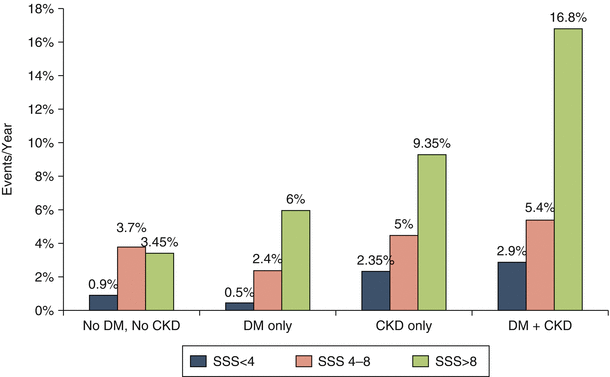
Patients with chronic kidney disease (CKD) are at particularly high risk for future ischemic coronary events [40]. The risk of extensive CAD without angina and cardiac mortality is substantial when the estimated glomerular filtration rate (eGFR) falls to <60. Patients on dialysis have a higher incidence of death due to CAD than from other causes. Several studies have shown that stress MPI may be valuable in cardiac risk assessment in patients with CKD. Hakeem et al. [41] reported a significant increase in cardiac death once the eGFR decreased to <60 and the summed stress score exceeded 8.0. Patients with an eGFR <60 and SPECT defect had an annual cardiac death rate of 9.5 % compared to 0.8 % in patients with an eGFR >60 and a normal SPECT MPI scan. In another study by the same group [38], the annual cardiac mortality was 16.8 % in CKD patients with a summed stress score of >8.0 and who also had diabetes. Figure 11.4 depicts this annual cardiac death rate on the basis of MPI findings and diabetes/chronic kidney disease status. Venkataraman et al. [42] showed that in patients on long-term hemodialysis, the larger the SPECT defect size on vasodilator MPI, the worse the prognosis. Risk stratification in these dialysis patients was better achieved with MPI than with coronary angiography. In this study, patients with no significant coronary artery stenosis on angiography had the same subsequent cardiac event rate as those with single and 2-vessel CAD. Although, the cardiac event rates are high in CKD patients with inducible ischemia, no randomized study has been performed in such patients to show that revascularization improves outcomes over medical therapy alone.


Fig. 11.4
Annual cardiac death rate versus increasing severity of perfusion defects quantified by the summed stress score (SSS) in patients with no diabetes (No DM) in patients with only diabetes (DM only), in patients with only chronic kidney disease (CKD only) and patients with the combination of diabetes and chronic kidney disease (DM + CKD). Note that patients in the DM = CKD group had the highest cardiac death rate for each level of perfusion abnormalities (From Hakeem et al. [41]. Reprinted with permission from Elsevier Science)
Elderly Patients
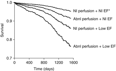
Coronary artery disease is the leading cause of death in the elderly and results in significant morbidity. SPECT MPI can adequately risk stratify elderly patients and is useful for prognostication and determining whether revascularization would be an effective approach to therapy compared to medical therapy alone. Nair et al. [43] performed a retrospective analysis from a database of 8,864 patients and examined the relationship of SPECT results and event rates in 1,093 patients ≥80 years of age. The very elderly patients with moderate or severe stress defects had a 9.6 % annual cardiac death or nonfatal infarction rate compared to 3.4 and 2.5 %, for those with mildly abnormal and normal scans, respectively. Across all categories of stress defect sizes, the very elderly had a higher cardiac event rate compared to younger patients. In another study by Hachamovitch et al. [44] in more than 5,000 patients ≥75 years of age, the cardiac death rate increased with worsening perfusion abnormalities. Figure 11.5 shows the risk-adjusted event-free survival in the elderly patients with normal or abnormal left ventricular ejection fraction and normal or abnormal scans in this study. The incremental value of SPECT MPI over other variables was also reported by the investigators.


