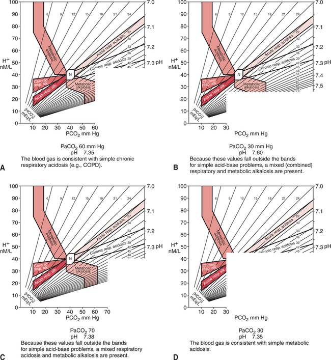
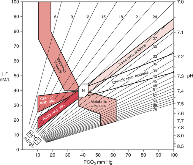
Mixed Acid-Base Disturbances and Treatment


Mixed Acid-Base Disturbances and Treatment


OVERVIEW


Three final aspects of clinical acid-base management are explored in this chapter. First, some of the major diseases and factors that tend to complicate the interpretation of clinical acid-base data are discussed. These factors include chronic lung disease, chronic renal disease, and therapeutic intervention. Blood gas and acid-base interpretation under these circumstances often requires special attention and skill.


Second, methods that can be used to differentiate simple (single) acid-base problems from mixed (complicated, multiple) acid-base disturbances are reviewed. In particular, the acid-base map and rules of thumb for compensation of simple acid-base disturbances are described. In addition, common settings and clues that may suggest a mixed acid-base disturbance are presented.


The final portion of this chapter deals with the supportive treatment of the four general acid-base disorders: respiratory acidosis, respiratory alkalosis, metabolic acidosis, and metabolic alkalosis. General guidelines are presented regarding the management of these generic disorders.


In addition, venous paradox during cardiopulmonary resuscitation or severe shock is described under metabolic acidosis. This phenomenon has important implica- tions regarding blood gas interpretation as well as the most appropriate use of NaHCO3 therapy.


FACTORS THAT MAY COMPLICATE CLINICAL ACID-BASE DATA


Respiratory/Renal Pathology


The primary organ systems involved in the maintenance of acid-base balance are the respiratory and renal systems. Disease, and in particular chronic disease, in either of these body systems can directly impair acid-base conditions or hamper the ability of the affected organ system to compensate for another acid-base disturbance. Thus, blood gas and acid-base interpretation in these chronic diseases requires special attention and understanding.


Chronic Obstructive Pulmonary Disease


Chronic obstructive pulmonary disease (COPD) is the classic example of a chronic respiratory disease. The typical acid-base picture in COPD is well known to most clinicians. Although respiratory alkalosis may possibly be seen at an early stage of the disease and in acute asthma, the characteristic picture in long-standing, severe, pulmonary disease is hypercapnia (e.g., PaCO2 > 50 mm Hg) with metabolic compensation (increased [BE], [HCO3]). Example 14-1 shows typical blood gases in long-standing COPD.


Example 14-1


The pH is often within the normal range (i.e., completely compensated) and may even be on the alkalotic side of the normal range.10 This finding is not consistent with rules that apply to compensation (i.e., overcompensation should not occur), however, and may be related to a mild concurrent primary metabolic alkalosis. The administration of steroids and diuretics with concomitant hypochloremia or hypokalemia is common in severe COPD.


Arterial blood gases are critical in the diagnosis and management of acute exacerbations of COPD. Nevertheless, blood gases in this group are often confusing and complex. Furthermore, they may be misleading if they are not clearly understood. Abnormal baseline values, unpredictable acute ventilatory changes, and the potential coexistence of lactic acidosis may all interact in a complex manner. The result may be misleading data when a single blood gas is considered in isolation. Some examples are given of how this result may occur.


Relative Hyperventilation

It is not uncommon for a patient with COPD to lower PaCO2 in response to acute hypo-xemia arising from an acute lung infection. Superimposing this acute change on the chronic (normal hypercapnic baseline) values shown in Example 14-1 results in blood gases approximating those shown in Example 14-2.


Similar results could occur if a patient with COPD were placed on a mechanical ventilator and ventilated to a PaCO2 of 40 mm Hg. Classification of this blood gas in isolation could result in the diagnosis of metabolic alkalosis. The underlying cause is, in fact, eucapnic (normal PaCO2) ventilation posthypercapnia.


Treatment for simple metabolic alkalemia here, however, would be inappropriate; optimal management requires an understanding of the disease process and the likely chain of events that have led to this point. It is probably more desirable to return this patient’s PaCO2 to their baseline level.


Relative Hyperventilation with Lactic Acidosis

Another important consideration in the patient with COPD is the potential for hypoxia and lactic acidosis. Individuals with COPD often have increased heart rates and elevated arterial blood pressure under chronic normal conditions to maintain adequate tissue oxygenation. In addition, right-sided heart failure is common in COPD secondary to increased pulmonary vascular resistance. When the acute stress of pneumonia and increasing hypoxemia is superimposed on an already compromised cardiovascular system, hypoxia may develop.


If lactic acidosis compounds the blood gas shown in Example 14-2, the result may appear as shown in Example 14-3. The net effect of these interactions is a relatively normal blood gas acid-base picture despite a severely compromised patient. Thus, serial blood gas measurements and other clinical findings are essential in understanding the significance of any isolated blood gas report.


Acute Hypercapnia

Many patients with severe COPD respond paradoxically to acute hypoxemia or oxygen therapy in that their PaCO2 increases instead of decreases. Reasons for this are unclear but are most likely related to worsening ventilation-perfusion mismatch and exhaustion secondary to the work of breathing. In addition, as described previously, excessive oxygen therapy may similarly precipitate acute hypercarbia. This affect has also recently been shown to occur during acute asthma exacerbations and the administration of FIO2 1.0.700 Excessive oxygen therapy may also be recognized by the concurrent presence of a PaO2 in excess of 60 mm Hg. When acute hypercapnia is superimposed on typical COPD chronic blood gases, the result may appear as shown in Example 14-4.


This particular blood gas picture is a common finding in acute exacerbation of COPD in the emergency department. The hallmark to recognition of this situation (acute exacerbation of COPD) is the surprisingly normal pH despite severe hypercapnia.


Patients with COPD who present with blood gas results such as those shown in Example 14-4 can often be treated successfully with low concentrations of oxygen therapy, noninvasive ventilation, and bronchial hygiene.543 544 Thus, mechanical ventilation, with related discomfort and the potential for complications, can often be avoided. Furthermore, a blood gas such as this may be the first clue that the patient has COPD. This finding, in turn, alerts the clinician to the potential for increasing hypercapnia with excessive oxygen therapy. Therefore, recognition of this situation may have great clinical importance.


Acute Hypercapnia with Lactic Acidosis

If lactic acidosis develops coincidentally with the acute hypoventilation shown in Example 14-4 (not an unlikely situation), COPD blood gases may appear as shown in Example 14-5.


When considered in isolation, this blood gas appears to show an acute hypoventilation (respiratory acidosis). Actually, this individual has two primary acid-base problems: respiratory acidosis and metabolic acidosis. Rather than immediately starting mechanical ventilation, a short, carefully controlled (and monitored) trial of oxygen therapy might mitigate gas exchange problems and obviate the need for mechanical ventilation. If this is unsuccessful, noninvasive ventilation should be attempted prior to full-blown mechanical ventilation.


Again, interpretation and treatment must be tailored to the specific case. These various examples have been provided to emphasize the complexity and the need for careful serial analysis of blood gases in COPD.


Therapeutic Intervention


Therapy given to a patient may sometimes distort the blood gas findings and may complicate the interpretation. The administration of diuretics, steroids, electrolytes, oxygen, bicarbonate, or mechanical ventilation may cause primary acid-base disturbances or may alter compensatory patterns. These factors must all be considered carefully during acid-base diagnosis, particularly in the critical care setting.


A specific area of application where blood gases may be measured frequently is during mechanical ventilation. The clinician must realize, however, that mechanical ventilation, by its very objective, controls at least a portion of ventilation in a set pattern. Therefore, compensation for metabolic acid-base disturbances cannot occur in exactly the same manner as it would in the patient who breathes spontaneously.


Example 14-6 shows blood gases that may be seen during mechanical ventilation in a patient with a metabolic acidosis. Note that this blood gas in isolation appears to be a completely compensated respiratory alkalosis. Actually, this patient has only a metabolic acidosis. Nevertheless, the rapid respiratory rate generated as a compensatory mechanism to the acidosis, in conjunction with the delivery of large tidal volumes via mechanical ventilation, has caused the apparent alkalosis. It would be inappropriate, however, to attempt to treat the respiratory alkalosis. The only true primary acid-base problem in this patient is metabolic acidosis. Mechanical ventilation has created the false impression of respiratory alkalosis.


If this same degree of metabolic acidosis developed in this individual during spontaneous breathing (i.e., not during mechanical ventilation), the blood gas picture might more closely resemble Example 14-7. Thus, the potential impact of respiratory assistance on the blood gas findings can be appreciated. The clinician must be mindful of the mode of mechanical ventilation and whether it may influence blood gas patterns.


The potential for mechanical ventilation to camouflage acid-base events has been described. Similarly, many of the other therapeutic measures mentioned earlier can lead to iatrogenic acid-base disturbances. Arterial blood gases and acid-base disturbances must always be interpreted within the context of therapeutic measures and long-standing pulmonary or renal disease.


MIXED ACID-BASE DISTURBANCES


Definition


The natural tendency of the body to compensate for primary acid-base disturbances was discussed in Chapter 8. Because of this natural phenomenon, whenever opposing respiratory and metabolic conditions were present, compensation was assumed. Although this initial assumption is logical, it may be incorrect. It is not uncommon to have two opposing primary acid- base disturbances that give the surface appearance of simple compensation. The coexistence of two primary acid-base disturbances is called a mixed acid-base disturbance.


Recognition of Mixed Disturbances


Acid-Base Map


How can simple compensation be differentiated from a mixed acid-base disturbance? Probably the most useful aid in this regard is the acid-base map that is shown in Figure 14-1. The labeled areas encompass with 95% confidence the range of pH, PaCO2, and bicarbonate that one would expect to find in patients who have only one simple acid-base disturbance. Separate bands are also given for both acute and chronic acid-base problems.


When a patient’s values fall outside these bands, it is very unlikely that the patient has just one disturbance. On the other hand, when a patient’s values fall within one of these bands it does not ensure that the patient has a single acid-base disturbance, it simply means that the data are compatible with this conclusion.


Figure 14-2 shows how the acid-base map can be used by simply aligning the two adjacent sides of a piece of paper with the patient’s respective PaCO2 (horizontal axis) and pH (vertical axis). The corner point of the paper represents where the values intersect on the map. The data in Figure 14-2,A are consistent with chronic simple respiratory acidosis. Remember, this does not mean that the elevated bicarbonate cannot be due to a primary problem, but only that the data are consistent with usual compensation for chronic respiratory acidosis.


Figure 14-2,B does not fall within any band, therefore the clinician can be relatively certain that there are two separate, primary, acid-base disturbances (respiratory alkalosis and metabolic alkalosis). Figure 14-2,C similarly represents two primary acid-base disturbances, although in this case they are in opposite directions (i.e., respiratory acidosis and metabolic alkalosis). Without an acid-base map, one might assume that these blood gas results are due to complete compensation. Finally, Figure 14-2,D is consistent with a simple metabolic acidosis.


The acid-base map is a simple, useful tool for the evaluation of mixed acid-base disturbances. Pocket versions of this map are available for use at the bedside.


Compensatory Patterns


The degree of compensation observed in the four simple primary acid-base disturbances, although quite similar, is not identical. For reasons that are unclear, some types of acid-base problems (e.g., metabolic alkalosis) result in more complete compensation than others. Nevertheless, in the absence of an acid-base map, it is useful to have some idea of the typical compensatory patterns that should accompany the four simple acid-base disturbances. This can help the clinician to evaluate the appropriateness of the degree of compensation observed in a given individual.


Respiratory Acidosis
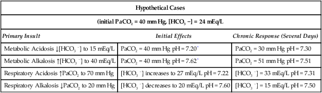
The pH falls approximately 0.06 unit for an acute 10-mm Hg increase in PaCO2. After maximal renal compensation, the change in pH associated with an increase of 10 mm Hg in PaCO2 is approximately 0.03 unit. Thus, the pH returns approximately 50% of the way back toward normal after maximal compensation.


Table 14-1 shows the typical compensatory response to respiratory acidosis. This table shows that when the PaCO2 increases to 70 mm Hg acutely, the pH drops immediately to approximately 7.22 (0.06 decrease/10 mm Hg PaCO2 increase). The immediate increase in bicarbonate is a result of the hydrolysis effect that was discussed in Chapter 8 and it does not represent renal compensation.


After maximal renal compensation (several days later), however, the pH returns approximately halfway back to normal (i.e., 7.31). Thus, complete compensation (i.e., pH in normal range) is not usually seen when respiratory acidosis is quite severe. Complete compensation for respiratory acidosis occurs only when the respiratory acidosis is not severe (i.e., PaCO2 < 60 mm Hg). In addition, because the mechanism of renal compensation for respiratory acidosis is bicarbonate retention, the chloride anion is typically low to preserve electroneutrality.


Respiratory Alkalosis

Compensation for respiratory alkalosis is similar in magnitude to compensation for respiratory acidosis. In general, the pH should return at least halfway back toward normal. Again, an example is shown in Table 14-1. Surprisingly, however, when the respiratory alkalosis persists for weeks, the pH may actually return completely to normal in some cases.515 Renal compensation for respiratory alkalosis requires the excretion of bicarbonate; therefore, hyperchloremia often develops to preserve electroneutrality.


Metabolic Acidosis

The major portion of the ventilatory response to metabolic acidosis usually begins quickly; however, the maximal compensatory response may take up to 24 hours.545 546 When metabolic acidosis develops in the plasma, it takes some time for the pH to fall in the cerebrospinal fluid owing to the limited permeability of ions across the blood-brain barrier. Lactic acidosis, however, may actually develop within the brain cells, and it is therefore associated with a more rapid ventilatory response.515


A very useful rule of thumb when an acid-base map is not at hand is that after maximal compensation, the PaCO2 generally approximates the last two digits of the pH.515 Thus, in simple chronic metabolic acidosis with a pH of 7.30, the PaCO2 is usually approximately 30 mm Hg (see Table 14-1).


Alerts to Mixed Disturbances


Mixed acid-base disturbances are far from uncommon in the hospital setting. When primary acid-base problems are camouflaged in mixed disturbances, they may easily be missed. In this setting, covert acid-base problems are untreated and are likely to lead to progressive deterioration. Furthermore, even use of the acid-base map does not identify those mixed disturbances that result in blood gas data that coincide with findings that normally accompany simple disturbances.


Therefore, the clinician must look for clues that suggest the presence of multiple (mixed) acid-base disorders. Box 14-1 suggests some situations that should alert the clinician to the likelihood of a mixed acid-base disturbance.


< div class='tao-gold-member'>

Only gold members can continue reading. Log In or Register to continue

Stay updated, free articles. Join our Telegram channel

Jul 10, 2016 | Posted by in RESPIRATORY | Comments Off on Mixed Acid-Base Disturbances and Treatment

Full access? Get Clinical Tree

Get Clinical Tree app for offline access